In selected neonates with hypoplastic left heart syndrome considered at high surgical risk, the staged hybrid procedure, consisting of banding of the right and left pulmonary arteries, stenting of the arterial duct, and, when necessary, either balloon dilation or placement of a stent in the atrial septal defect, has been demonstrated to be a safe and less invasive alternative.Reference Akintuerk, Michel-Behnke and Valeske1, Reference Galantowicz and Cheatham2 While there is relatively little experience with placement of stents to relieve restrictive interatrial communications, a recent study reported no major complications.Reference Rupp, Michel-Behnke, Valeske, Akintürk and Schranz3 We have now encountered recurrent and progressive atrioventricular block as a complication of stenting the atrial septum. Normal atrioventricular conduction was restored following removal of the stent and surgical septectomy.
Case report
A neonate weighing 2.8 kilograms, in whom hypoplastic left heart syndrome had been diagnosed prenatally, developed necrotising enterocolitis on the third day of life while awaiting the planned first stage of surgical palliation following the Norwood protocol. In view of this complication, it was decided to undertake a hybrid approach, as pioneered in Giessen.Reference Akintuerk, Michel-Behnke and Valeske1 Accordingly, on the 11th day of life, we performed banding of the right and left pulmonary arteries to 3 millimetres in diameter on each side, followed on the 16th day by transcatheter placement of a stent of 7 millimetres diameter in the arterial duct.
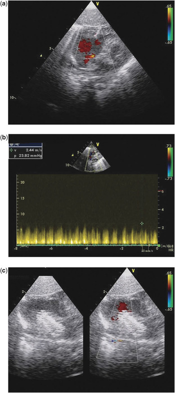
At serial follow-up thereafter, the patient developed progressive systemic desaturation, with saturations of oxygen measured transcutaneously dropping intermittently to values of less than 75%. We deemed this to be due to progressive restriction of flow through the interatrial communication. This had been unrestrictive directly after birth, but a progressive increase in the transseptal gradient was noted during serial echocardiographic follow-up (Fig. 1a, b). At 35 days of age, when we determined that the interatrial defect needed to be enlarged, we chose to implant a stent rather than opting for balloon septostomy, believing that stenting would provide a more predictable result. Accordingly, we implanted precutaneously a Genesis stent, 19 millimetres long and premounted on a balloon of 10 millimetres diameter, to create an unrestrictive interatrial communication (Fig. 1c). The procedure was uncomplicated, and resulted in an immediate increase in systemic saturations of oxygen to 85%.
Colour flow Doppler interrogation from subcostally at 30 days of age (a) shows a restrictive interatrial communication. Continuous wave Doppler interrogation of the interatrial septum (b) confirms a peak instantaneous pressure gradient of more than 20 millimetres mercury. As shown in Figure 1c, a stent was been positioned across the atrial septal defect, and expanded to a diabolo shape to ensure stability. The left panel shows the stent, while the right panel, with colour flow, demonstrates unobstructed flow into the right atrium.

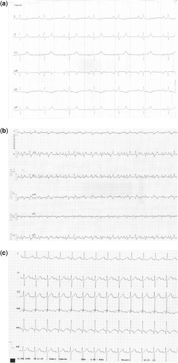
Within six hours following implantation of the stent, the patient developed second degree atrioventricular block, with constant atrioventricular conduction at a rate of 2 to 1 (Fig. 2a). This was managed with intravenous infusions of isoproterenol, atropine, and a course of intravenous hydrocortisione. The stent appeared to be stable, and far away from the atrioventricular junction. Normal atrioventricular conduction returned within 24 hours.
The surface electrocardiogram six hours following implantation of the stent (a) demonstrates 2 to 1 atrioventricular conduction. Four days later (b), the surface electrocardiogram shows complete atrioventricular dissociation. Following surgical removal of the stent (c), the electrocardiogram shows return to 1 to 1 atrioventricular conduction.

Four days later, the patient suddenly developed complete atrioventricular dissociation, which was associated with low cardiac output and metabolic acidosis (Fig. 2b). This did not respond to pharmacologic therapy. The patient was, therefore, temporarily paced using a transvenous lead implanted via the femoral vein into the systemic ventricle. The following day, the atrial septum was resected under cardiopulmonary bypass, and the stent removed. The stent was not in proximity to the left atrial wall, nor to any coronary arterial branch. Although normal atrioventricular conduction returned immediately following surgery, temporary pacing wires were inserted, and left in place for five days (Fig. 2c). The subsequent postoperative course was uncomplicated.
Discussion
It is important to establish an unrestrictive interatrial communication as part of the surgical or hybrid palliation of patients with hypoplastic left heart syndrome. Beyond one month of age, standard techniques, such as balloon septostomy or balloon dilation of the atrial septum, may be suboptimal. For selected patients, stenting of the atrial septum has been recommended as an alternative approach.Reference Rupp, Michel-Behnke, Valeske, Akintürk and Schranz3–Reference Daehnert, Hennig, Walther and Kostelka5 The mechanism by which atrioventricular block developed in our patient is unclear. The event was related temporally to implantation of the stent within the oval fossa, and resolved after removal of the stent and surgical septectomy. We postulate that implantation of a rigid stent across a thickened septum, with pushing away of septal tissue, may have produced pressure on the atrioventricular node and the atrioventricular conduction axis. That this was not due to transient oedema may be surmised from the observation that the atrioventricular block resolved initially, but recurred four days later in a more severe form. Direct observation of the atrial septum and the stent at surgery confirmed that the stent was indeed far away from the atrioventricular junction. Intuitively, it is difficult to presume that the stent had a direct effect on the atrioventricular conduction system, yet the patient did develop atrioventricular dissociation. It is important, therefore, to be aware of this potential complication when considering implantation of stents to relieve restrictive interatrial communications in young infants.