Globally, approximately 357 000 individuals are diagnosed with bladder carcinoma each year, with about 145 000 people dying from the disease (Ref. Reference Parkin1). The most common histological type of bladder cancer in Western countries has been termed urothelial carcinoma (UC) by the World Health Organization 2004 classification of bladder tumours (Ref. Reference Montironi and Lopez-Beltran2); it was formerly known as transitional cell carcinoma. Squamous cell carcinoma (SCC) of the bladder is also common worldwide, and other less common tumours include adenocarcinoma and small-cell carcinoma. It is estimated that there were approximately 71 000 new cases of UC and 14 300 deaths in the USA in 2009 as a result of the disease (Ref. Reference Jemal3). There are multiple risk factors for UC, which include exposure to tobacco and industrial chemicals, ingestion of arsenic-laced water, radiation therapy to organs adjacent to the bladder, therapeutic use of alkylating agents in chemotherapy regimens, and infection with the trematode Schistosoma haematobium. The latter risk factor is closely associated with SCC of the bladder (Ref. Reference Sonny and Samuel4).
Approximately 80% of patients with UC present with non-muscle-invasive urothelial carcinoma (NMIUC; cancer stages Ta, Tis and T1), which can be successfully treated with local endoscopic resection, although a significant number of these cases exhibit recurrence with or without progression to invasive disease (Refs Reference Kamat and Lamm5, Reference Pasin6). The remainder of patients present with muscle-invasive urothelial carcinoma (MIUC; cancer stages T2–4), which is commonly treated with a radical cystectomy or chemoradiotherapy. Despite aggressive treatment, approximately 50% of these MIUC patients develop subsequent metastatic disease, with most succumbing within two years of diagnosis (Refs Reference Sengelov, Kamby and von der Maase7, Reference Steinberg, Trump and Cummings8, Reference Liebert and Seigne9). Each tumour stage within the NMIUC and MIUC subtypes can be associated with a grade (G1, G2, G3) that corresponds to cellular differentiation of the tumour cells in question. G1 tumours are morphologically well differentiated, whereas G3 tumours are morphologically poorly differentiated. G3 tumours are more aggressive and have a greater tendency to progress to a higher stage cancer.
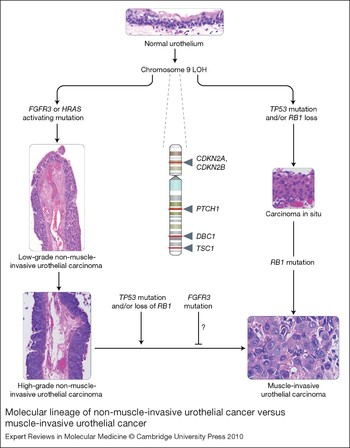
Evidence suggests that NMIUCs have a different aetiology to that of their invasive counterparts (Refs Reference Koss10, Reference Wu11, Reference Bakkar12, Reference Lamy13, Reference Hernandez14, Reference Karsten15). NMIUCs are thought to arise from localised urothelial hyperplasia, while MIUCs are thought to arise de novo from high-grade carcinoma in situ (CIS) lesions without necessarily having antecedent urothelial hyperplasia. These two types of tumours have a very different set of genetic lesions, which include activation of the intracellular GTPase HRAS and the receptor FGFR3 (fibroblast growth factor receptor 3) in the noninvasive subtype, and defects in the p53 (TP53) and RB (RB1; retinoblastoma 1) tumour suppressor pathways in the invasive subtype (Fig. 1). Loss of heterozygosity (LOH) of chromosome 9 is frequently found in both tumour types (Fig. 1). In this review, the genetic alterations that correspond to the noninvasive subtype of UCs are discussed with an emphasis on their functional relevance to pathogenesis, their value in assessment of prognosis, and their promise as therapeutic targets. For common genetic alterations in MIUC see the excellent recent review by Wu (Ref. Reference Wu11).
Molecular lineage of non-muscle-invasive urothelial cancer versus muscle-invasive urothelial cancer. The figure summarises the major molecular alterations that are responsible for the onset of non-muscle-invasive urothelial carcinomas (NMIUCs) and muscle invasive urothelial carcinomas (MIUCs), along with providing representative haematoxylin and eosin micrographs of these disease states. Starting from a normal urothelium five to seven cells thick (top), loss of heterozygosity (LOH) of chromosome 9 (arrows indicate relative sites of potential tumour suppressor gene loss; names of genes are given) has been associated with the majority of urothelial cancers. This is often coincident with a mutation in either FGFR3 or HRAS, which have been implicated in the genesis of NMIUCs (low- and high-grade papillary NMIUCs indicated on the left show examples of the increasing degree of architectural disorder and increasing cytologic atypia). FGFR3 and HRAS mutations are usually not present within the same cancer. By contrast, from a molecular standpoint, the MIUC class of tumours is defined by mutations in the tumour suppressor genes TP53 and RB1, which frequently coincide in the same tumours. Generally, MIUCs are thought to arise de novo from carcinoma in situ (shown on the right). Histologically, marked cytologic atypia (high ratio of nuclei to cytoplasm, mitoses, and total loss of tissue architecture) are present (bottom right) in a context of observation of definitive invasion of the muscularis propria of the bladder, in contrast to NMIUCs. As a class, NMIUCs have a high tendency to recur and rarely progress to a higher-grade carcinoma or MIUC. When this progression does occur, it is usually coincident with loss of function of the tumour suppressors TP53 or RB1. Some studies have shown that the presence of activating FGFR3 mutations correlate to a reduced frequency of progression to a higher-grade/stage cancer, but these events have not been definitively shown to be causal. CDKN2A, cyclin-dependent kinase inhibitor 2A (also known as P16); CDKN2B, cyclin-dependent kinase inhibitor 2B (also known as P15); DBC1, deleted in bladder cancer 1; FGFR3, fibroblast growth factor receptor 3; HRAS, Harvey rat sarcoma viral gene homologue; PTCH1, patched homologue 1; RB1, retinoblastoma tumour suppressor; TP53, p53 tumour suppressor; TSC1, tuberous sclerosis 1 gene. [Images: S.C. Smith. Originally photographed at 40× (NMIUCs) and 60× (MIUC, carcinoma in situ and urothelium)].

Alterations on chromosome 9
LOH is common in cancer. In NMIUC, LOH of chromosome 9 is present at the earliest stages of development and is the most frequent chromosomal deletion. One of the first studies noting this phenomenon identified that a large number of human bladder cancer specimens contained allelic loss of chromosome 9q independent of tumour grade (Ref. Reference Tsai16). Subsequently, multiple groups sought to identify the specific regions of mutation on chromosome 9. After analysing 252 bladder specimens, one group identified that 50% of Ta tumours contained LOH on either 9p or 9q and speculated that these regions were important in early UC development and likely to harbour tumour suppressor gene loci (Ref. Reference Cairns, Shaw and Knowles17). Later studies that employed restriction fragment length polymorphism (RFLP) mapping and microsatellite analysis of chromosome 9 implicated the region between 9p12 and 9q34.1 as potential sites for tumour suppressor genes (Refs Reference Miyao18, Reference Llnnenbach19, Reference Orlow20, Reference Habuchi21, Reference Keen and Knowles22, Reference Simoneau23). Finer mapping of chromosome 9 led to the identification of specific tumour suppressor genes lost as a result of these chromosomal changes.
Chromosome 9p
The 9p chromosomal arm contains the best-characterised genetic lesions of bladder cancer, and its importance in the molecular pathogenesis of the disease is supported by similar lesions occurring in both UC and SCC despite these forms being strongly associated with different environmental risk factors (Ref. Reference Sonny and Samuel4). Among the most common lesions are those in the gene encoding P15 (CDKN2B; also known as INK4B and MTS2) and P16 (CDKN2A; also known as INK4A and MTS1). Both P15 and P16 are important potential tumour suppressors because of their respective roles as negative regulators of the cell cycle: they inhibit the activity of the complex of cyclin D and cyclin-dependent kinase 4 (CDK4) to stop cell proliferation at the G1 phase of the cell cycle (Refs Reference Serrano, Hannon and Beach24, Reference Hannon and Beach25). Mechanistically, this function is important because the CDK4 complex has been shown to catalyse the phosphorylation of RB; this in turn releases the transcription factor E2F, which is involved in regulating transcriptional targets essential for the transition from the G1 to S phase of the cell cycle (Refs Reference Schulze26, Reference Johnson27).
In the mid 1990s, several independent studies identified alterations in CDKN2A and CDKN2B in UC. A comparative multiplex polymerase chain reaction (PCR) approach revealed homozygous deletions and sequence mutations in CDKN2A in SCC that were present at three times the frequency seen in UC (Ref. Reference Gonzalez-Zulueta28). In addition, hemizygous and homozygous deletions were shown to be present in 92% of SCCs on chromosome 9p but in only 39% of UCs, and deletions in 9p without concomitant mutation in 9q were found at a rate of 92%, compared with only 10% of UCs having this pattern. Around the same time that the association of CDKN2A with SCC was being established, another group noted significant de novo methylation of the 5′ CpG islands at the CDKN2A locus in 67% of a cohort of 18 UC tissue samples, and suggested that this methylation might be responsible for inactivating the gene (Ref. Reference Gonzalez-Zulueta29). In addition, statistically significant frequencies of homozygous and hemizygous deletions in both CDKN2A and CDKN2B were reported for a cohort of patients with UC (Ref. Reference Orlow30). Homozygous deletions in both these genes were also found in a cohort of 13 human bladder cancer cell lines (Ref. Reference Southgate31), providing support for the clinical relevance of these lines in laboratory experiments as models of UC. Finally, CDKN2B was found to have decreased mRNA expression levels in NMIUC (Ref. Reference Le Frere-Belda32). Taken together, these findings implicate the 9p21 locus, including the CDKN2A and CDKN2B tumour suppressor genes, in both SCC and UC, albeit with different frequencies of alteration in each histological subtype.
Chromosome 9q
Identification of candidate tumour suppressor genes on 9q initially proved difficult because of a lack of high-frequency subchromosomal deletions on this arm. The first indication of chromosome 9q as a possible site of tumour suppressor gene inactivation in UC was provided when a common partial deletion was found on this chromosome in a small portion of cases (5 of 70 cases) (Ref. Reference Habuchi21). It was later shown using single-strand conformation polymorphism analysis (SSCP) that there are multiple nonsense and missense mutations in 9q34, a region that encompasses the TSC1 (tuberous sclerosis 1) gene and is frequently deleted (Ref. Reference Hornigold33). TSC1 has been shown to be silenced in cases of tuberous sclerosis harmatomas and is speculated to act as a tumour suppressor (Refs Reference Hornigold34, Reference van Slegtenhorst35). When the mutations found on chromosome 9q were analysed, 90% were found to be novel and specific to bladder cancer in that they were not found in previous germline tuberous sclerosis cases (Ref. Reference Knowles36). It was later shown that missense mutations in the TSC1 gene induced mislocalisation of the TCS1 protein in bladder cancer cells (Ref. Reference Pymar37). This study also identified that when these same missense mutations were present in tuberous sclerosis, which was infrequent, they played no role in causation of tuberous sclerosis.
LOH at chromosome 9q34 was associated with decreased expression of P27, a CDK inhibitor protein encoded by CDKN1B, in a cohort of four tumours, suggesting that there might be a relationship between loss-of-function mutations in TSC1 and CDKN1B in UC (Ref. Reference Adachi38). In addition to TSC1, the 9q32–q33 locus was identified as having frequent partial deletions in UC (Ref. Reference Habuchi, Yoshida and Knowles39). This region was later found to contain a potential tumour suppressor gene, DBC1 (‘deleted in bladder cancer gene 1’; originally named DBCCR1), by searching for expressed sequence tags (ESTs) that map to yeast artificial chromosomes (YACs) spanning the region (Ref. Reference Habuchi40). Expression of DBC1 mRNA was not detected in 50% (5 of 10) of bladder cancer cell lines evaluated. It was also found that the DBC1 gene is a frequent target for hypermethylation, suggesting that hypermethylation at this locus could be important in the early development of bladder tumour formation even in cases where deletion does not occur (Ref. Reference Habuchi40).
An initial report of frequent deletion mutations on chromosome 9q in the region spanning q22.3–q31 (Ref. Reference Simoneau23) was later validated using microsatellite analysis following a primer-extension preamplification technique (Ref. Reference Simoneau41). Specifically, the 9q22.3 locus was found to harbour deletions in the region encompassing PTCH1 (patched homologue 1) and MSSE (multiple self-healing squamous epithelioma) genes, both of which are known to play important roles in diseases characterised by the development of skin tumours. These findings suggest that mutations in the q22.3–q31 region of chromosome 9 are important in UC development.
Prognostic significance of LOH on chromosome 9 for recurrence following NMIUC
As the tumour suppressor role of chromosome 9 in NMIUC became more established, a question arose of whether LOH on chromosome 9 could be used as a prognostic factor regarding the disease course of patients with NMIUC. Comparison of recurrent UCs with nonrecurrent UCs found that 54% of recurrent versus 12% of nonrecurrent tissue samples harboured LOH on chromosome 9 (Ref. Reference Joanne42). In this study, microsatellite markers for regions on chromosome 9p and 9q that encompass putative tumour suppressor genes CDKN2A, DBC1 and TSC1 were analysed. A significant association between risk of recurrence and LOH at the region encompassing TSC1 was noted, whereas LOH at CDKN2A or DBC1 did not show any significant relation to tumour recurrence. In addition, a later study showed that LOH of the region of chromosome 9p21 encompassing CDKN2A was not significantly associated with recurrence-free interval in NMIUC (Ref. Reference Friedrich43). In contrast, another group, which utilised PCR analysis, identified that 33 of 56 (58.9%) patients with low-grade (G1–G2) UC had loss of CDKN2A at the 9p21 locus, and that loss was significantly associated with reduced recurrence-free probability (Ref. Reference Bartoletti44). Furthermore, a separate study noted that 12 of 33 (36%) patients who had recurrent UC demonstrated LOH on chromosome 9q12 while only one patient with nonrecurrent UC showed LOH of chromosome 9 (Ref. Reference Bartlett45). Additional support for the positive correlation between LOH of chromosome 9 and tumour recurrence included a recent study that showed LOH at the DCB1 locus (9q32–q33) was correlated positively with tumour recurrence of NMIUC only if the LOH was located in the normal urothelium adjacent to the tumour and not in the tumour itself. This suggests that when LOH occurs at the DBC1 locus in the NMIUC itself the recurrence of that tumour is less likely (Ref. Reference Lopez-Beltran46). In this study, it was shown that histologically normal urothelium with LOH at the DCB1 locus was associated with larger tumours than in cases without this anomaly.
In summary, the body of data regarding chromosome 9 seems to suggest that LOH at chromosome 9q has more prognostic value than LOH at chromosome 9p, although different studies employing varying technologies have made sometimes disparate findings. The presence of candidate tumour suppressor genes on each of these chromosome regions warrants further investigation into their potential use as prognostic factors for tumour recurrence following an initial diagnosis of NMIUC, and we suggest that such studies employ standardised technologies for detection of genetic lesions as well as large interinstitutional cohorts to favour findings with general applicability.
Fibroblast growth factor receptor 3
Mutation and overexpression of FGFR3 in urothelial cancer
One of the most compelling findings in recent years regarding the molecular pathogenesis of UC has been the role discovered for FGFR3. FGFR3 belongs to a five-member family of structurally related tyrosine kinase receptors, where each protein in the family is encoded by a different gene. The basic structure of the FGFR family is a glycoprotein that contains three immunoglobulin (Ig)-like domains (extracellular ligand-binding domain), a transmembrane helix domain, and an intracellular tyrosine kinase domain (Refs Reference Eswarakumar, Lax and Schlessinger47, Reference Karuppiah and David48, Reference L'Hôte and Knowles49). FGFR3 is known to have two major splice isoforms, FGFR3b and FGFR3c, with the former being most prevalent in epithelial tissue and the latter being associated with mesenchymal tissue (Ref. Reference Murgue50). FGFR3 first became relevant to human disease when specific point mutations in different domains of the FGFR3 gene were discovered in various autosomal dominant human skeletal diseases including achondroplasia, hypochondroplasia, thanatophoric dysplasia I and II, and severe achondroplaisa with developmental delay and acanthosis nigrans (SADDAN) (Refs Reference Rousseau51, Reference Shiang52, Reference Bellus53, Reference Superti-Furga54, Reference Ikegawa55, Reference Tavormina56, Reference Tavormina57, Reference Fuu-Jen58, Reference Bonaventure59).
Interestingly, the mutations found to be responsible for the aforementioned debilitating autosomal dominant human skeletal diseases were found to be prevalent in human bladder cancer. In 1999, somatic missense mutations (R248C, S249C, G370/372C, K650/652E) were reported in 35% (9 of 26) of UCs and 25% (3 of 12) of cervical carcinomas examined (Ref. Reference Cappellen60). All mutations identified in the UC and cervical carcinoma samples were identical to the mutations found in the autosomal dominant human skeletal disease thanatophoric dysplasia. It was later shown that the mutations in FGFR3 that correspond to the mutations in the autosomal dominant human skeletal diseases occur at a much higher rate in UCs than in cervical carcinomas and multiple myeloma and are virtually nonexistent in carcinomas of the upper aerodigestive tract, oesophagus, stomach, lung, rectum, colon, prostate, ovarian, breast, brain or kidneys (Refs Reference Karoui61, Reference Sibley, Stern and Knowles62).
Interestingly, the R248C and K650/652E mutations had previously been implicated in constitutive activation of FGFR3 (Refs Reference Webster and Donoghue63, Reference Naski64), thus hinting at a mechanism of the oncogenic role these mutations might play in bladder cancer. The R248C mutation has been shown to constitutively activate FGFR3 through exposure of a cysteine residue that can participate in an intermolecular disulfide bond, resulting in ligand-independent dimerisation of the receptor (Ref. Reference Naski64). The K650/652E mutation is thought to constitutively activate the FGFR3 receptor by altering the conformation of the activation loop in the kinase domain of the receptor (Refs Reference Webster and Donoghue63, Reference Naski64). It was later shown that the oncogenic role of mutated FGFR3 might be working through the Ras–MAPK/ERK (mitogen-activated protein kinase or extracellular-signal-regulated kinase) pathway. Constitutively active FGFR3 was able to transform NIH 3T3 cells with corresponding activation of the Ras–MAPK and phosphoinositide 3-kinase (PI3K) signalling pathways (Ref. Reference Agazie65); in addition, FGFR3 was shown to interact with the Ras-coupling molecules SHC1, GRB2, 80K-H (PRKCSH), SOS1 and an unidentified 66 kDa protein to modulate Ras signalling through RAF1 (Ref. Reference Kanai66). In fact, a recent report also indirectly hinted at the importance of the Ras pathway downstream of FGFR3 by finding that Ras mutations, also common in NMIUC (see section below), and mutations in FGFR3 are mutually exclusive in this tumour type, suggestive of functional equivalence (Ref. Reference Jebar67). Taken together, these findings implicate FGFR3 as an oncogene in UC that likely acts through the Ras–MAPK signalling pathway.
The association of FGFR3 alterations and tumour stage in urothelial carcinoma
The discovery of somatic missense mutations in the FGFR3 gene in human bladder cancer spawned the question of where in the pathogenesis of UC were these mutations relevant. One study found that 34 of 53 TaG1–2 bladder cancer samples had somatic missense mutations leading to amino acid changes that were identical to those found in the autosomal dominant skeletal diseases (Ref. Reference van Rhijn68). Furthermore, none of the 19 higher-stage tumours had such mutations. Another study found that the frequency of mutations was significantly higher in Ta tumours (74%; 37 of 50) than Tis (0%; 0 of 20), T1 (21%; 4 of 19) and T2–4 (16%; 7 of 43) (Ref. Reference Billerey69). Furthermore, when stratifying by grade, FGFR3 mutations were more prevalent in G1 (84%; 27 of 32) tumours than G2 (55%; 16 of 29) or G3 (7%; 5 of 71) tumours.
These differences in expression of mutant FGFR3 highlight an important point concerning NMIUCs. Multiple studies have shown that Ta tumours tend to exhibit a different natural history and clinical behaviour than Tis or T1 tumours: in particular, the Ta group, especially low-grade Ta, have very low rates of progression to higher-stage cancers (Refs Reference Spruck70, Reference Reznikoff71, Reference Lee and Droller72). In tandem, genomic and transcriptomic studies of the Ta and Tis/T1 tumours suggest they have contrasting gene expression profiles (Refs Reference Sanchez-Carbayo73, Reference Sanchez-Carbayo74, Reference Dyrskjøt75). Taken together these findings support the assumption that the Ta tumours have a distinct aetiology that is separate from that of the higher-stage urothelial tumours, even when considering tumours that are technically non-muscle-invasive (i.e. Tis, T1).
Similar to the previous study that showed a higher frequency of FGFR3 mutation in noninvasive tumours, another group showed that in a cohort of 81 patients there was an inverse correlation between FGFR3 mutations and grade/stage of bladder cancer (Ref. Reference Takahiro76). A study that looked at FGFR3 mutations in urothelial papillomas, considered the least aggressive of urothelial tumours, found that 75% (9 of 12) of urothelial papillomas evaluated harboured FGFR3 mutations (Ref. Reference van Rhijn77). This frequency was similar to the frequency of FGFR3 mutations in papillary urothelial neoplasms of low malignant potential (85%) and low-grade papillary UCs (88%). Thus, several studies validate that the FGFR3 mutation is important in the pathogenesis NMIUC, but also suggest that the FGFR3 mutation is not essential or is possibly inversely associated with progression to high-stage/grade bladder cancer.
FGFR3 and p53: divergent oncogenic pathways
As FGFR3 somatic mutations are the most common somatic mutation in NMIUC while TP53 and RB1 mutations were noted to be frequent in MIUC (Refs Reference Spruck70, Reference Cordon-Cardo78), the idea of a dual-track pathway implicating alterations in FGFR3 in NMIUC versus TP53 in MIUC arose (Ref. Reference Spiess and Czerniak79). When the mutational status of FGFR3 and TP53 in a cohort of 81 newly diagnosed UCs was evaluated, a strong association of FGFR3 mutations with low-stage/grade cancers was found while mutation in TP53 was almost exclusively associated with high-stage/grade bladder cancer (Ref. Reference Bakkar12). In a separate study that examined DNA from 260 UC tissue samples, only 5.7% had concomitant mutations in FGFR3 and overexpression of TP53 (which is generally presumed to reflect missense mutation of the TP53 gene conferring supraphysiological protein stability), while 72.7% of the samples were positive for either FGFR3 or TP53 individually (Ref. Reference van Rhijn80). Furthermore, mutations in FGFR3 and TP53 overexpression were found to be dependent on stage and grade. Mutations in FGFR3 were present in 77% of Ta, 31% of T1, and 15% of ≥T2 tumours; in relation to grade, mutations were found in 85% of urothelial neoplasia of low malignant potential, and in 71% of low-grade and 26% of high-grade cases. By contrast, overexpression of TP53 was present in 11% of Ta, 51% of T1, and 56% of ≥T2 tumours; in relation to grade, overexpression was present in 1% of LMP, and in 15% of low-grade and 54% of high-grade cases. This evidence further suggested that FGFR3 and TP53 are on two separate tracks in UC. Other work has also found an inverse relationship between mutations in FGFR3 and bladder tumour stage/grade, and a positive correlation between mutations in TP53 and bladder tumour stage/grade (Ref. Reference Lamy13). In 2006, a study looking at expression of FGFR3 in bladder tumours showed that loss of FGFR3 expression was positively correlated with tumour stage (Ref. Reference Mhawech-Fauceglia81). In addition, FGFR3+/TP53− tumours had a slower recurrence rate than the other tumours studied (FGFR3+/TP53+, FGFR3−/TP53− and FGFR3−/TP53+). Furthermore, FGFR3 mutations were found in 45% (9/20) of examined inverted papillomas (which are usually benign) while there were no TP53 mutations in this cohort (Ref. Reference Lott82). Taken together, these data support distinct pathways leading to NMIUC versus MIUC (Fig. 1).
Prognostic significance of FGFR3 alterations
The inverse correlation between the presence of FGFR3 mutations and UC grade and stage that was established by various independent studies raised an intriguing question: can the mutational status of FGFR3 predict disease recurrence or progression in patients with low-stage/grade bladder cancer? One of the first studies to address this important question looked at disease recurrence in a cohort of 57 patients with bladder cancer (Ref. Reference van Rhijn68). It was noted that 14 of 23 patients with wild-type FGFR3 developed recurrent tumours within a year while only 7 of 34 patients with mutant FGFR3 developed recurrent tumours. The mean recurrence rates per year for tumours harbouring wild-type FGFR3 and mutant FGFR3 were 1.12 and 0.24, respectively. In 2003, this same group found that evaluation of FGFR3 mutational status is a better and more reproducible predictor than pathological grade of tumour recurrence rate, progression, and disease-specific survival (Ref. Reference van Rhijn83). A later study showed that in a cohort of 772 patients having NMIUCs (including TaG1, TaG2, TaG3 and T1G3 tumours) those tumours with mutations of FGFR3 trended towards reduced rates of progression but increased rates of recurrence when compared with tumours expressing wild-type FGFR3 (Ref. Reference Hernandez84), contrasting with prior studies. Another study showed that while the mutational status of FGFR3 was generally not reliable in predicting disease recurrence in a cohort of 221 patients, in a particular subset of these patients (high-grade NMIUC) mutant FGFR3 corresponded with decreased progression when compared with high-grade NMIUC cases with wild-type FGFR3 (P = 0.009) (Ref. Reference Burger85). An immunohistochemistry study reported that in papillary urothelial neoplasm of low malignant potential (PUNLMP) there was strong staining intensity of FGFR3 in nonrecurrent PUNLMP (80.5%) and weaker staining intensity in recurrent PUNLMP (56.4%), which suggests that expression of FGFR3 without regards to mutational status of the protein might be useful in defining low-grade/stage and nonrecurring tumours (Ref. Reference Francesca86). Interestingly, no correlation between mutational status of FGFR3 and ability to predict disease recurrence and survival was found in T1G3 tumours, leading to the suggestion that T1G3 tumours are at a crossroads between the FGFR3 and p53 oncogenic pathway and that the role of FGFR3 in this subset of tumours is diminished and less significant (Ref. Reference Hernandez14). In summary, the preponderance of data suggests that FGFR3 mutation status might serve as a predictor of reduced progression of NMIUCs, although large, prospective interinstitutional studies will be necessary to resolve this conundrum definitively. There has been no study to date that has definitively developed a relationship between mutant FGFR3 and tumour recurrence.
These findings could potentially set the stage for evaluation of FGFR3 mutational status as a precursor to therapeutic intervention. For example, as has been noted for other receptor tyrosine kinases such as EGFR (epidermal growth factor receptor) (Ref. Reference Gazdar87), mutational status might correlate to exquisite sensitivity of the tumour to pharmacological inhibition of the receptor, through a process that has been termed oncogene addiction (Ref. Reference Weinstein, Joe and Felsher88). In such a scenario, even though a mutant-FGFR3-harbouring tumour carries a relatively favourable prognosis, it could be treated and exhibit dramatic responses to a noninvasive, targeted modality like a small-molecule kinase inhibitor. However, given the positive prognostic associations between FGFR3 mutation and nonprogression, one could also imagine a scenario where tumours that harbour FGFR3-activating mutations might be contraindicated for FGFR3-targeted therapy, and where such therapy could paradoxically induce tumour aggression. These issues beg the question of whether FGFR3's role in this setting is that of a marker or a mediator – that is, does it simply serve as a biomarker on low-risk tumours, or does it actually induce a signalling pathway that prevents tumour invasion that could be reactivated upon its inhibition? Clearly a deeper knowledge of this key molecule, from a clinicopathological and a molecular cell biological standpoint, will be necessary as a justification and prerequisite of implementation of future FGFR3-targeted therapies, the development of which is discussed below.
Targeted therapy against FGFR3
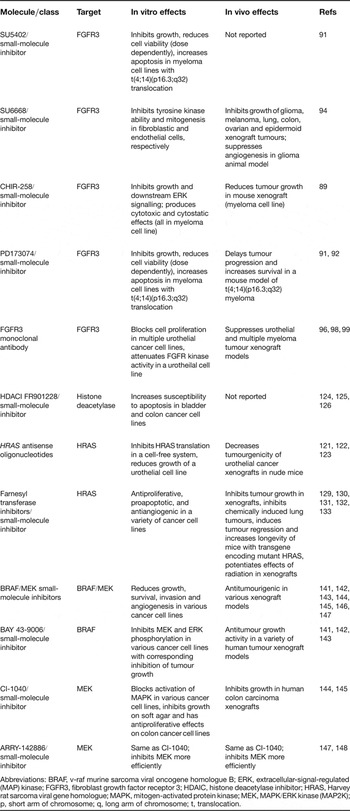
As the importance of FGFR3 in bladder cancer becomes better understood, recent efforts have been devoted to finding ways to disrupt FGFR3 signalling in order to treat NMIUC. There have been several in vitro studies using small-molecule inhibitors designed against FGFR3 (SU5402, SU6668, CHIR-258, PD173074) that have shown strong in vitro inhibition (Refs Reference Trudel89, Reference Mohammadi90, Reference Grand91, Reference Trudel92, Reference Mohammadi93, Reference Laird94) (Table 1). Many of these studies have used multiple myeloma cell lines as they have been shown to harbour activating FGFR3 mutations, although these mutations are different from those generally in NMIUC. Nevertheless, these findings suggest a potential role for the use of FGFR3 inhibitors in UC.
Targeted therapeutics for FGFR3, HRAS and the MAPK signalling pathway

Abbreviations: BRAF, v-raf murine sarcoma viral oncogene homologue B; ERK, extracellular-signal-regulated (MAP) kinase; FGFR3, fibroblast growth factor receptor 3; HDAIC, histone deacetylase inhibitor; HRAS, Harvey rat sarcoma viral gene homologue; MAPK, mitogen-activated protein kinase; MEK, MAPK/ERK kinase (MAP2K); p, short arm of chromosome; q, long arm of chromosome; t, translocation.
Using the human combinatorial antibody library Fab 1 (HuCAL Fab-1) to screen for antibodies that complexed with FGFR3, 25 antibodies out of 37 were identified that complexed with and inhibited proliferation of FDCP-1 cells (mouse myeloid progenitor cell line) transfected with FGFR3 expression vectors (Ref. Reference Rauchenberger95) (Table 1). This study was the first to show that monoclonal antibodies against FGFR3 could have significant functional blocking activity. Another group utilised human single-chain Fv antibody fragments that were generated against the extracellular domain of FGFR3α (IIIc) and showed that these antibodies blocked proliferation of RT112 cells (UC cell line) in a dose- and FGF-dependent fashion (Ref. Reference Martínez -Torrecuadrada96). A recent study using antibodies that were generated from hybridoma clones recognised bacterially expressed fragments of FGFR3 corresponding to loops II–III of the extracellular domain (region that encompasses the S249C oncogenic mutation) (Ref. Reference Gorbenko97). These antibodies could detect bacterially expressed wild-type FGFR3 and the mutant S249C FGFR3, implying a use for this methodology in detection or therapeutics in bladder cancer. Another recent study utilising a monoclonal antibody (designated R3Mab) against FGFR3 showed, through high-resolution crystallography, blocking of ligand-binding activity and inhibition of receptor dimerisation (Ref. Reference Qing98); furthermore, the R3Mab was shown to have significant antitumour activity through interruption of FGFR3 signalling and antibody-dependent cell-mediated cytotoxicty (ADCC).
In addition to the use of monoclonal antibodies directed against FGFR3 in UC, one group has attempted to enhance the effect of antibody fragments directed against FGFR3 (3C) (Ref. Reference Martínez -Torrecuadrada96) by fusing the antibodies with recombinant gelonin toxin (rGel) (Ref. Reference Martínez-Torrecuadrada99). These antibodies, designated 3C/rGel, were injected into SCID (severe combined immunodeficient) mice with RT112 tumour xenografts and resulted in a delay in tumour growth and a reduction in tumour size of 55–70% when compared with the control group. Furthermore, immunohistochemistry analysis showed that treatment with the 3C/rGel antibodies resulted in significant apoptotic damage when compared with controls. Although these studies are preliminary, the use of antibodies directed against FGFR3 in order to treat NMIUC might prove beneficial for the prevention of recurrence. The antibody approach is especially attractive for UC since intravesical treatment can be applied repeatedly with a reduced risk of immunity against the antibody being generated.
HRAS
Role of HRAS in urothelial cancer
HRAS is a cellular proto-oncogene that encodes a member of the Ras GTPase family. When Ras family members become activated, through physiological stimuli or through mutation, they signal to key pathways involved in cell growth, survival, differentiation and migration, including the RAF–MAPK (Ref. Reference Herrera and Sebolt-Leopold100), PI3K (Ref. Reference Jiang101), and the RALGEF (RALGDS; ral guanine-nucleotide-dissociation stimulator) (Ref. Reference Rusanescu102) pathways. In NMIUC, HRAS has been shown to be the most commonly mutated Ras family oncogene (Refs Reference Feinberg103, Reference Fujita104, Reference Fujita105). Several point mutations have been identified in the HRAS oncogene in UC, with the most frequent mutations occurring at codons 12, 13 and 61 (Refs Reference Visvanathan, Pocock and Summerhayes106, Reference Joyce107, Reference Saito108, Reference Boulalas109), causing an inability to hydrolyse GTP to GDP (Refs Reference Tabin110, Reference Reddy111, Reference Taparowsky112). This mutation is responsible for constitutive activation of the HRAS protein and the ability to transform NIH 3T3 cells.
Historically, characterisation of the exact role of HRAS mutation in UC, both in terms of epidemiology of the mutation in tumours and its functional phenotypic consequence, has proven complex, despite the early isolation of the mutant HRAS oncogene from the classic UC cell line model T24 (Ref. Reference Reddy111). Initial studies on the status of HRAS in bladder cancer tended to argue that its expression or mutational status was positively correlated to the invasive potential of the UC. One early study of HRAS in UC showed that by transforming a NMIUC cell line (RT4) with either normal or mutated HRAS and then orthotopically inoculating nude mice transurethrally, one could cause RT4 cells to acquire a phenotype that was more similar to cell lines derived from tumours of high grade or stage (Ref. Reference Theodorescu113). It was argued that either deregulation of HRAS expression (overexpression in this case) or expression of a mutant form of HRAS manifested as a ‘second hit’ in a two-hit pathway could lead to invasive cancer. A combination of PCR amplification and computerised analysis later identified the point mutation G to T at codon 12 in 30 of 67 tumour samples examined (Ref. Reference Czerniak114), and when these tumour samples were stratified by grade, the overwhelming majority of HRAS mutations were found in higher-grade tumours. Furthermore, another study showed that the highly tumourigenic T24T cell line had higher expression of HRAS than its less aggressive parent T24 (Ref. Reference John115). In contrast, evaluation of 152 bladder tumours of varying grade or stage showed that the presence of HRAS mutations had no statistically significant correlation as verified by chi-squared analysis to either grade or stage of the UC studied (Ref. Reference Knowles and Williamson116). Interestingly, another study that evaluated DNA fragments present in urine for the most common HRAS mutations in UC found that HRAS mutation status was better able to identify low-grade (1–2) lesions (47%) than cytology alone (16%) in a cohort of 100 patients (Ref. Reference Fitzgerald117).
One of the first studies to clearly define the role of HRAS in human UC evaluated the development of bladder tumours in transgenic mice expressing mutant HRAS under the regulation of the urothelial-specific uroplakin promoter (Ref. Reference Zhang118). The study showed that mice with low copy numbers of chromosomally integrated, mutant, uroplakin-driven rabbit HRAS developed urothelial hyperplasia followed by the onset of superficial papillary tumours. In addition, mice that had high copy numbers of mutated HRAS also exhibited urotheilal hyperplasia with accelerated-onset superficial papillary tumours. None of these tumours, in the 26 month time period in which they were evaluated, progressed to invasive tumours. Interestingly, this same group later found that mice harbouring mutant HRAS formed these superficial papillary tumours independent of the loss of function of TP53, a tumour suppressor that is frequently mutated or deleted in a large proportion of MIUCs (see above section) (Ref. Reference Gao119). In this study, the group crossed mice that harboured mutant TP53 (dominant-negative mutant, driven by the mouse uroplakin II gene promoter, that lacks the DNA-binding domain but retains the tetramerisation domain, interfering with the function of the normal TP53 protein) with mice expressing activated HRAS. These mice did exhibit urothelial hyperplasia and dysplasia, but were not able to develop frank tumours. In contrast, when the mice expressing HRAS-activating mutations were crossed with TP53-knockout mice, the progeny went on to develop either low- or high-grade superficial papillary tumours, suggesting that complete loss of TP53, rather than reduction of expression or function, is requisite to synergise with mutant HRAS. Another group found that progression to papillary tumours of the bladder with regards to mutant HRAS expression was dose dependent (Ref. Reference Mo120): mice that were homozygous for the activated HRAS mutation were able to develop early-onset superficial papillary tumours, while mice that were heterozygous for the activating HRAS mutant were not able to develop superficial papillary tumours, regardless of mutational status of potential tumour suppressor genes on chromosome 9. Taken together, these findings implied that activating mutations in HRAS are important in the development and progression of early urothelial tumour oncogenesis.
Targeted therapeutics for HRAS
As activated HRAS gained prominence as an early contributor in the oncogenic pathway of superficial papillary tumours of the bladder there have been efforts to disrupt its function. One of the first studies aimed at disrupting HRAS signalling involved construction of a short modified antisense nucleotide that targeted the region encompassing the activating point mutation on the 12th codon (Ref. Reference Saison-Behmoaras121) (Table 1). This antisense nucleotide caused 50% inhibition of translation in a cell-free system and also a 60% reduction in growth of T24 cells over five days. In another study, using synthetic oligonucleotides (ribozymes designed against HRAS mRNA that are capable of hydrolysing its phosphodiester bonds) it was shown that EJ cells (more aggressive variant of T24 cells) that were transfected with plasmids containing the anti-HRAS ribozymes were less effective at forming invasive tumours than EJ cells that did not express the ribozyme (Ref. Reference Eastham and Ahlering122). A similar study using a different vehicle for delivery of the anti-HRAS ribozyme (adenovirus) also showed significant reduction in the tumourigencity of EJ cells injected subcutaneously into nude mice (Ref. Reference Irie123). While the studies on anti-HRAS oligonucleotides have been intriguing, the use of antisense nucleotides to treat human disease has found little footing in the clinic as of yet.
Recently, there have been connections made between the acquisition of oncogenic HRAS in cells and increased susceptibility to histone deacetylase inhibitors (HDACIs) (Table 1). In one study, J82 cells (malignant urothelial cancer cell line) that had been transfected with vectors expressing activated HRAS were much more susceptible to death by apoptosis in the presence of the HDACI FR901228 than cells that had not been transfected (Ref. Reference Choudhary and Wang124). A similar result was shown in the colon cancer cell line HT-29 when activated HRAS was overexpressed (Ref. Reference Choudhary and Wang125). A later study by the same group showed that the potential mechanism for apoptotic cell death in mutant HRAS transgenic J82 cells is an increase in reactive oxygen species, which induces both intrinsic and extrinsic caspase pathways (Ref. Reference Choudhary and Wang126). In addition, suppression of the reactive oxygen species axis via the use of the antioxidant NAC (N-acetylcysteine) resulted in decreased effectiveness of the HDACI FR901228 to induce apoptosis. Taken together, these data show that HDACIs might potentially play an important role in stopping the growth of bladder tumours that harbour activating HRAS mutations. In vivo studies will need to be completed to validate the potential clinical effectiveness of this class of drugs in human bladder cancer. Very recently, genome-wide siRNA (small interfering RNA) screens have successfully identified synthetic lethal interactions with the closely related Ras-family oncogene KRAS2. In cells with mutant KRAS2, inhibition of PLK1 (polo-like kinase 1) and depletion of STK33 (serine/threonine kinase 33) result in a robust induction of cell death that does not occur in normal cells (Refs Reference Luo127, Reference Scholl128). Such synthetic lethal screens suggest that targeting this class of genes could provide an exquisitely specific way to target mutant tumour cells with minimal toxicity to wild-type cells. This type of screening could potentially prove useful in development of modalities to target the HRAS axis in UC.
Targeting the MAPK signalling pathway
Both HRAS and FGFR3 converge on the MAPK signalling pathway (Refs Reference Kanai66, Reference Jebar67, Reference Herrera and Sebolt-Leopold100). For this reason, studies have aimed at inhibiting various steps in this pathway in order to reduce the growth or recurrence of cancer (Table 1). Farnesylation is a post-translational requirement for the Ras protein. This modification leads to attachment of Ras to the plasma membrane since the protein does not have a transmembrane domain. Several farnesyl transferase inhibitors (FTIs) spanning distinct chemical classes have been developed and have shown promising anticancer effects (Refs Reference Zhu, Hamilton and Sebti129, Reference Sebti and Hamilton130). Unfortunately, many of these anticancer effects did not hold up in clinical trials, where it was shown that when using FTIs as a single agent there was no therapeutic effect on lung, pancreatic, colorectal or bladder tumours (Ref. Reference Sebti and Adjei131). Reasons that have been given for these poor results are that the FTIs might be inhibiting proteins other than farnesyl transferases or that inhibition of HRAS by these drugs could be causing a compensatory activation of other Ras family members, ultimately resulting in treatment failure (Refs Reference Wu11, Reference Brunner132). Despite the poor results in clinical trials, in an animal model of urothelial tumours with mutationally active HRAS, treatment with FTIs significantly sensitised the tumours to radiation (Ref. Reference Cohen-Jonathan133). This finding suggests that FTIs used in combination with radiation therapy have the potential for clinical use in UC.
The Raf (ARAF, BRAF and RAF1) and MEK (MEK1/MAP2K1 and MEK2/MAP2K2) family of proteins are critical downstream mediators of the Ras and other oncogenic signalling pathways (Refs Reference Davies134, Reference Singer135, Reference Kimura136, Reference Mansour137, Reference Hoshino138). The Raf family of proteins are responsible for phosphorylating MEK, which results in its activation (Refs Reference Katz and McCormick139, Reference Williams and Roberts140). Hence there was an impetus to determine if inhibition of Raf/MEK activity would have anticancer benefits. With regards to Raf and UC, clinical data have been limited because the Raf mutation, specifically in BRAF, is not a common occurrence in UC. The cancers in which BRAF mutations are prevalent (i.e. melanoma, colorectal, ovarian and thyroid) have shown some response to the small-molecule BRAF inhibitor BAY 43-9006, but it has shown limited clinical success due to its lack of specificity (Refs Reference Lee and McCubrey141, Reference Hotte and Hirte142, Reference DeGrendele143). MEK inhibitors (CI-1040, ARRY-142886), in contrast, have proven to be better at specifically inhibiting their target and have shown efficacy against tumours. For example, CI-1040 showed great promise in inhibition of colon carcinomas in vivo and was demonstrated to have a negative impact on the growth, survival, invasion and angiogenesis profiles of cancer cells (Refs Reference Sebolt-Leopold144, Reference Allen, Sebolt-Leopold and Meyer145). However, despite promising antitumour activity in Phase I trials, CI-1040 showed limited tumour activity in Phase II trials, ultimately leading to its discontinuation (Refs Reference Allen, Sebolt-Leopold and Meyer145, Reference Rinehart146). This result prompted development of the MEK inhibitor ARRY-142886, which is much more specific than CI-1040 (Refs Reference Yeh147, Reference Davies148). This drug is currently being used in clinical trials. At this time there are no specific clinical trials that are solely focused on the impact of Raf/MEK inhibitors with regards to UC. As the mechanism and impact of these inhibitors are better understood in the cancer models in which they are currently being studied, it is likely that these inhibitors will find some use in UC given the crucial role of Ras signalling in the pathogenesis of UC.
Summary and future directions
NMIUC is a disease with low mortality but significant morbidity due to a high recurrence rate. Understanding the various molecular signalling mechanisms that underpin this disease is crucial in developing therapies to prevent its onset and progression. Loss of heterozygosity of chromosome 9, activating mutations in FGFR3, and activating mutations in the oncogene HRAS have all been shown to be prevalent and important in NMIUC, with the latter two alterations being infrequent or absent in the invasive subtype of UC. Currently, there is much work being done in the field of inhibiting the activity and signalling of aberrant FGFR3 and HRAS, whether targeting these oncogene products themselves or their downstream mediators. Each of these tools has shown some success in vitro and in vivo. Several small-molecule inhibitors have been developed that can disrupt the function of either FGFR3 or HRAS directly or disrupt signalling at various points in the MAPK signalling pathway. Many of these inhibitors have shown some promise in clinical trials in other tumours types, suggesting that they might have some relevance in slowing UC progression. Unfortunately, no single drug has been approved for use in bladder cancer that targets the MAPK signalling pathway, FGFR3 or HRAS, although the possibility of using such drugs alone or in combination with standard of care therapies is attractive. In addition to the therapeutics discussed in this review, there are several novel therapeutic modalities that are currently being investigated, such as gene therapy using viral and nonviral vectors and direct tumouricidal viruses (Ref. Reference Lojo Rial, Wilby and Sooriakumaran149). Further investigation of these therapeutics might prove beneficial to NMIUC patients in an adjuvant setting. Finally, NMIUC is among the most expensive tumours to treat, per person, primarily due to its frequent recurrence and necessary surveillance (Refs Reference Jemal3, Reference Botteman150). Hence, any understanding of the molecular pathogenesis of this disease that can lead to future therapies would be a great benefit to society.
Acknowledgements and funding
We thank members of the Theodorescu lab for their suggestions and assistance and we also thank the reviewers of this manuscript for their insightful comments. This work was supported by NIH grants CA142163 to Courtney Pollard and CA075115 to Dan Theodorescu.