Introduction
Multiple myeloma (MM) is a B-cell neoplasm characterized by clonal proliferation of plasma cells in the bone marrow (BM), the production of a monoclonal protein detectable in serum and/or urine, and the presence of organ damage: osteolytic lesions, renal failure, anaemia and/or hypercalcaemia. Although MM remains an incurable disease, in recent years the introduction of new drugs such as immunomodulators or proteasome inhibitors have significantly changed the paradigm of the disease.
Epidemiology
MM represents 1% of all cancers and 10% of haematological malignancies,Reference Moreau, San Miguel, Ludwig, Schouten, Mohty, Dimopoulos and Dreyling1 thereby being the second most common blood cancer behind lymphoma. It is said to be a disease associated with ageing, with a median age at diagnosis of 65–70 years. At the time of diagnosis, more than 70% of patients are over 65 years of age.Reference Kyle and Rajkumar2,Reference Palumbo and Anderson3 The incidence in Europe is 4.5–6.0/100,000/year, but increases with age, with an annual incidence of >40/100,000 over 80 years and a prevalence of 46/100,000 in patients over 75 years. It is expected that both the incidence and prevalence of the disease increase with age due to, on one hand, increased life expectancy of the general population and, on the other, the higher life expectancy in these patients due to the introduction of more effective treatments.
There are differences in the incidence by sex and race:Reference Jemal, Siegel and Ward4 it is greater in men, and African Americans have an incidence almost twice that of American Caucasians.
Pathophysiology
Myeloma arises from an asymptomatic premalignant proliferation of monoclonal plasma cells that are derived from post-germinal-centre B-cells. There are multiple genetic factors and changes in the bone marrow environment that cause the transformation of these cells into neoplastic malignant cells. It is believed that myeloma often progresses in monoclonal gammopathy of undetermined significance, progressing to asymptomatic myeloma and ultimately to symptomatic myeloma.Reference Palumbo and Anderson3,Reference Kuehl and Bergsagel5
The malignant plasma cells have a morphology identical to normal plasma cells, but phenotypically they are not equal. They express CD38, CD138 and CD56.
Myelomatous cells may remain for a time in a pre-malignant state. Subsequently, they sequentially acquire genetic alterations. First primary translocations (occurring on chromosome 14 where the Ig heavy chain gene is found), then secondary translocations and other genetic mutations (deletions of chromosome 13, chromosome 17, etc.), in addition to epigenetic alterations (hypomethylation of genes, post-translational alterations, etc.).Reference Kuehl and Bergsagel5
Essential in the pathogenesis of MM are, in addition, interactions between the malignant plasma cells with cells of the bone marrow microenvironment, where the tumour transformation and progression of the disease occurs.Reference Mitsiades, Mitsiades, Munshi, Richardson and Anderson6 Various cytokines and adhesion molecules are critical in these interactions: cytokines such as IL-6, IGF-1, VEGF, BAFF, TNF-α, TGF-β, MIP-1α, IL-5 and IL-21, which through metabolic pathways such as MAPK, PI3-K/Akt, Jak/STAT and NF-κβ, cause an increase in tumour growth, survival and drug resistance. Adhesion molecules, such as CD44, VLA-4, VLA-5, etc. are responsible for the migration of myelomatous cells to BM and plasma cell adhesion to the bone marrow microenvironment. Disruption of the interactions between the myelomatous plasma cell and the bone marrow microenvironment can therefore stop the growth and proliferation of these cells, this being one of the targets of new drug research.Reference Podar, Kenneth, Anderson and Ghobrial7,Reference Podar, Tai, Hideshima, Vallet, Richardson and Anderson8
Diagnosis
MM is situated within the so-called monoclonal gammopathies (MG),9 a group of disorders characterized by the proliferation of monoclonal B-lymphoid cells in the final maturation stages (plasma and/or lymphoplasmacytic cells), which produce a monoclonal immunoglobulin (Ig), called the M component or monoclonal component (MC), and can be of different types: IgG, IgA, IgM, IgD and IgE. Sometimes (20% of cases) myelomatous plasma cells secrete only free light chains, kappa or lambda; this is a Bence-Jones multiple myeloma.
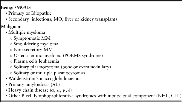
MG may be benign, such as malignant monoclonal gammopathy of undetermined significance (MGUS) or malignant, which includes MM (Table 1). The risk of progression from MGUS to malignant MG is 1% per annum. Risk factors for progression are non-IgG serotypes (IgA and IgM), an abnormal ratio of free light chains (FLC) in serum (kappa/lambda <0.26 or >1.65); the plasma cell phenotype, its presence in peripheral blood, and the most important predictor, the level of monoclonal component in serum at diagnosis.
Monoclonal gammopathies

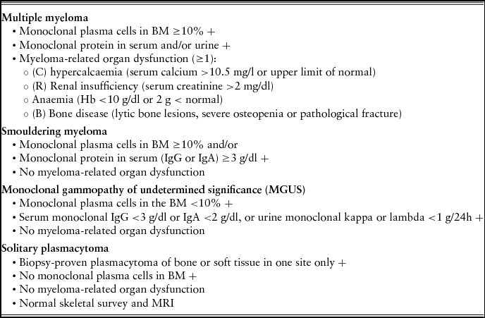
Diagnosis of MM must meet the following criteria:Reference Kyle and Rajkumar10 presence of ≥10% plasma cells in the bone marrow and presence of monoclonal peak in serum and/or urine (in case of non-secretory MM, the percentage of plasma cells in BM must be ≥30%). Myeloma can be asymptomatic or symptomaticReference Palumbo and Anderson3,11 depending on if there is organ dysfunction attributable to the proliferation of plasma cells, which is determined by the CRAB criteria (C: serum calcium >10.5 mg/l; R: renal failure with serum creatinine >2 mg/dl; A: anaemia <10 g/dl or 2 g < normal; B: bone involvement with lytic lesions, pathological fractures or severe osteopenia) (Table 2).Reference Palumbo and Gay12
Plasma cell-related disorder diagnostic criteria
The study of the monoclonal component should be performed in serum and urine. For quantification of serum peak a monoclonal proteinogram is performed (by electrophoresis, EP) and nephelometry is performed to quantify Ig, which must be assessed jointly. Immunofixation (IF) or immunosubtraction in serum confirms the monoclonal character and allows an understanding of the structure of the light and heavy chain. In cases in which a monoclonal component is detected by EP and IF is negative or positive for only a light chain, this should suggest IgD or IgE MM. Monoclonal component detection must also be performed in 24-hour urine, using protein electrophoresis (EP) and IF. The most common type of Ig detected is IgG, in 65–70% of patients.9
Measurement of free light chains (FLC) in serum is recommended to be performed at diagnosis,Reference Snozek, Katzmann and Kyle13 since they are useful in cases of non-secretory MM or oligosecretory MM (with low production of monoclonal component) in plasmacytomas or smouldering MM (an abnormal value is associated with a high risk of progression to symptomatic MM) and in AL. However, currently there are insufficient data to justify its use in monitoring the treatment of symptomatic MM.
Other biochemical parameters studied in all patients with MM are: creatinine and renal function tests, calcium, albumin, β2 microglobulin, LDH, ESR, CRP, liver function tests and electrolytes. The blood count and examination of peripheral blood smear (typical of RBCs Rouleaux) are fundamental.
Examination of the BM by performing a bone marrow aspiration (BMA) is necessary not only for determining the percentage of plasma cells, but also for morphological, immunophenotypic and cytogenetic examination.Reference Kyle and Rajkumar10 Bone biopsy (BB) is not routinely required, unless a non-readable aspirate is obtained. If both BMA and BB are performed, the higher infiltration rate of plasma cells will be selected. Cytogenetic and immunophenotypic studies are currently required.Reference Fonseca, Barlogie and Bataille14,Reference Sawyer16 Cytogenetics is mainly based on FISH (fluorescence in situ hybridization), which allows the identification of the number of abnormal cells by detection of deletions and translocations. It is essential to always conduct p53 (chromosome 17) and IgH translocations, especially t(4;14) and t(14;16), which carry a very poor prognosis. Other abnormalities such as gains and losses from short and long arms of chromosome 1 (poor prognosis) and hyperploidism (good prognosis) are useful, but not mandatory to analyse. The karyotype is performed when there are more than 20% plasma cells in the BM sample. It detects additions, deletions, translocations or other less frequent rearrangements that could not be identified with the usual FISH probes.Reference Fonseca, Barlogie and Bataille14,Reference Smadja, Fruchart and Isnard15
The main usefulness of flow cytometry (FCM) is its ability to monitor response to treatment and detect minimal residual disease (MRD) after treatment. In addition, its cell count is more accurate than that done visually due to morphology and its allowing the identification of the immunophenotyping profile of plasma cells. The phenotypic profile of the pathological plasma cell is CD38+, CD138+, CD19–, CD56+, IgS–, IgC+, as opposed to the normal plasma cell, which is CD19+, CD56–.
For the study of bone lesions, conventional X-ray must be performed, including a bone series (BS) that comprises the skull, rib cage, spine, pelvis and long bones.Reference Dimopoulos, Terpos and Comenzo17 In certain situations it is necessary to perform other imaging tests such as magnetic resonance imaging (MRI): solitary plasmacytoma (to rule out other locations), suspected spinal cord compression, vertebral collapse, the assessment of areas of bone pain where there is no alteration evident in the BS and before performing a kyphoplasty. It can also be useful in smouldering myeloma and non-secretory myeloma.Reference Walker, Brown, Jones-Jackson, De Blanche and Bartel18
Other imaging tests such as CT or PET-CT are currently being evaluated;Reference Regelink, Minnema and Terpos19 they are not routinely used and their use is not currently standardized in the diagnosis and outcome of patients with multiple myeloma. They may be useful in some situations, however, such as differentiating between active disease vs. old lesions or benign disease.
Prognostic factors and impact of age
Multiple myeloma remains an incurable disease, but advances in recent years, with the development of new drugs, have significantly improved survival. Treatment with high-dose chemotherapy followed by autologous transplant achieved an overall survival of 35% at 12 years in patients who achieved a CR, with late relapse beyond 10 years from diagnosis.
In elderly patients the results are not as satisfactory.Reference Palumno, Bringhen and Ludwig20 On the one hand, the deterioration of physiological functions exhibited by this group of patients (impaired renal, liver, gastric and cardiovascular function; reduced blood flow; decreased bone marrow reserve, etc.) entails an alteration of the pharmacokinetics and pharmacodynamics of anti-myeloma drugs, altering their clinical efficacy and enhancing their toxicity. On the other hand, due to the elderly patient's increased susceptibility to adverse drug effects they cannot tolerate full doses of such drugs. This group of patients also has the increased risk of associated malignancies and second neoplasms.
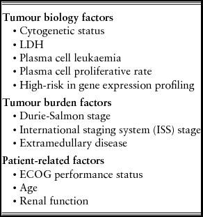
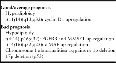
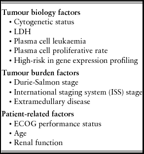
Numerous factors that influence the prognosis and the risk of progression must be taken into account in the choice of treatment. These are shown in Table 3. The main factor for stratification of prognostic risk are genetic alterations,Reference Bladé, Teresa Cibeira, Fernández de Larrea and Rosiñol25 which are summarized in Table 4. Importantly, deletion of chromosome 13 alone does not imply a poor prognosis. The 13q14 deletion is the most frequent mutation (40%). Patients with non-hyperdiploid mutations present IgH gene translocations and are associated with more aggressive disease and lower survival rates. Mutations of the p53 gene (deletion 17p–) confer worse prognosis.
Prognostic factors in multiple myeloma
Cytogenetic prognostic subgroups in multiple myeloma
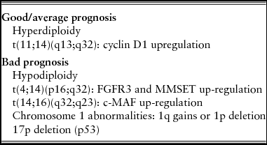
FGFR3: fibroblastic growth factor receptor 3; MMSET: multiple myeloma SET domain.
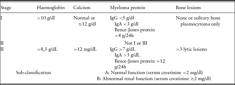
Over the years numerous prognostic staging systems have been developed. The classic system, which is most commonly used in daily clinical practice, is the Durie-Salmon,Reference Durie and Salmon21 which assesses the degree of anaemia, serum calcium, type of immunoglobulin (Ig) present and the presence or absence of bone lesions (Table 5).
Durie-Salmon staging system for multiple myeloma
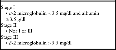
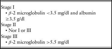
In 2005 the International Myeloma Working Group developed a systemReference Greipp, San Miguel and Durie22 based on serum β-2 microglobulin and albumin levels, the International Staging System (ISS) (Table 6). Measurement of β-2 microglobulin is considered a determining factor in risk stratification, with a median survival of 62 months for patients with an ISS of I and 29 months for those with ISS III. This classification is reproducible in all patients of all age groups who have been treated with either conventional chemotherapy, or intensive chemotherapy and autologous bone marrow transplant.
International staging system (ISS) criteria for multiple myeloma
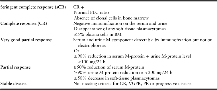
Response to treatment is another crucial factor to consider in the prognosis of patients; it should be maintained for a minimum of 6 months. The depth of response that is achieved after treatment correlates with an improvement in disease-free survival (DFS), overall survival (OS), progression-free survival (PFS), long-term survival and duration of remission.Reference Durie, Harousseau and Miguel23–Reference Bladé, Teresa Cibeira, Fernández de Larrea and Rosiñol25 Response criteria are summarized in Table 7.
Response criteria for multiple myeloma
FLC: free light chain.
Symptoms
Clinical manifestations of MM are varied. Most patients at diagnosis have some type of symptoms attributable to the disease; only 30% of patients are asymptomatic at diagnosis.
Bone lesions are the result of an imbalance between bone formation due to osteoblasts and bone destruction due to osteoclasts.Reference Oyajobi26 Bone pain is the most common symptom at diagnosis, present in 58% of patients,Reference Kyle, Gertz and Witzig27 and the most disabling, especially in older patients. The typical pain is triggered by movement and often is secondary to pathological bone fractures caused by lytic lesions. The bones that are most affected are those where haematopoiesis occurs, by frequency: vertebrae, hips, skull, pelvis and proximal region of the long bones of the limbs. Up to 67% of patients have lytic lesions,Reference Snowden, Ahmedzai and Ashcroft28 osteoporosis and pathological fractures, and up to 5% of patients exhibit symptoms of spinal cord compression, due to a myelomatous mass invading the medullary channel due to fractured vertebral bone fragments. Almost 85% of patients diagnosed with multiple myeloma will present bone involvement throughout the course of their disease.
Asthenia and weakness are also common and are usually secondary to anaemia, present in 60% of patients at diagnosis, with an Hb figure of less than 12 g/dl.
Anaemia in MM is frequently a multifactorial process:Reference Kyle, Gertz and Witzig27 myelomatous cells have a pro-apoptotic effect on erythroid precursors, as well as producing a displacement of normal bone marrow cellularity; endogenous erythropoietin production and the response to it decreases and there is a shortening of the half-life of circulating red cells and reduction in their use of iron; on the other hand, paraprotein produced has a dilutional effect on the plasma.
Renal failure is present in 20–40% of patients at diagnosis. The damage produced by free light chains, hypercalcaemia, hyperuricaemia, infections and neurotoxic treatments contribute to kidney damage.Reference Burnette, Leung and Rajkumar29
Hypercalcaemia, which is the most common metabolic complication, also contributes to kidney damage.Reference Oyajobi26,Reference Kyle, Gertz and Witzig27 Hypercalcaemia is accompanied by nausea, altered level of consciousness (ranging from confusional state to coma), constipation, polyuria and fatigue. The development of an acute form of hypercalcaemia requires rapid intervention. It is caused by the increase of calcium in the circulation due to increased bone destruction, which produces myeloma cells by various cytokines.
Infections in patients with MM are frequent,Reference Nucci and Anaissie30 especially those caused by encapsulated bacteria, such as pneumococcus, and viral infections. The risk of infection is determined by several factors: the activity of the disease (the incidence of infection is highest in the first months after diagnosis in patients in relapse or with refractory active disease), the type and degree of immunodeficiency, treatments administered (high-dose steroids, bortezomib, lenalidomide, etc.), specific epidemiological factors and patient age and co-morbidity (renal failure, iron overload, etc.).
Immunodeficiency presented by patients with MM is primarily humoral, affecting B-lymphocytes, resulting from abnormal production of immunoglobulins. Cellular immunodeficiency affecting T-, NK-lymphocytes and dendritic cells, which is less frequent, can be potentiated by the treatment such as the use of corticosteroids or bortezomib.Reference Nucci and Anaissie30 The B-lymphocyte dysfunction favours encapsulated bacteria infections, especially Streptococcus pneumoniae or Haemophilus influenza. Reference Rapezzi, Sticchi and Racchi31,Reference Robertson, Nagesh and Jowitt32 Staphylococcus aureus and Gram-negative bacilli are more frequent in patients with advanced disease and kidney failure.
These patients also have a high risk of developing thromboembolic events Reference Kristinsson, Fears and Gridley33 from various causes related to the disease itself, the type of anti-myeloma drugs (the use of thalidomide and lenalidomide with high-dose dexamethasone or with other drugs in combination with chemotherapy increase the risk of thrombosis) and patient-related factors (age, obesity, surgery, immobilization, history of thromboembolic disease).
The neurological complications these patients may present are caused by multiple factors:Reference Snowden, Ahmedzai and Ashcroft28 spinal cord compression due to vertebral collapse, medullary plasmacytoma, metabolic cause (hypercalcaemia), hyperviscosity due to immunoglobulin production and treatment (mainly bortezomib). The neurological symptoms range from mild, such as parasthesiae, especially in the distal part of the limbs, headache, and confusion, etc. to severe, in spinal cord compression situations or in cases of acute onset hypercalcaemia requiring urgent treatment. Less common is hyperviscosity, produced by increased blood Ig. Various organs can be affected, producing a wide range of symptoms: altered level of consciousness, cardiorespiratory symptoms, kidney damage and even mucosal and retinal bleeding.
Treatment
It is important to note that age alone should not be regarded as the sole criterion when choosing a treatment.Reference Palumno, Bringhen and Ludwig20 The choice of treatment must be made carefully and individually, assessing the patient's physical and medical condition, taking into account several factors:Reference Palumbo and Mina34
(1) Biological age prevails over chronological age; these are not always correlated.
(2) Co-morbidity, frailty and disability are factors to take into account when choosing a treatment, since they add a negative connotation to treatment outcomes. Co-morbidity is defined as the presence of ≥2 concomitant diseases and frailty as the presence of ≥3 of the following: weakness, weight loss, poor physical activity, poor endurance and poor gait speed. Disability refers to limitations, both physical and mental, in the basic activities of daily life.
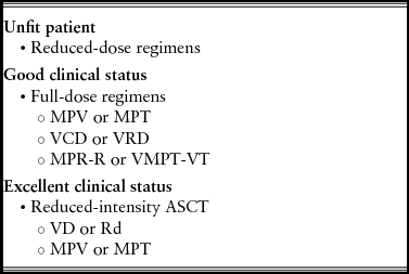
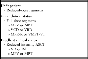
Generally, patients over 65 are not considered candidates for transplantation of haematopoietic stem cells. However, the above-mentioned factors, along with a detailed clinical assessment, will allow us to determine which patients are candidates to receive one treatment or another (Table 8). Those who do not have risk factors receive more aggressive treatment, while those who present as few as one will be candidates for a reduced-intensity regimen.Reference Palumbo and Gay35–Reference Klepin, Rizzieri, Palumbo, Magarotto and Eichhorst37 Nowadays, treatment is considered necessary for the patient diagnosed with symptomatic multiple myeloma (with organ damage) or rapid progression in asymptomatic (MC doubling in 2 months), so those with smouldering myeloma would require close monitoring (re-evaluation every 3 months) without need for treatment.
Elderly patients with multiple myeloma: status assessment and treatment
There are multiple options in the scheme of treatment of MM, because of the different combinations that can be used with different drugs, well tolerated and more effective than standard treatment in this group of patients available until now, as was the use of melphalan combined with prednisone. The most common are alkylating agents (melphalan and cyclophosphamide), anthracyclines (adriamycin~and doxorubicin), corticosteroids (dexamethasone and prednisone), immunomodulators (thalidomide and lenalidomide) and proteasome inhibitors (bortezomib). Various studies have shown the desirability, in elderly patients with the presence of one or more risk factors, of reducing treatment doses in order to limit the toxicities associated with treatment without sacrificing its effectiveness.
The standard first-line treatment in elderly patients, not transplant candidates, is based on a three-drug regimen: bortezomib/melphalan/prednisone (VMP),Reference San Miguel, Schlag and Khuageva38 melphalan/prednisone/thalidomide (MPT)Reference Mateos, Oriol and Martinez39 or bortezomib/lenalidomide/dexamethasone (VRD), with VMP and MPT currently considered the gold standard. VRD is used in the United States as a treatment of choice. The combination of four drugs (VMPT)Reference Palumbo, Bringhen and Rossi40 is another alternative: bortezomib/melphalan/prednisone/thalidomide followed by maintenance with bortezomib/thalidomide (VMPT-VT) has shown a higher CR compared with bortezomib/melphalan/prednisone (VMP).
In frail patients not candidates for the above schemes of treatment, a regimen based on two drugs, such as bortezomib/dexamethasone (VD) or lenalidomide/low-dose dexamethasone (Rd),Reference Rajkumar, JAcobus and Callander41 is recommended. These combinations have proven to be less toxic and more effective than bortezomib/melphalan/prednisone (VMP) and bortezomib/thalidomide/dexamethasone (VTD) and lenalidomide/consolidation with prednisone (RD), respectively. If after a few months of treatment, the disease is not controlled or treatment was well tolerated, the addition of a new drug or increasing the dose would be an option to consider. Furthermore, these patients are more likely to present secondary treatment toxicity and other adverse effects and hence discontinuation of the treatment, which results in less efficacy.
Patients with myeloma require treatments directed at their disease activity but also a wide range of supportive and palliative measures to optimize quality of life at all stages of their disease,Reference Snowden, Ahmedzai and Ashcroft28 like treatment of anaemia (blood transfusion or treatment with erythropoiesis-stimulating agents), pain management (analgesics, radiotherapy), skeletal complications (bisphosphonates, radiotherapy, percutaneous vertebroplasty), with multidisciplinary collaboration.
Conflicts of interest
None declared.