Dextro-transposition of the great arteries is a congenital anomaly with ventriculoarterial discordance. Mustard and colleagues first reported on atrial switch surgery, the so-named “Mustard procedure”, which converts the parallel circulations of dextro-transposition of the great arteries into a circulation in series, thereby correcting cyanosis.Reference Mustard1 Pulmonary venous obstruction is one of the baffle-associated complications after a Mustard procedure, which induces pulmonary congestion and pulmonary hypertension.Reference Champsauer, Sokol and Trusler2, Reference Hagler, Donald and Douglas3 Real-time three-dimensional trans-oesophageal echocardiography (3D-TOE) provides useful information for recognising complex intra-cardiac abnormalities. To the best of our knowledge, this is the first case report that demonstrates the use of real-time 3D-TOE as guidance for the balloon dilation of pulmonary venous pathway obstruction after a Mustard procedure.
Case
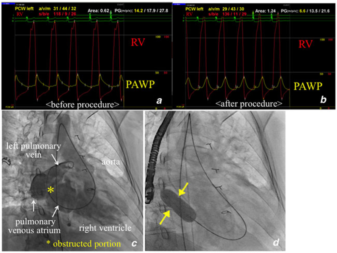
The patient was a 40-year-old male who experienced dyspnoea upon exertion. Soon after birth he was diagnosed with dextro-transposition of the great arteries and at the age of 3 months underwent a Mustard procedure. Cardiac catheterisation demonstrated a 14.2 mmHg mean pressure gradient across the pulmonary-venous-obstructed site. A cross-sectional area of the pulmonary venous pathway obstruction was measured as 0.62 cm2 by Gorlin’s equation and a cardiac index of 1.65 L/min/m2 (Fig 1a). In addition, his systemic right ventricular (RV) function was impaired at 38% of ejection fraction. Surgery seemed too invasive, so the patient was offered percutaneous transcatheter balloon dilation of the pulmonary venous pathway obstruction.
(a) Simultaneous measurement of both PAWP and RV pressure revealed 14.2 mmHg of mean diastolic pressure gradient across the obstructed portion in the pulmonary venous atrium. A cross-sectional area of the obstructed pulmonary venous portion was measured as 0.62 cm2 by Gorlin’s equation. (b) After balloon dilation, the mean pressure gradient across the pulmonary venous obstruction site decreased to 6.6 mmHg and the cross-sectional area enlarged to 1.24 cm2. (c) This angiography shows the right-anterior oblique view, in which contrast medium was injected from the left upper pulmonary vein. A pigtail catheter was passed through the aortic valve, tricuspid valve, and pulmonary venous obstruction site sequentially into the left upper pulmonary vein. (d) A 20-mm VACS balloon was inflated at the obstructed portion in the pulmonary venous atrium. PAWP= pulmonary artery wedge pressure; RV= right ventricular.
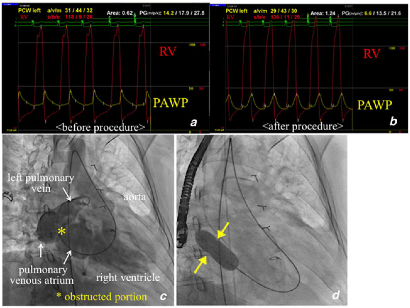
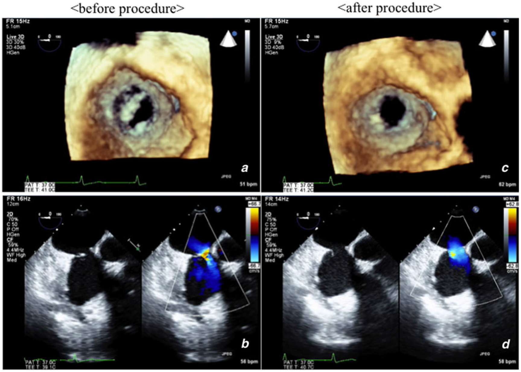
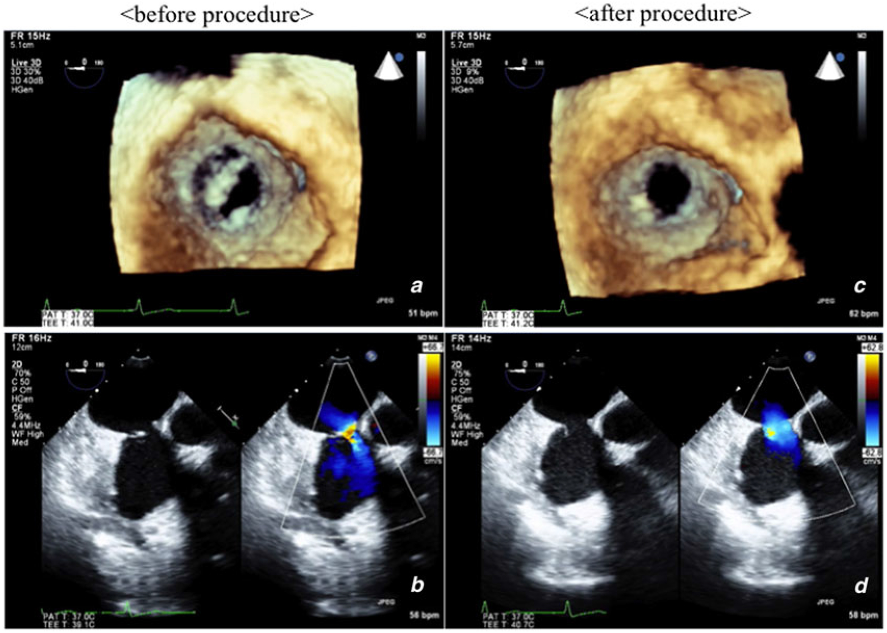
The procedure was performed under general anaesthesia with fluoroscopy and a TOE guide. Because of the occlusion of the bilateral femoral veins, a retrograde arterial approach via the right femoral artery was used to access the pulmonary venous atrium. A 10F sheath was inserted and a guide wire passed sequentially through the aortic valve, tricuspid valve, and pulmonary-venous-obstructed portion into the left pulmonary vein (Fig 1c, Movie 1). Real-time 3D-TOE provided a clear image of the pulmonary-venous-obstructed section as a circle-like shape, the right half of which was covered with a membranous tissue (Fig 2a, Movie 2). The membranous tissue seemed very thin and floppy, and therefore could easily be broken and enlarged by balloon dilation. In addition, real-time 3D-TOE clearly demonstrated the spatiality of the membranous tissue, which two-dimensional trans-oesophageal echocardiography (2D-TOE) could not provide. The minimum diameter was 5.7 mm with a reference diameter of 21 mm (Fig 2b); therefore, a 20-mm VACS (Valvuloplasty Balloon Catheters) balloon (OSYPKA AG, Germany) was inflated. After the procedure, the mean pressure gradient decreased to 6.6 mmHg and the cross-sectional area enlarged to 1.24 cm2 (Fig 1b). The patient’s cardiac index improved to 2.10 L/min/m2. Real-time 3D-TOE after the procedure showed an enlarged pulmonary venous atrium in which the membranous tissue was broken (Figs 2c, D, Movie 3).
(a) Real-time 3D-TOE before the procedure provided an antero-posterior view towards the stenotic portion of the pulmonary venous atrium via the tricuspid valve. It revealed a circle-like shape, the right half of which was covered with a membranous tissue. (b) 2D-TOE before the procedure revealed a 5.7 mm of minimum diameter of the obstructed portion. (c) Real-time 3D-TOE after balloon dilation showed an enlarged pulmonary venous atrium in which the membranous tissue was broken. 3D-TOE= three-dimensional trans-oesophageal echocardiography; 2D-TOE= two-dimensional trans-oesophageal echocardiography.
Discussion
In Japan, the Mustard procedure – an atrial switch surgery for dextro-transposition of the great arteries – was first reported on in 1969 and Jatene surgery, an arterial switch procedure, was initially reported on in 1976. The incidence of pulmonary venous pathway obstruction after a Mustard procedure is reported as being 3–18%.Reference Champsauer, Sokol and Trusler2, Reference Hagler, Donald and Douglas3 In general, it is treated by additional surgery.Reference Cooper, Sullivan and Bull4 However, for some patients considered a high surgical risk, a catheter-based treatment would be indicated.Reference Abdulhamed, Alyousef and Mullins5 A transeptal puncture through the femoral vein is another option for accessing the pulmonary venous atrium.Reference Abdulhamed, Alyousef and Mullins5 Our patient was at high risk for further surgery because of low systemic RV function and occlusion of his bilateral femoral veins would make transeptal puncture difficult.
In previous reports, the use of real-time 3D-TOE for the assessment of dextro-transposition of the great arteries, such as baffle leak or systemic venous obstruction, has been demonstrated.Reference Ahmed, Nekkanti and Nanda6, Reference Cua, Kollins and Roble7 In general, when dealing with baffle obstruction after a Mustard procedure, interventional cardiologists usually use fluoroscopy or intra-cardiac echocardiography not equipped with a three-dimensional function. Compared to the conventional two-dimensional image, our three-dimensional image revealed the characteristics of the stenosis in the pulmonary venous atrium more precisely, which could be easily dilated with balloon.
Conclusion
Real-time 3D-TOE is useful for evaluating the morphology and character of pulmonary venous pathway obstruction and guiding transcatheter intervention in patients with dextro-transposition of the great arteries after atrial switch surgery.
Supplementary Material
To view supplementary material for this article, please visit https://doi.org/10.1017/S1047951119001045.
Author ORCIDs
Manabu Nitta, 0000-0003-2834-1248
Acknowledgements
None.
Financial Support
This report received no specific grant from any funding agency, commercial or not-for-profit sectors.
Conflict of Interest
None.
Ethical Standards
The authors assert that all procedures contributing to this work comply with the ethical standards of the relevant national guidelineson human experimentation (please name) and with the Helsinki Declaration of 1975, as revised in 2008, and has been approved by the institutional committees (Yokohama City University Graduate School of Medicine).