PROPOSED ‘EVERYDAY’ FUNCTIONS FOR MICROGLIA
Microglia, the resident myeloid cells in the central nervous system (CNS) parenchyma, constitute a key and unique part of the cellular composition of the CNS (Ransohoff and Cardona, Reference Ransohoff and Cardona2011). The functional significance of microglia has been increasingly studied over the last 20 years (Graeber, Reference Graeber2010), with a focus on their roles in CNS development (Streit, Reference Streit2001) and immune defense in conditions of infection and injury (Perry et al., Reference Perry, Nicoll and Holmes2010). However, even in the healthy, uninjured adult animal, microglia likely play important constitutive roles in the day-to-day function in the CNS. What these roles may be are gradually coming to light from recent studies.
The functions of microglia in the adult healthy CNS have largely been imputed from observations about their morphology, distribution, behavior and their relationships with other CNS cells. Distinct from extra-CNS macrophages, ‘resting’ microglia possess a typical ramified morphology with radial, branching processes that radiate symmetrically from the soma. Unlike astrocytes, they are not in a syncytial network but are spaced out ‘individualistically’ in close proximity with each other, but with little overlap between the processes of neighboring cells (Graeber, Reference Graeber2010). Most uniquely, microglia in the cerebral cortex (Davalos et al., Reference Davalos, Grutzendler, Yang, Kim, Zuo and Jung2005; Nimmerjahn et al., Reference Nimmerjahn, Kirchhoff and Helmchen2005), spinal cord (Davalos et al., Reference Davalos, Grutzendler, Yang, Kim, Zuo and Jung2005; Dibaj et al., Reference Dibaj, Steffens, Nadrigny, Neusch, Kirchhoff and Schomburg2010, Reference Dibaj, Steffens, Zschuntzsch, Nadrigny, Schomburg and Kirchhoff2011) and retina (Lee et al., Reference Lee, Liang, Fariss and Wong2008) demonstrate marked, rapid and constitutive motility in their processes. These random but balanced movements, occurring without significant migration of microglial cell bodies, are estimated to cover all available extracellular space in the CNS over a time scale of hours (Nimmerjahn et al., Reference Nimmerjahn, Kirchhoff and Helmchen2005). These microglial features are evocative of a function of dynamic and thorough immune surveillance of the CNS parenchyma that assures the comprehensive occupation of the entire extracellular milieu by at least one microglial processes over a short period of time (Raivich, Reference Raivich2005).
In addition, recent studies involving in vivo live imaging and electron microscopy have proposed that dynamic microglial processes, in addition to seeking out immunological signals, also make meaningful direct contact with synapses (Wake et al., Reference Wake, Moorhouse, Jinno, Kohsaka and Nabekura2009; Tremblay et al., Reference Tremblay, Lowery and Majewska2010). The consequence of these microglia–synapse contacts appear to favor the elimination of synapses (Tremblay et al., Reference Tremblay, Lowery and Majewska2010), with prolonged contacts resulting in greater rates of synapse loss (Wake et al., Reference Wake, Moorhouse, Jinno, Kohsaka and Nabekura2009), possibly by the phagocytosis of synapses (Paolicelli et al., Reference Paolicelli, Bolasco, Pagani, Maggi, Scianni and Panzanelli2011). Imaging experiments in the visual cortex also indicated that decreases in neuronal activity induced by visual deprivation reduced microglial process motility and altered the nature of microglia–synapse associations. These observations posit that additional constitutive function of ‘resting’ microglia involves the dynamic monitoring of the functional status of synapses and influencing synapse maintenance and turnover in the adult CNS.
Lastly, a constitutive trophic role has also been suggested. Microglia express neurotrophins (Elkabes et al., Reference Elkabes, DiCicco-Bloom and Black1996; Harada et al., Reference Harada, Harada, Kohsaka, Wada, Yoshida and Ohno2002), and may therefore help provide trophic support to neurons and macroglia. Dystrophic microglia with abnormal morphologies and fragmented processes have been described in brains of Alzheimer's patients in association with neurodegeneration, giving rise to the notion that a loss in the normal delivery of trophic support by dystrophic microglia contributes to the demise of nearby neurons (Streit et al., Reference Streit, Braak, Xue and Bechmann2009).
NEUROTRANSMITTER SIGNALING AS A REGULATORY INFLUENCE ON MICROGLIA IN THE UNINJURED CNS
The efficiency at which microglia carry out these proposed functions is arguably a function of how well their processes occupy the CNS parenchyma, which is in turn influenced by their cellular distribution in the CNS, the size and complexity of their ramified morphology, and their degree of process dynamism. There is evidence that these microglial features are not ‘hard-wired’ in a cell-autonomous manner but are modulated by a combination of extracellular influences that include neuronal activity (Wake et al., Reference Wake, Moorhouse, Jinno, Kohsaka and Nabekura2009; Tremblay et al., Reference Tremblay, Lowery and Majewska2010), chemokine signaling (Liang et al., Reference Liang, Lee, Wang, Ma, Fontainhas and Fariss2009) and purinergic signaling (Davalos et al., Reference Davalos, Grutzendler, Yang, Kim, Zuo and Jung2005; Dibaj et al., Reference Dibaj, Steffens, Nadrigny, Neusch, Kirchhoff and Schomburg2010; Fontainhas et al., Reference Fontainhas, Wang, Liang, Chen, Mettu and Damani2011; Ohsawa and Kohsaka, Reference Ohsawa and Kohsaka2011). As highly dynamic and constitutive process motility is likely to be energetically costly (Dibaj et al., Reference Dibaj, Steffens, Nadrigny, Neusch, Kirchhoff and Schomburg2010), regulation and direction by signals that appropriately communicate the requirements of the surrounding CNS parenchyma are likely to be beneficial. Information about the nature of these regulatory influences can also provide insight into the mechanisms of action for these functional roles.
Neurotransmitter signaling is a prominent candidate for being one such regulator of microglial physiology. As the primary form of rapid communication among neurons and macroglia, neurotransmission directly influences and reflects the level and locus of neuronal activity in the CNS, and is therefore directly relevant to the microglial functions of synapse maintenance and trophic factor delivery. The negative regulatory influence of decreasing overall neuronal activity through tetrodotoxin application, reduction of body temperature or by visual deprivation on microglial process motility has suggested the role of neurotransmission in regulating the constitutive physiology of microglia (Wake et al., Reference Wake, Moorhouse, Jinno, Kohsaka and Nabekura2009).
ENDOGENOUS NEUROTRANSMISSION AS A REGULATORY SIGNAL FOR MICROGLIAL MORPHOLOGY AND BEHAVIOR
Does neurotransmitter signaling exert an influence on the maintenance of microglial morphology and process motility in the healthy CNS? If so, do different neurotransmitter signals convey different kinds of regulatory effects? In our recent work, we addressed the question of whether endogenous ionotropic neurotransmission in the CNS serves as a rapid regulatory signal to microglia. We employed in our studies an ex vivo explant preparation of the mouse retina in which spontaneous patterns of electrical activity in neurons (Wong, Reference Wong1999) and macroglia (Newman, Reference Newman2004), driven by various forms of ionotropic neurotransmission, are preserved (Wong et al., Reference Wong, Myhr, Miller and Wong2000; Kurth-Nelson et al., Reference Kurth-Nelson, Mishra and Newman2009). The retina has a flat geometry which allows it to be explanted ex vivo as a single sheet without the need for tissue slicing and represents an accessible system for pharmacological and electrophysiological investigations. Retinal microglia, which demonstrate a ramified ‘dendritic’ morphology and dynamic process motility like other CNS microglia, are also arrayed in regular horizontal mosaic arrays that respect the laminar organization of the retina (Hume et al., Reference Hume, Perry and Gordon1983; Santos et al., Reference Santos, Calvente, Tassi, Carrasco, Martin-Oliva and Marin-Teva2008).
Our experiments showed that endogenous neurotransmission does regulate both microglial morphology and resting motility. We used measures of dendritic area (i.e. area of the ‘territory’ covered by the arbor of a single cell), number of branch points (to reflect morphological complexity) and total dendritic length to characterize the morphology in individual cells. We found that antagonists to AMPA and kainate-gated ionotropic glutamate channels (NBQX and GYKI) decreased microglial morphology and process motility (Fontainhas et al., Reference Fontainhas, Wang, Liang, Chen, Mettu and Damani2011). Conversely, bath application of AMPA and kainate rapidly increased these parameters within minutes. Similar experiments with modulators of NMDA-gated channels did not demonstrate a comparable effect. Interestingly, experiments on ionotropic GABAergic neurotransmission had the opposite effect to AMPA and kainate-mediated transmission, with bicuculline, a GABAA receptor antagonist, increasing morphology and motility, and bath application of GABA decreasing morphology and motility. Bicuculline has similarly been demonstrated to increase microglial process motility in recordings in the mouse cortex in vivo (Nimmerjahn et al., Reference Nimmerjahn, Kirchhoff and Helmchen2005). Therefore, endogenous excitatory ionotropic glutamatergic signaling and inhibitory ionotropic GABAergic signaling appear to reciprocally regulate microglia, possibly by influencing the overall level of activity in the CNS. These data show that the constitutive features of ‘resting’ microglia (of ramified morphology and process motility) are responsive to ongoing levels of glutamatergic and GABAergic neurotransmission. The reduction in overall levels of activity that follow from decreased excitatory neurotransmission and/or increased inhibitory neurotransmission induced a smaller and less complex microglial morphology with slower-moving processes which translate to a diminished microglial ‘coverage’ capacity, possibly commensurate with decreased functional requirements under these circumstances.
There is also speculation as to the range and nature of neurotransmitter regulation on microglial activities. While microglia appear to be responsive to overall alterations of activity, such as that induced by bath applications of neurotransmitter agonists or antagonists (Fontainhas et al., Reference Fontainhas, Wang, Liang, Chen, Mettu and Damani2011) or by sensory deprivation (Wake et al., Reference Wake, Moorhouse, Jinno, Kohsaka and Nabekura2009; Tremblay et al., Reference Tremblay, Lowery and Majewska2010), they are interestingly much less responsive to local delivery of neurotransmitter agonists over a small area of the CNS. Wu and Zhuo (Reference Wu and Zhuo2008) had found that microglia in hippocampal slices were unaltered in morphology or motility following local applications of glutamate or GABA, or following strong tetanic stimulation of Schaffer-collateral pathways. Similarly, local applications of a wide range of neurotransmitters, neuromodulators and chemokines were unable to influence microglial morphology and motility in the spinal cord (Chen et al., Reference Chen, Koga, Li and Zhuo2010). Although it is tempting to envision a mechanism in which local neurotransmission at or near a particular synapse may signal to nearby microglia to influence microglial interactions relevant to that specific synapse, evidence for this hypothesis has not yet been uncovered, and is instead contraindicated by current findings. It may be more likely that the locus of microglial regulation by activity/neurotransmission is a much broader one, in which overall levels of neuronal activity in a particular CNS compartment modulate microglial physiology across a more ‘global’ scale.
INDIRECT NATURE OF NEUROTRANSMITTER REGULATION OF MICROGLIA
Given that overall levels of ionotropic neurotransmission can influence microglia, are these regulatory effects mediated directly or do they occur through another intermediary signal? The functional expression of ionotropic receptors on microglia has been the subject of some controversy. While previous studies reported the expression of ionotropic neurotransmitter receptors on microglial cell surfaces (Pocock and Kettenmann, Reference Pocock and Kettenmann2007; Iglesias et al., Reference Iglesias, Dahl, Qiu, Spray and Scemes2009; Kettenmann et al., Reference Kettenmann, Hanisch, Noda and Verkhratsky2011), they have been primarily performed in cultured microglia (Noda et al., Reference Noda, Nakanishi, Nabekura and Akaike2000; Hagino et al., Reference Hagino, Kariura, Manago, Amano, Wang and Sekiguchi2004; Yamada et al., Reference Yamada, Sawada and Nakanishi2006; Liu et al., Reference Liu, Nagarajah, Banati and Bennett2009), and in some cases involve only a minority of cells assayed (Noda et al., Reference Noda, Nakanishi, Nabekura and Akaike2000). Further, ultrastructural evidence for the localization of neurotransmitter receptors is also lacking in many studies. In one in vivo study, microglial expression of ionotropic glutamate receptor subunits was only detected following the induction of ischemic injury (Gottlieb and Matute, Reference Gottlieb and Matute1997), suggesting that receptor expression detected in in vitro studies may have occurred only in microglia that had been activated in culture conditions or in response to injury. However, on the other hand, GABAA receptors were detected in microglia in human brain sections fixed 8–72 h post-mortem (Lee et al., Reference Lee, Schwab and McGeer2011). In immunohistochemical studies in the retina, we were unable to colocalize glutamate receptor subunits to the surface of adult uninjured microglia (Fontainhas et al., Reference Fontainhas, Wang, Liang, Chen, Mettu and Damani2011). In patch-clamp recordings of microglia in the hippocampus (Wu and Zhuo, Reference Wu and Zhuo2008), the spinal cord (Chen et al., Reference Chen, Koga, Li and Zhuo2010) and the retina (Fontainhas et al., Reference Fontainhas, Wang, Liang, Chen, Mettu and Damani2011), electrical responses to direct applications of glutamatergic or GABAergic agonists could not be detected. Rather, evidence for indirect effects was indicated in patch-clamp recordings of microglia in acute slice preparations of the postnatal corpus callosum. While a transient increase in conductance could be induced by the bath application of muscimol, a GABAa receptor agonist, in microglia located at the slice surface, this response disappeared when the microglia cell attached to the end of a patch pipette was moved some distance away from the slice surface. These findings indicate that the microglia did not respond directly to muscimol but to a secondary signal produced by neighboring cells in response to muscimol (Cheung et al., Reference Cheung, Kann, Kohsaka, Faerber and Kettenmann2009).
Taken together, these studies indicate that unactivated microglia in the uninjured CNS may not directly respond to ionotropic glutamatergic and GABAergic neurotransmitter agonists. As a result, constitutive neurotransmitter regulation of microglia would then necessarily involve one or more secondary signals produced in response to neurotransmission by neurons or macroglia. The question of whether other ionotropic neurotransmitters (e.g. acetylcholine, glycine) may also play a role in the regulation of microglia in the uninjured CNS is one that has not been extensively studied, and one for which studies involving cultured microglia may be inadequate in addressing. As these other neurotransmitters also have excitatory and inhibitory effects, it is possible that they can modulate overall neuronal/macroglial activity, and thereby indirectly influence microglial process motility in the uninjured CNS.
ATP AS AN INDIRECT SIGNAL FOR NEUROTRANSMITTER-BASED MICROGLIAL REGULATION
ATP is a ubiquitous transmitter that facilitates multiple modes of neuron–glia and glia–glia communications (Butt, Reference Butt2011; Verderio and Matteoli, Reference Verderio and Matteoli2011). Microglia express ionotropic P2X and metabotropic P2Y purinergic receptors, which mediate multiple cellular functions including chemotaxis (Honda et al., Reference Honda, Sasaki, Ohsawa, Imai, Nakamura and Inoue2001), activation (Xiang et al., Reference Xiang, Chen, Ping, Dunn, Lv and Jiao2006), cytokine release (Ferrari et al., Reference Ferrari, Pizzirani, Adinolfi, Lemoli, Curti and Idzko2006) and phagocytosis (Koizumi et al., Reference Koizumi, Shigemoto-Mogami, Nasu-Tada, Shinozaki, Ohsawa and Tsuda2007). In surveying microglia, ATP also appears to play a significant role in modulating the dynamic process motility; local applications of ATP induce prominent and directed extensions of microglial processes (Davalos et al., Reference Davalos, Grutzendler, Yang, Kim, Zuo and Jung2005; Chen et al., Reference Chen, Koga, Li and Zhuo2010), while reduction of extracellular ATP by apyrase slows down process motility (Davalos et al., Reference Davalos, Grutzendler, Yang, Kim, Zuo and Jung2005; Fontainhas et al., Reference Fontainhas, Wang, Liang, Chen, Mettu and Damani2011).
Contrary to what has been found for ionotropic glutamatergic and GABAergic neurotransmission, there is evidence that ATP regulation of ‘resting’ microglial morphology and motility is directly mediated. Patch-clamp recordings of microglia in brain (Wu and Zhuo, Reference Wu and Zhuo2008) and in retina (Fontainhas et al., Reference Fontainhas, Wang, Liang, Chen, Mettu and Damani2011) demonstrate direct responses to ATP application in the form of a large inward current. These data suggest that glutamatergic and GABAergic neurotransmission may indirectly regulate microglial morphology and motility by regulating ATP release which then acts directly on microglia. This possibility was supported by combinatorial pharmacological experiments in which application of exogenous ATP was able to induce positive changes even in the presence of glutamatergic blockade, suggesting that ATP signaling occurs ‘downstream’ of glutamatergic signaling (Fontainhas et al., Reference Fontainhas, Wang, Liang, Chen, Mettu and Damani2011). The negation of the positive effect of exogenous application of AMPA on microglial morphology and motility by the application of suramin, a P2 receptor antagonist also supports this mechanism (Fontainhas et al., Reference Fontainhas, Wang, Liang, Chen, Mettu and Damani2011).
The cellular source of regulatory ATP and the mechanism of its release however remain incompletely elucidated. ATP is known to be released by both neurons and glia through a variety of mechanisms, including vesicle exocytosis (Stout et al., Reference Stout, Costantin, Naus and Charles2002; Pankratov et al., Reference Pankratov, Lalo, Verkhratsky and North2006), hemichannels/connexons (Cotrina et al., Reference Cotrina, Lin, Alves-Rodrigues, Liu, Li and Azmi-Ghadimi1998) or pannexin channels/pannexons (Dahl and Locovei, Reference Dahl and Locovei2006; Iglesias et al., Reference Iglesias, Dahl, Qiu, Spray and Scemes2009). There is evidence from a number of sources that the mechanism of ATP release relevant to microglial regulation is likely to involve pannexin or pannexin-like channels. Pannexin channels are large pore, ATP-permeable channels that are expressed in a variety of cells capable of exhibiting calcium waves, including macroglia (Bao et al., Reference Bao, Locovei and Dahl2004; Locovei et al., Reference Locovei, Wang and Dahl2006), which can be activated by depolarization (Bruzzone et al., Reference Bruzzone, Barbe, Jakob and Monyer2005). It is possible that depolarization of neurons and/or macroglia, as regulated by ionotropic neurotransmission, regulates pannexin channel opening and ATP release. In the leech, blockade of innexins, a pannexin-like channel, with carboxolone decreased ATP release and reduced microglial migration and accumulation (Samuels et al., Reference Samuels, Lipitz, Dahl and Muller2010). In the mouse retina, blockade of pannexin-1 channels with probenecid (Silverman et al., Reference Silverman, Locovei and Dahl2008) also decreased ‘resting’ microglia morphology and motility. Probenecid was effective even in the presence of exogenously applied AMPA, suggesting that the effect of AMPA on microglia physiology may be through the opening of pannexin-1 channels (Fontainhas et al., Reference Fontainhas, Wang, Liang, Chen, Mettu and Damani2011). The macroglial source of ATP in this context was indicated in experiments in the leech in which specific ‘knockdown’ of innexons in glial cells, induced by the micro-injection of RNAi for innexin-1 into glia cells, effectively decreased microglial migration (Samuels et al., Reference Samuels, Lipitz, Dahl and Muller2010). However, the cellular source of ATP for microglial regulation in the vertebrate CNS remains to be definitely determined.
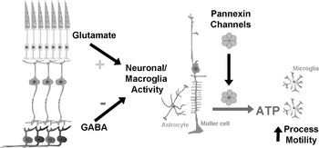
Figure 1 presents a schematic that summarizes some possible mechanisms relating neurotransmitters to microglial motility in the uninjured CNS.
Possible relationships between endogeneous neurotransmission in the retina and ‘resting’ microglial process motility. Ionotropic excitatory glutamatergic neurotransmission and inhibitory GABAergic neurotransmission may directly influence the level of electrical activity in intermediate neuronal or macroglial elements of the retina. The levels of activity may positively regulate the release of intracellular ATP, at least partially through the opening of pannexin-1 channels. Increases in extracellular ATP can then act directly onto microglia via P2 receptors on their surfaces to modulate microglial process motility.

CONCLUSIONS
Recent observations have revealed that microglia in the uninjured CNS have highly suggestive patterns of distribution, morphology, behavior and cellular associations that reflect important constitutive functions of immune surveillance, synapse maintenance and trophic support. Additional data also indicate that these microglial features are not cell-autonomously regulated but are subject to constant regulation by different external signals, including ionotropic neurotransmission. These regulatory mechanisms, although still incompletely understood, can provide insight into the ongoing communications that microglia have with other CNS cell types, and confer further information about constitutive microglial functions.
Currently, it appears that different forms of ionotropic neurotransmission are able to provide bidirectional regulation of ‘resting’ microglial morphology and motility. Interestingly, these effects are indirectly mediated, and may act through modulating the overall level of activity in particular regions of the CNS. It also appears that overall levels of activity, as opposed to local effects at particular loci or synapses, may be more influential. Electrical activity, perhaps reflected in calcium waves propagating through a macroglial syncytium, induces ATP release through pannexin-1 channels which then act to influence microglia directly.
The evidence accumulated so far suggests that microglia in the CNS are not ‘silent sentinels’, passively awaiting a pathological stimulus, but are instead integrated into an ongoing and functionally significant conversation involving the neuronal, macroglial and the immune components of the CNS. The notion that constitutive microglial process motility may underlie synapse maintenance gives rise to the idea that neuronal activity and neurotransmitter signaling may indeed be coupled to the health and even the plasticity of synapses through the microglial actions. In this schema, microglia may be a cellular mediator of activity-dependent scaling of synaptic strength in brain circuits, possibly through tumor necrosis factor-α signaling (Stellwagen and Malenka, Reference Stellwagen and Malenka2006). Indeed, modulation of microglial physiology through the CX3CR1 receptor can lead to maturational changes of brain circuits (Paolicelli et al., Reference Paolicelli, Bolasco, Pagani, Maggi, Scianni and Panzanelli2011), and the elimination of a putative microglial target, C1q, which is found on developing synapses, can result in surplus synapses that give rise to epileptic activity (Stevens et al., Reference Stevens, Allen, Vazquez, Howell, Christopherson and Nouri2007; Chu et al., Reference Chu, Jin, Parada, Pesic, Stevens and Barres2011). Conversely, CNS pathologies that result in imbalances of neurotransmission and neural activity (Schaeffer and Gattaz, Reference Schaeffer and Gattaz2008; Luchetti et al., Reference Luchetti, Huitinga and Swaab2011) may potentially perturb microglial physiology and thereby exacerbate pathological synaptic losses and derangements. Studies clarifying the relationship between microglia physiology, neuronal activity and synaptic structure will be of significant interest in the next steps forward.
Statement of interest
None.
ACKNOWLEDGEMENTS
We thank Benjamin Chaon for helpful comments on the manuscript. This work is supported by the National Eye Institute Intramural Research Program.