Implications
The use of drugs in livestock and public perceptions about drug residues in animal products has prompted studies of alternative approaches to support efficient animal production. In this study, antibodies raised in cattle against insulin-like growth factor-1 were purified and injected into rats in order to determine whether they may be protective against weight loss under nutrient-limited conditions. This passive immunisation approach provides a drug-free model to inform our understanding of the underlying biology. It also provides a preview of the potential of the approach to reduce weight loss in cattle during the dry season in tropical production systems.
Introduction
Dry season weight loss is a problematic aspect of cattle production in the dry tropics. It arises due to a general paucity of quality feed during the dry winter months. Sensitivity of the insulin-like growth factor (IGF) axis to nutritional status is well established and its role in catabolic states, both nutritionally induced and otherwise (hypermetabolism associated with trauma, sepsis and thermal injury), have been widely investigated. During the catabolic response, characterised by weight loss, negative nitrogen balance and muscle wasting, circulating IGF-1 levels are rapidly depressed, and commonly become refractory to growth hormone (GH) stimulation. That is, circulating IGF-1 is depressed, despite increased basal levels or exogenous administration of GH (Botfield et al., Reference Botfield, Ross and Hinds1997; Nass and Thorner, Reference Nass and Thorner2002). Depression of circulating IGF-1 has consistently been associated with fasting, restricted feed, protein/nitrogen and caloric intake in both monogastric and ruminant species. In such situations, provision of a nutritionally adequate diet will restore circulating IGF-1 levels (Hossner et al., Reference Hossner, McCusker and Dodson1997; O’Connor et al., Reference O’Connor, Bush, Suryawan, Nguyen and Davis2003). However, nutrition alone is not sufficient to protect lean body mass or reverse protein catabolism during severe catabolic illness (Cerra et al., Reference Cerra, Siegel, Coleman, Border and McMenamy1980). The characteristic depression of circulating IGF-1 has been widely indicated as a contributing factor to the net protein catabolism associated with the sustained catabolic response (Gibson and Hinds, Reference Gibson and Hinds1997; Thissen et al., Reference Thissen, Underwood and Ketelslegers1999).
IGF-1 administration has been reported to attenuate the catabolic response to nutritional restriction in a range of species. Improvements in muscle protein synthesis rates were observed in fasted rats (12 to 15 h) when IGF-1 was administered concomitantly with either amino acids or insulin (Jacob et al., Reference Jacob, Hu, Niederstock, Hasan, McNulty, Sherwin and Young1996). Infusion (90 min) of IGF-1 into 24-h fasted rats resulted in decreased circulating amino acids, but this was independent of increased protein synthesis. Thus, it appears that exogenous IGF-1, under these conditions, inhibited protein degradation (Jacob et al., Reference Jacob, Barrett, Plewe, Fagin and Sherwin1989). Furthermore, human subjects in states of moderate catabolism induced by dietary restriction exhibited improved nitrogen balance and decreased fasting blood glucose in response to rhIGF-1 administration (Clemmons et al., Reference Clemmons, Smith-Banks and Underwood1992).
In cattle, retained nitrogen (Elsasser et al., Reference Elsasser, Rumsey, Hammond and Fayer1988) and body weight (BW) gain (Breier et al., Reference Breier, Gluckman and Bass1988) have been correlated with circulating IGF-1 during feed restriction. Ogawa et al. (Reference Ogawa, Breier, Bauer, Gallaher, Grant, Walton, Owens and Gluckman1996) observed a negative correlation between net protein catabolism and plasma IGF-1 levels during GH treatment of fasting lambs. We have also reported the protein sparing effects of an intravenous infusion of the IGF-1 analogue Long(R3)-IGF-1 in beef heifers, which were slowly losing live-weight due to restricted feeding (Hill et al., Reference Hill, Hunter, Lindsay and Owens1999). Long(R3)-IGF-1 administration also markedly reduced the plasma concentrations of all amino acids measured and glucose. In addition, net protein catabolism in fasted lambs was decreased by the infusion of IGF-1 alone, and totally reversed with concomitant parenteral nutrition (Koea et al., Reference Koea, Douglas, Breier, Shaw and Gluckman1992). Oddy and Owens (Reference Oddy and Owens1996) also showed that IGF-1 infusion increased protein gain in the hindlimb of fasted and feed restricted lambs.
Thus, across multiple species, including ruminants and rodents, circulating IGF-1 is implicated as a controlling factor of BW and nitrogen balance during times of nutritional restriction. Furthermore, infusion of exogenous IGF-1 enhances protein retention during catabolic stress.
Previously, growth-promoting actions of antibodies raised against IGF-1 have been reported in nutrient sufficient dwarf rats (Stewart et al., Reference Stewart, Bates, Calder, Woodall and Pell1993), dwarf mice (Stewart et al., Reference Stewart, Bates, Calder, Woodall and Pell1993; Hill and Pell, Reference Hill and Pell1998) and cattle (Hill et al., Reference Hill, Smith and Holmes1998a and Reference Hill, Gazzola, Herd and Oddy1998b). Circulating IGF-1 was significantly increased by administration of ovine anti-IGF-1immunoglobulin (Ig) in dwarf rats, which have reduced endogenous serum IGF-1 (Stewart et al., Reference Stewart, Bates, Calder, Woodall and Pell1993). These increases in circulating IGF-1 were associated with a significant increase in whole BW gain and muscle mass (gastrocnemius) compared with non-immune Ig treated controls. The authors proposed that anti-IGF-1 Ig was protecting circulating IGF-1 from degradation in a manner analogous to that of the IGF binding proteins (IGFBPs). It was later demonstrated by Hill et al. (Reference Hill, Flick-Smith, Dye and Pell1997) that clearance of IGF-1 from the serum of dwarf rats was impeded by pre-treatment with anti-IGF-1 Ig.
With evidence for IGF-1 to enhance protein retention in catabolic states, and the potential for combined exogenous IGF-1 plus anti-IGF-1 Ig to enhance growth in IGF insufficient rodent models, we investigated whether treatment of genetically normal rats with bovine anti-IGF-1 Ig would enhance growth and if anti-IGF-1 Ig treatment would ameliorate live-weight loss in genetically normal rats offered a severely protein-restricted diet. We hypothesised that anti-IGF-1 antibody treatment of genetically normal rats offered a protein-restricted diet would improve protein retention and decrease weight loss.
Material and methods
Anti-IGF-1 Ig production, purification and determination of anti-IGF-1 Ig titres
Polyclonal antibodies were raised against recombinant human IGF-1 in six adult Brahman cross steers. IGF-1 was conjugated to ovalbumin using glutaraldehyde as follows: Ovalbumin (5 mg/ml in 0.01 M phosphate buffered saline (PBS), pH 7.4; Sigma Chemical Co., St Louis, USA) was added to IGF-1 (10 mg/ml in 10 mM HCl; GroPep Pty Ltd, Adelaide, Australia) to give a 2 : 1 molar ratio of IGF-1 : ovalbumin. Glutaraldehyde (0.2%; Sigma Chemical Co.) was slowly added to obtain a 50 : 1 molar ratio of glutaraldehyde : IGF-1 and then constantly agitated for 1 h at room temperature. Protein concentration of the conjugate solution (typically between 0.4 and 2.3 mg/ml) was determined using the micro bicinchoninic acid (BCA) protein assay (Pierce, Rockford, USA) and bovine serum albumin (BSA; Sigma Chemical Co.) as the standard. Aliquots were stored at −80°C until use. Before administration, conjugate was diluted (150 μg/ml) with sterile PBS, 150 μg/ml diethylaminoethyl-dextran hydrochloride (Fluka, Buchs, Switzerland) added and the final volume emulsified in an equal volume of Freund’s incomplete adjuvant (Pierce) using an Ultra Turrax T25 homogeniser (IKA Labortechnik, Selangor, Malaysia). Control immunogen was similarly prepared using an equivalent volume of sterile PBS. Monthly subcutaneous injections were given bilaterally in the neck and shoulder region. Test steers received 150 μg IGF-1 per challenge, while sham-immunised animals received an equivalent volume of control.
Igs were partially purified from non-immune and anti-IGF-1 antisera (from the best responding animal) by two incubations with saturated ammonium sulphate (0.54 and 0.25×original serum volume), followed by centrifugation (1000×g for 15 min at 4°C), re-suspension in a minimum of distilled water and dialysis against PBS at 4°C. The protein concentration of the Ig preparations (49 to 62 mg/ml) was determined by BCA protein assay (Pierce) using BSA (Sigma Chemical Co.) as the standard. Indirect enzyme-linked immunosorbent assay (ELISA) was used to determine anti-IGF-1 Ig titres. Titration to negative absorbance levels (typically <0.1) allowed interpolation of titres, which were defined as the serum dilution at which the sample absorbance was 0.2 absorbance units above that of the negative control (Hill et al., Reference Hill, Flick-Smith, Dye and Pell1997). Plates were coated with 100 μl IGF-1 (5 μg/ml in 0.1 M carbonate, pH 9.6). After incubation with the Ig fractions, bound antibody was detected using peroxidase conjugated rabbit anti-bovine IgG (Sigma Chemical Co.), 0.55% 2,2′-azino-bis(3-ethylbenz-thiazoline-6-sulfonic acid) in 0.05 M citric acid/sodium phosphate buffer, pH 4.0 and hydrogen peroxide (0.01%). Absorbance at 405 nm was measured.
Determination of specificity and affinity of anti-IGF-1 Ig
The binding specificity of anti-IGF-1 Ig was investigated by comparing the titres obtained when LR3IGF-1 (GroPep, Adelaide, Australia), [Gly1] IGF-2 (Media grade; GroPep, Adelaide, Australia) and insulin (Boehringer Manheim, North Ryde, Australia) were applied as the coating antigens in the previously described indirect ELISA protocol.
Affinity of the anti-IGF-1 Ig for IGF-1 and LR3IGF-1 was determined using saturation (IGF-1) and competitive (LR3IGF-1) radioligand binding techniques. In saturation binding studies, total binding was determined using 10 concentrations (31.70 to 53.07 pm) of 125I-labelled IGF-1 (specific activity 137.7 to 277.8 Ci/g) incubated with 3 μg protein of the anti-IGF-1 Ig preparation in buffer (1% BSA in PBS). Non-specific binding was defined by 100 nM unlabelled IGF-1 (Receptor grade, GroPep, Australia). Separation of bound from free radioligand was achieved by polyethylene glycol precipitation and centrifugation at ∼2000×g (30 min, 4°C). Scatchard analysis of saturation isotherms and the fitting of data to the appropriate binding model was by non-linear regression analysis of untransformed data using the EBDA-LIGAND computer programs (Munson and Rodbard, Reference Munson and Rodbard1980; McPherson, Reference McPherson1985).
Competitive binding studies were performed using 3 μg protein/tube of Ig preparation and ∼0.5 nM 125I-IGF-1 (∼250 000 cpm, specific activity 58.37 to 150.00 Ci/g) in competition with 19 concentrations of unlabelled LR3IGF-1 and IGF-1 (1 × 10⁻¹¹ M to 1 × 10⁻⁵ M). Similar studies were conducted using IGF-2. However, complete displacement of 125I-IGF-1 was not attained despite the use of high concentrations of IGF-2 (up to 1 × 10⁻⁴ M). The EBDA-LIGAND computer program (Munson and Rodbard, Reference Munson and Rodbard1980; McPherson, Reference McPherson1985) was used to estimate the affinity of anti-IGF-1 Ig for LR3IGF-1 from the resultant inhibition curves. Iodination did not alter the affinity of the Ig for IGF-1.
Animals and treatments
Thirty female, JC Lewis rats (3 to 4 weeks old) were purchased from the University of Queensland Central Animal Breeding House (St. Lucia, Australia). Rats were housed individually and allowed ad libidum access to food and water. During a 6-day acclimatisation period, all rats were offered a standard pelleted rodent diet (minimum 20% crude protein). Live-weight and dietary intake were recorded (0900) throughout. Animals (mean initial live-weight 100.6 g) were stratified by live-weight and randomly allocated to one of the six treatment groups (n = 5 per group) as follows: From day 1 of the treatment period, rats were offered one of the two diets: diet 1 (15 rats) continued to be offered standard rat pellets, while for diet 2, 15 rats were offered a pelleted, isocaloric diet containing 4% protein. Within each dietary group, rats were administered either diluent (sterile PBS), non-specific Ig (5 mg/ml) or anti-IGF-1 Ig (5 mg/ml). All treatment volumes were 1.5 ml/day and administered as two equal i.p. injections (0930 and 1630). Animals were sacrificed by stunning and cervical dislocation on day 13. Animals were decapitated and blood collected. Extracted plasma was stored at −20°C until analysis. Tissues: small and large intestine, stomach, caecum, liver, kidneys, skin, visible fat and carcase were dissected and weighed. All portions of the gut were flushed with PBS before weighing.
Plasma from all rats was screened (1 : 100 dilution) to confirm the presence or absence of biologically active, specific anti-IGF-1 Ig using the previously described indirect ELISA protocol.
Size exclusion chromatography of plasma
In vitro binding of 125I-IGF-1 in pooled end point plasma from each of the treatments was characterised using size exclusion chromatography, by the method of Ballard et al. (Reference Ballard, Knowles, Walton, Edson, Owens, Mohler and Ferraiolo1991). Plasma (300 μl) was incubated with ∼300 000 cpm (0.75 ng) of 125I-IGF-1 for 18 h at 4°C. Duplicate plasma samples (100 μl) were fractionated at room temperature on a 16 × 200 mm Sephacryl S-300-HR column (Sigma Chemical Co.) equilibrated with a buffer containing sodium phosphate (50 mM), sodium chloride (150 mM), sodium azide (0.02%; w/v) and heparin (10 U/ml) at pH 7.2. The flow rate was 0.5 ml/min and 0.5 ml fractions were collected and counted. Elution of protein was constantly recorded at 280 nm and the following standards used, in addition to radio-labelled IGF-1 (7.6 kDa), to regularly calibrate the column: thyroglobulin (669 kDa), apoferritin (443 kDa), bovine gamma globulin (160 kDa), BSA (66 kDa), ovalbumin (45 kDa), myoglobin (17 kDa) and cytochrome C (12 kDa).
Statistical treatment of results
Values are presented as means or least squares means ±s.e. for each treatment period or treatment day. All analyses were performed using SAS (SAS, 1999). A two-way ANOVA was used to test for effects of diet and treatment and their interaction. The interaction between diet and treatment was tested, and omitted from the analysis when non-significant. The multiple comparisons involved necessitated the use of a Tukey–Kramer adjustment for multiple comparisons of means. To analyse the effect of diet and treatment upon growth rate (g/day) and the change in feed intake per day (g/day), a regression of weight or feed intake was fitted to the daily measurements for each rat throughout the study. These predicted rates were analysed by an ANOVA using Proc GLM of SAS. Change in dietary intake (rate) was used to describe whether it was increasing, decreasing or unchanging throughout the experimental period. Initially, cumulative dietary intake was fit as a covariate in the analysis of growth rate, and final BW as a covariate in the analysis of dietary intake. Neither were significant (P > 0.05) and therefore omitted from further analysis. To assess within rat variation in the rate of the dietary intake and growth rate regressions, an analysis of the root mean square error of the individual rat regressions was performed.
Results
Antibody characteristics
The titre of the bovine anti-IGF-1 antiserum was 22 266 (±1334) before Ig precipitation, and 16 755 (±814) in the Ig preparations used in these studies. When assessed using saturation radioligand binding techniques, this preparation yielded an almost linear Scatchard plot, with a Hill co-efficient of 0.951 ± 0.012, indicating a single class of IGF-1 binding sites. The affinity of anti-IGF-1 Ig for IGF-1 was 2.14 ± 0.66 × 10⁹ l/mol. Neither the non-immune serum nor Ig preparation exhibited any IGF-1 binding when similarly assayed.
Indirect ELISA using insulin, IGF-1 and LR3IGF-1 as the coating antigens was used to investigate the specificity of anti-IGF-1 Ig binding. Initial screening (1 : 100 and 1 : 200) of the anti-IGF-1 Ig preparation indicated that, in comparison to absorbance values achieved using IGF-1 as the coating antigen; the antibodies similarly bound LR3IGF-1, bound IGF-2 with a reduced affinity and did not bind insulin. Subsequent calculation of titres using IGF-1, IGF-2 and LR3IGF-1 as the coating antigens were in agreement; there was no significant difference between titres calculated using LR3IGF-1 and IGF-1 as the coating antigens, but anti-IGF-1 Ig binding to IGF-2 was significantly reduced (10 755 ± 349, P < 0.01).
Competitive radioligand binding techniques were used to more accurately compare anti-IGF-1 Ig affinity for 125IGF-1, IGF-2 and LR3IGF-1. Binding to IGF-2 was of an extremely low affinity; complete displacement of 125I-IGF-1 was not attained despite the use of high concentrations of IGF-2 (up to 1 × 10⁻⁴ M). Anti-IGF-1 Ig exhibited ∼20-fold decrease in affinity for LR3IGF-1 in comparison to IGF-1 (0.11 ± 0.02 v. 2.14 ± 0.66 × 10⁹ l/mol, P < 0.01). There was no significant difference between the maximum densities of binding sites calculated for each ligand.
Growth rate and organ mass
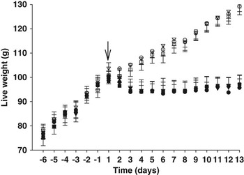
As might be expected, markedly divergent growth rates were observed between groups offered diets containing 4% or 20% protein (P < 0.01, Figure 1). Rats offered the 20% protein diet continued to grow at an average rate of 2.37 g/day, whereas rats offered the 4% protein diet lost, on average 0.13 g/day during the treatment period.
Live weights of rats including 6 pre-treatment days and 13 days after commencement (arrow) of twice-daily injections (1.5 ml/day; i.p.) of either diluent (○, •), non-immune Ig (∇, ▾), 5 mg/ml or anti-IGF-1 Ig (□, ▪) 5 mg/ml. Rats either remained on a 20% protein diet (open symbols) as in the pre-treatment period (days −6 to −1) or were transferred to a 4% protein diet (closed symbols) on day 1. Values are means ± s.e.

Treatment with anti-IGF-1 Ig had no effect on growth rate in either dietary group, even when differences in dietary intake were taken into account. There appeared to be a trend towards attenuation of weight loss during protein restriction (4% protein diet) as a result of treatment with anti-IGF-1 Ig (diluent −0.183, non-immune Ig −0.203 and anti-IGF-1 Ig 0.061 g/day, P = 0.08). Unlike dietary intake, day-to-day variation in growth rate was unaffected by diet and treatment.
Dietary protein restriction (4% protein diet) resulted in a significant decrease in the mass of all tissues measured (P < 0.01). However, as there was a significant effect of diet upon live-weight, no significant differences in tissue weights remained when live-weight was fitted as a covariate. This indicated that the decrease in live-weight associated with protein restriction was not associated with disproportionate wasting of any tissues measured. Treatment with anti-IGF-1 Ig did not affect the mass of any tissues measured in either analysis.
Dietary intake
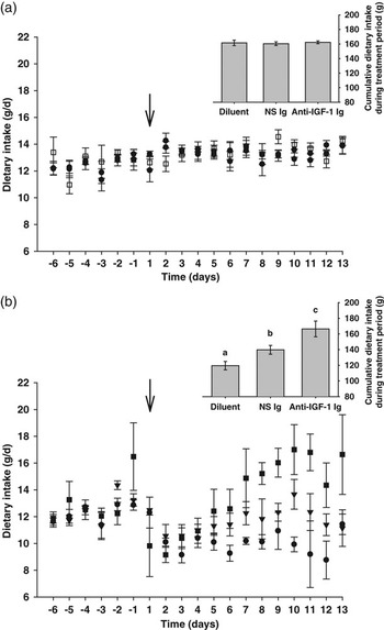
Dietary treatment had marked effects on both cumulative dietary intake (P < 0.01) and the rate of dietary intake per day (P = 0.011, Figure 2). Treatment with bovine anti-IGF-1 Ig had no effect on the rate of dietary intake per day or cumulative dietary intake for animals offered a 20% protein diet. However, treatment with bovine anti-IGF-1 Ig prevented the significant depression of cumulative dietary intake observed in diluent, and non-specific Ig treated groups offered the 4% protein diet. The cumulative dietary intake of the anti-IGF-1 Ig treated, 4% dietary protein group did not differ significantly from that of the groups offered the 20% protein diet. In addition, after an initial decrease when transferred to the low protein diet, this group exhibited a constantly increasing dietary intake per day (P < 0.001) during treatment. In addition, within the 4% dietary protein group, rats treated with non-immune Ig exhibited a cumulative feed intake that was intermediate between that of the diluent treated and anti-IGF-1 Ig treated groups (P < 0.05).
(a) and (b) Dietary intakes (g/day) of rats including 6 pre-treatment days and 13 days after commencement (arrow) of twice-daily injections (1.5 ml/day, i.p.) of either diluent (○, •), non-immune IgG (∇, ▾; NI; 5 mg/ml) or anti-IGF-1 Ig (□, ▪; 5 mg/ml). Rats either remained on a 20% protein diet (a, open symbols) or were transferred to a 4% protein diet at this time (b, closed symbols). Bar graphs display the cumulative dietary intake (g) of each group during the treatment period. Values are means ± s.e.

When day-to-day variation in the rate of dietary intake was considered, it was found that rats which were offered a 4% protein diet exhibited greater variation in dietary intake per day than the rats that were offered a 20% protein diet. This was not associated with an increased variation in growth rate. In addition, anti-IGF-1 Ig treated rats offered a 4% diet exhibited a significantly greater variation in change in dietary intake per day than all other groups (P < 0.05).
Size exclusion chromatography: characterisation of in vitro binding of 125I-IGF-1 in rat plasma
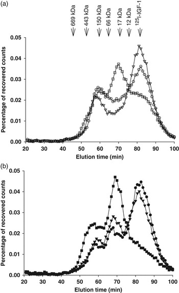
Figure 3 illustrates the molecular mass distribution of 125I-IGF-1 after in vitro incubation in pooled plasma from each treatment group. Three major peaks of radioactivity were eluted at ∼150 kDa, between 30 and 40 kDa and at 7.6 kDa (free IGF-1). Within each dietary treatment, diluent and non-immune Ig treated control groups exhibited similar 125I-IGF-1 elution profiles. However, 125I-IGF-1 elution profiles appeared to differ between the two dietary treatments: a peak of radioactivity in the 30 to 40 kDa region was only observed after protein restriction. In comparison to control groups, anti-IGF-1 Ig treatment resulted in apparently increased 125I-IGF-1 binding in the 30 to 40 kDa region and a concomitant reduction in elution of free 125I-IGF-1. Although a similar 125I-IGF-1 elution profile was observed in anti-IGF-1 Ig treated animals in both dietary groups, the reduction in free 125I-IGF-1 appeared to be of a greater magnitude when dietary protein was restricted. Protein restriction appeared to depress binding at ∼150 kDa in the plasma of diluent and non-immune Ig treated groups. Anti-IGF-1 Ig treatment was effective in preventing this apparent decrease in ∼150 kDa binding. All protein sufficient groups exhibited similar peaks in the 150 kDa region.
Binding of 125I-IGF-1 in rat plasma. Rats (n = 5 per group) were offered either a 20% (a) or 4% (b) protein diet and administered (1.5 ml/day, i.p.) either diluent (○, •), non-immune Ig (∇, ▾; 5 mg/ml) or anti-IGF-1 Ig (□, ▪; 5 mg/ml) for 13 days. Plasma was collected on day 13 and a sub-sample pooled by treatment group. Pooled plasma samples (300 μl) were incubated with 0.75 ng of 125I-IGF-1 for 18 h at 4°C. Duplicate plasma samples (100 μl) were subjected to size exclusion chromatography on a Sephacryl S300-HR column. Values are expressed as the percentage of recovered radioactivity in each 1-min fraction (0.5 ml) and represent duplicate means of the pooled plasma samples from each group. Arrows indicate molecular weight standards.

Discussion
To date, the enhancement of growth by anti-IGF-1 antibodies has been demonstrated only in animals possessing an abnormal IGF axis. Potentiation of IGF-1 action by passively administered ovine anti-IGF-1 Ig has been demonstrated only in dwarf rats and mice, in which suppressed GH secretion results in reduced endogenous IGF-1, IGFBP-3 and acid-labile subunit (ALS) (Stewart et al., Reference Stewart, Bates, Calder, Woodall and Pell1993; Hill and Pell, Reference Hill and Pell1998; Pell et al., Reference Pell, Hill, Stewart, Weston and Flick-Smith2000). Similarly, a moderate growth response achieved in cattle actively immunised against IGF-1 was observed after a period of feed restriction, which is also known to suppress circulating IGF-1 and perturb normal IGFBP concentrations. These cattle were grazed under field conditions and it is of interest that cattle under feedlot conditions, and presumably possessing an unperturbed IGF axis, did not exhibit a similar growth response to active immunisation against IGF-1. There was evidence, however, of a repartitioning of nutrient deposition from fat to lean (Hill et al., Reference Hill, Gazzola, Herd and Oddy1998b). It was therefore of importance to investigate whether antibodies raised against IGF-1 were capable of enhancing growth in animals with an intact IGF axis. Furthermore, given the marked depression of circulating IGF-1 during catabolism (regardless of the cause) and in light of previous positive responses to IGF-1 administration in such states, it was of interest to investigate whether anti-IGF-1 Ig administration could ameliorate wasting associated with catabolic conditions using a model in which catabolism was induced by protein-restriction.
An important difference between the studies of Pell and co-workers and this study was that we administered anti-IGF-1 Ig alone, rather than concomitantly with exogenous IGF-1. Previously, growth potentiation has predominantly been observed when anti-IGF-1 Ig was co-administered with IGF-1, although studies by Hill et al. in cattle (Reference Hill, Smith and Holmes1998a) and Stewart et al. in dwarf rats (Reference Stewart, Bates, Calder, Woodall and Pell1993) indicated that anti-IGF-1 Ig may also elicit growth-promoting effects via modulation of endogenous IGF-1 action. Certainly, it has been demonstrated that passively administered ovine anti-IGF-1 Ig can form complexes with endogenous IGF-1 and that pre-incubation of IGF-1 and anti-IGF-1 Ig was not necessary to achieve potentiation of weight gain (Hill and Pell, Reference Hill and Pell1998). Thus, this study also investigated whether any growth response to administration of anti-IGF-1 Ig to protein-sufficient and protein-restricted normal rats could be obtained via modulation of endogenous IGF-1.
In this study, administration of bovine anti-IGF-1 Ig did not significantly affect growth rate in protein-sufficient or protein-restricted rats. The most outstanding effect observed was the severe reduction in growth rate and dietary intake and as a result of protein deprivation. Previous studies have also reported inhibitory effects of low-protein diets on food intake and BW (Miura et al., Reference Miura, Kato and Noguchi1992; Du et al., Reference Du, Higginbotham and White2000; Katsumata et al., Reference Katsumata, Kawakami, Kaji, Takada and Dauncey2002), while others have reported no effect, or an increase in these parameters (VandeHaar et al., Reference VandeHaar, Moats-Staats, Davenport, Walker, Ketelslegers, Sharma and Underwood1991; Du et al., Reference Du, Higginbotham and White2000; Katsumata et al., Reference Katsumata, Kawakami, Kaji, Takada and Dauncey2002). In this connection, Forbes (Reference Forbes2000) outlined the general relationship between the concentration of an essential nutrient in food and the rate of voluntary food intake, which is associated with concomitant changes in growth rate and other production parameters (e.g. milk production). Severe nutrient deficiencies suppress dietary intake while marginal deficiencies are stimulatory. Accordingly, in rats, Du et al. (Reference Du, Higginbotham and White2000) demonstrated increased dietary intake with moderate protein restriction (at or just below growth requirements), but decreased intake and BW in response to more severe protein restriction. This pattern of response, in addition to other factors that affect protein requirements and therefore intake (e.g. age, gender, psychological stress), may explain the varied reported dietary intake and growth responses to dietary protein content (Du et al., Reference Du, Higginbotham and White2000).
Depression of dietary intake during protein-restriction was prevented by anti-IGF-1 Ig administration. Although some of this effect can be attributed to daily administration of extra protein (7.5 mg/day), demonstrated by the significant elevation of cumulative dietary intake of the non-immune Ig treated group, this does not fully account for the observed response. Voluntary intake is influenced by many factors, including a series of negative feedback signals from the digestive tract, liver and other organs in response to the presence of nutrients. Integration of signals to determine what food to eat, and whether feeding should start or stop, is coordinated by the central nervous system and involves diverse pathways including the hindbrain and hypothalamus as important components (Forbes, Reference Forbes2000). Factors known to affect hunger and satiety include neuropeptide Y, leptin, serotonin, cholecystokinin, enterostatin and ghrelin. Some of these factors, such as leptin, are known to be influenced by, or influence, components of the GH-IGF-1 axis (LaPaglia et al., Reference LaPaglia, Steiner, Kirsteins, Emanuele and Emanuele1998; Böni-Schnetzler et al., Reference Böni-Schnetzler, Hauri and Zapf1999; Villafuerte et al., Reference Villafuerte, Fine, Bai, Zhao, Fleming and DiGirolamo2000; Leury et al., Reference Leury, Baumgard, Block, Segoale, Ehrhardt, Rhoads, Bauman, Bell and Boisclair2003). Thus, administration of bovine anti-IGF-1 Ig may have modulated any of these factors to prevent reduction of dietary intake. Spontaneous reduction of food intake, such as that observed in rats offered a protein-restricted diet, results in protein-energy malnutrition. Energy absorbed from extra food ingested by anti-IGF-1 Ig treated rats was not stored in detectable quantities as no effect on body or organ masses was detected. Such an observation may suggest an increase in metabolic rate. Still, increased rate of passage through the digestive system (possibly associated with reduced absorptive capacity of the small intestine) cannot be discounted.
In contrast to the studies of Stewart et al. (Reference Stewart, Bates, Calder, Woodall and Pell1993) and Hill et al. (Reference Hill, Flick-Smith, Dye and Pell1997), in this study, the in vitro plasma binding profile of 125I-IGF-1 in bovine anti-IGF-1 Ig treated groups was characterised by; (a) an apparent increase in the proportion of 125I-IGF-1 eluting at a peak molecular mass of ∼30 to 40 kDa, presumably representing low molecular mass IGFBPs, and (b) a concomitant decrease in the 7.6 kDa peak, presumably representing free hormone (Figure 3). These changes were observed in both protein-sufficient and protein-restricted groups, although were more marked in the latter, and suggest that bovine anti-IGF-1 Ig either facilitates binding of 125I-IGF-1 by low molecular mass IGFBPs, or in some way increases the quantity of these IGFBPs in plasma. Low molecular mass IGFBPs have a defined role in regulating transfer of IGF-1 from the circulation to the tissues (Butler and LeRoith, Reference Butler and LeRoith2001; Clemmons, Reference Clemmons2009). Facilitation of IGF-1 binding by these IGFBP may be a result of their similar affinity for IGF-1 as bovine anti-IGF-1 Ig, or an antibody-induced conformational change in IGF-1 that increases its affinity for the low molecular mass binding proteins. Thus, IGF-1 bound by bovine anti-IGF-1 Ig would become more accessible to the low molecular mass IGFBPs and more available to the tissues. Bovine anti-IGF-1 Ig facilitated transfer of IGF-1 to low molecular mass IGFBPs as an explanation for enhanced 125I-IGF-1 binding in the ∼30 to 40 kDa region is consistent with the existing literature as Hill et al. (Reference Hill, Flick-Smith, Dye and Pell1997) showed that ovine IGF-1 Ig increased 125I-IGF-1 binding in a 50 kDa peak from dwarf rat plasma over time. However, it is possible that at least a portion of this peak consists of anti-IGF-1 Ig degradation fragments capable of binding 125I-IGF-1. It must be noted that heparin (albeit at a low concentration, 10 U/ml) was included in the elution buffer to prevent column fowling. Moller et al. (Reference Moller, Jorgensen, Chen, Larnkjaer, Ledet, Flyvbjerg and Frystyk2006) have shown that a substantially higher concentration of heparin (50 U/ml) is required to displace IGF-1 from the IGFBPs though interactions with their heparin binding domains. Thus, it is possible that there was a small fractional heparin-induced displacement of IGF-1-IGFBP interactions in this study. However, this would have a similar effect on all treatments and is unlikely to have affected our conclusions.
During dietary protein restriction, transfer of circulating IGF-1 to the low molecular mass IGFBPs (a consequence of depressed circulating IGFBP-3 and ALS and elevated IGFBP-1 and IGFBP-2) results in net movement of IGF-1 from the circulation to the tissues (Thissen et al., Reference Thissen, Davenport, Pucilowska, Miles and Underwood1992 and Reference Thissen, Ketelslegers and Underwood1994). Accordingly, Thissen et al. (Reference Thissen, Davenport, Pucilowska, Miles and Underwood1992) demonstrated a significant shift in the predominant 125I-IGF-1 binding species from a 150 kDa complex to a 30 kDa complex as a result of dietary protein restriction in normal rats. Similarly, in this study, dietary protein restriction in control groups resulted in an apparent reduction in the proportion of IGF-1-bound fraction eluting in the ∼150 to 300 kDa and 30 to 40 kDa peaks, without concomitant changes in the proportion of 125I-IGF-1 eluting as free hormone (Figure 3). Similar enhancement of the 30 to 40 kDa 125I-IGF-1 binding species was also observed in the bovine anti-IGF-1 Ig treated group during protein restriction.
Furthermore, administration of bovine anti-IGF-1 Ig during protein restriction completely prevented the depression of high molecular mass binding observed in control groups, but had no effect upon this peak during protein sufficiency. This lack of effect tends to suggest either that (i) at the applied dosage, anti-IGF-1 Ig could not effectively complete with the quantity of ternary IGFBP-3 complex present in normal rat serum, given that soluble IGFBP-3 exhibits an approximate 10- to 20-fold greater affinity (∼1.7 × 10¹⁰ l/mol) for IGF-1 than do bovine anti-IGF-1 Ig, IGFBP-1 and IGFBP-2 (Conover, Reference Conover1992; Wong et al., Reference Wong, Fong and Yang1999) and/or (ii) the epitope recognised by bovine anti-IGF-1 Ig is located within a region of IGF-1 involved in IGFBP-3 binding, or is proximal enough for steric hindrance to effect antibody binding. In this respect, it is perhaps relevant that all stimulatory effects of anti-IGF-1 Ig reported to date have been observed in animals with an impaired IGF-1 axis (including protein-restricted normal rats), characterised by low levels of circulating IGF-1, IGFBP-3 and ALS. Furthermore, almost all of the growth-promoting effects of ovine anti-IGF-1 Ig in dwarf rodent models (Stewart et al., Reference Stewart, Bates, Calder, Woodall and Pell1993; Hill and Pell, Reference Hill and Pell1998; Pell et al., Reference Pell, Hill, Stewart, Weston and Flick-Smith2000) have been achieved with co-administration of IGF-1. As circulating IGF-1 was likely to have been in excess of the ternary complex in these studies, it becomes difficult to assess the impact of these observations.
The findings of this study suggest that the level of circulating IGF-1 and IGFBP-3/ALS may be of importance in determining the effects of anti-IGF-1 Ig administration. The only significant indications of an effect of bovine anti-IGF-1 Ig on growth of normal rats were observed in protein-restricted groups, which exhibited depressed levels of circulating IGF-1 and IGFBP-3/ALS, analogous to the dwarf rodent models. Significant changes in the 125I-IGF-1 binding profile in plasma of both protein-sufficient and protein-restricted normal rats indicate that treatment with bovine anti-IGF-1 Ig may facilitate a shift to low molecular mass IGFBPs, which function to transfer IGF-1 from the circulation to the tissues. In accordance with the above conclusions, these changes were more marked in protein-restricted groups. Furthermore, only during protein-restriction did treatment with bovine anti-IGF-1 Ig increase 125I-IGF-1 binding in the ∼150 to 300 kDa region, relative to control groups. Thus, it would seem that (i) rats with depressed IGF-1 and/or IGFBP-3 are more sensitive to administration of anti-IGF-1 Ig and (ii) a greater dosage of anti-IGF-1 Ig may be required to influence growth of normal rats in a similar manner.
Previously Hill et al. (Reference Hill, Smith and Holmes1998a) indicated the importance of the effective dosage of anti-IGF-1 Ig when reporting a significant relationship between the sum of antibody titres and weight gain in actively immunised cattle. In this connection, Kerr et al. (Reference Kerr, Laarveld and Manns1990) observed that administration of a high affinity (1.3 × 10¹¹ l/mol) anti-IGF-1 monoclonal antibody, which had demonstrated inhibitory activity in vitro, had no effect on any measured growth parameter. In that study, antibody was not administered in excess of circulating IGF-1.
In conclusion, this study has shown that anti-IGF-1 Ig administration in dietary protein-restricted rats has a beneficial effect, increasing voluntary intake. Anti-IGF-1 Ig treatment was associated with a shift in plasma binding profiles of IGF-1. Thus, studies of the underlying mechanism have the potential to inform future technologies aimed at reducing dry-season weight loss of ruminants raised in tropical environments.
Acknowledgements
The authors specially thank Ms Marianne Reich, Dr Senka Margetic and Ms Debra White for their assistance in these studies. The authors thank the Australian Research Council and the Co-operative Research Centre for the Beef and Cattle Industry for funding support. NNS was supported by an Australian Post-graduate Award.