Hypoxic-ischaemic encephalopathy has been associated with mortality and adverse neurodevelopmental outcomes. Therapeutic hypothermia has been the standard-of-care in newborns with moderate to severe hypoxic-ischaemic encephalopathy and has been shown to decrease mortality without increasing major disability in survivors. Reference Jacobs, Berg, Hunt, Tarnow-Mordi, Inder and Davis1,Reference Shankaran, Laptook and Ehrenkranz2 Despite therapeutic hypothermia, some newborns continue to suffer various degrees of cerebral injury. Cardiac dysfunction may be an important contributing factor to residual neurologic compromise.
Perinatal distress leading to hypoxic-ischaemic encephalopathy may also be associated with altered cardiovascular transition, characterised by a persistence of increased pulmonary vascular resistance and circulatory maladaptation, potentially disturbing myocardial function. Reference Lakshminrusimha, Shankaran and Laptook3 Right ventricular dysfunction in neonatal hypoxic-ischaemic encephalopathy has been linked with mortality or cerebral anomalies on imaging. Reference Giesinger, Bailey, Deshpande and Mcnamara4,Reference Giesinger, El Shahed and Castaldo5 We recently described a cohort of hypoxic-ischaemic encephalopathy newborns undergoing therapeutic hypothermia concerning the cardiac risk factors associated with death or significant brain injury. Reference Altit, Bonifacio and Guimaraes6 In our cohort, both right ventricle and left ventricular functions were different in those with adverse outcomes. In the context of post-natal management, disturbed myocardial performance may contribute to abnormal cerebral recovery due to impaired ventricular outputs. Furthermore, our understanding regarding the interplay between cardiac performance and the impact of hypoxia-ischaemia on cerebral vascular tone is limited.
Although markers of cardiac dysfunction have been reported in hypoxic-ischaemic encephalopathy cohorts, Reference Giesinger, El Shahed and Castaldo5,Reference Breatnach, Forman and Foran7 a comprehensive understanding of post-natal cardiovascular transition by echocardiography has not been described. A better understanding of systolic and diastolic biventricular function is of importance to identify potential future therapeutic targets to promote cardiovascular management approaches and improve neurological recovery. As such, we aim to describe the right and left ventricular function by echocardiography in newborns with hypoxic-ischaemic encephalopathy undergoing therapeutic hypothermia. Specifically, we compared systolic and diastolic echocardiographic parameters to a control population of newborns without hypoxic-ischaemic encephalopathy.
Methods
Study population and study design
This retrospective case–control study assessed a formerly described cohort of hypoxic-ischaemic encephalopathy newborns at a single institution. Reference Altit, Bonifacio and Guimaraes6 Clinical and echocardiographic data of hypoxic-ischaemic encephalopathy and control newborns were collected retrospectively between 2008 and 2017. A portion of the outlined control population was previously presented as a comparative group in publications by our team. Reference Altit, Bhombal, Chock and Tacy8,Reference Altit, Bhombal, Van Meurs and Tacy9 Newborns with moderate to severe hypoxic-ischaemic encephalopathy were treated with therapeutic hypothermia at Lucile Packard Children’s Hospital Stanford and had echocardiography performed during therapeutic hypothermia. All the controls were retrieved from the Lucile Packard Children’s Hospital Stanford echocardiographic database and were born during the 2008–2017 time period. Control patients included newborns born at term and referred for evaluation of a benign murmur, as well as those with an antenatal suspicion of coarctation ruled out in the post-natal period.
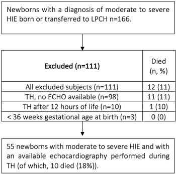
A number of hypoxic-ischaemic encephalopathy newborns were excluded if they were born at <36 weeks estimated gestational age at birth, had therapeutic hypothermia initiated >12 hours of life, or if no echocardiography was available during therapeutic hypothermia (Fig 1). Therapeutic hypothermia eligibility was ascertained according to the National Institute of Child Health and Human Development Whole-Body Hypothermia trial entry criteria. Reference Shankaran, Laptook and Ehrenkranz2 Level of encephalopathy was determined as per the modified Sarnat scoring system. Reference Shankaran, Laptook and Ehrenkranz2,Reference Sarnat and Sarnat10 The decision to perform echocardiography was at the discretion of the clinical team. This study was approved by the Stanford University Institutional Review Board.
Flowchart of HIE newborns included for analysis. ECHO = echocardiography; HIE = hypoxic-ischaemic encephalopathy; LPCH = Lucile Packard Children’s Hospital at Stanford; TH = therapeutic hypothermia.

Echocardiography data collection
Echocardiography methodology was carried out from previous studies done by our group. Reference Altit, Bonifacio and Guimaraes6,Reference Altit, Bhombal, Chock and Tacy8,Reference Altit, Bhombal, Van Meurs and Tacy9,Reference Altit, Bhombal, Feinstein, Hopper and Tacy11,Reference Altit, Bhombal, Van Meurs and Tacy12 Initial echocardiographic images obtained for clinical purposes were used for analysis. Echocardiography was performed according to the American Society of Echocardiography standards. Reference Lai, Geva and Shirali13 Images were acquired using the Philips iE33, Philips EPIQ 7 (Philips Medical Systems, Bothell, United States of America), Siemens SC2000 or Siemens Sequoia C512 (Siemens Medical Solutions, Mountain View, California, United States of America) systems. Individual parameters were remeasured using offline analysis of images on a Syngo Dynamics workstation (Siemens Medical Solutions, California, United States of America) by an expert evaluator (GA). Blood pressure was measured either invasive or non-invasively at the beginning of the echocardiogram. The left ventricle end-diastolic and end-systolic volumes, as well as the ejection fraction were estimated by Simpson’s disc method using the apical 4-chamber view. The detailed echocardiography protocol and data extraction are presented as Supplementary material.
Data analysis
Descriptive statistics were used including mean (with standard deviation) and median (interquartile range) for continuous variables, and counts (proportions) for categorical variables. We used the Fisher’s exact and chi-square tests to assess differences in categorical characteristics. Student t-test or Wilcoxon–Mann–Whitney test were used for continuous variables with parametric or non-parametric distribution to compare hypoxic-ischaemic encephalopathy newborns to controls. Linear regression analysis with R2 values was calculated to assess for association between right and left ventricular deformation. We were a priori interested in exploring the association between markers of function by speckle-tracking echocardiography and by conventional echocardiographic measurements in different physiological states, by combining the markers of the entire cohort of hypoxic-ischaemic encephalopathy and controls. Receiver operating characteristics curves (with area under the curve) were generated for parameters related to cardiac performance and right ventricular afterload. The level of significance was set at 0.05 for all comparisons. Statistical analysis was done with Stata/SE 14.2 (Texas, United States of America).
Results
From 2008 to 2017, 166 newborns with a diagnosis of moderate to severe hypoxic-ischaemic encephalopathy were born at or transferred to our institution within the neonatal period for therapeutic hypothermia. Of these, 111 patients met at least 1 exclusion criteria for this study (Fig 1). Fifty-five newborns were identified with moderate to severe hypoxic-ischaemic encephalopathy undergoing therapeutic hypothermia, with available echocardiography performed during therapeutic hypothermia, and were compared to 28 newborns with normal clinical course. Hypoxic-ischaemic encephalopathy and control newborns were similar in terms of birth weight and singleton status (Table 1), with a slightly higher but non-clinically significant difference in gestational age in the control group. Hypoxic-ischaemic encephalopathy patients had lower APGAR scores at 5 minutes, as well as were more likely to be males or to be delivered by caesarean section. Echocardiography occurred at a similar age in controls and hypoxic-ischaemic encephalopathy newborns (around the first day of life). In newborns with hypoxic-ischaemic encephalopathy, the echocardiography was performed at a median of 20 hours of life (interquartile range: 9–38). As such, these echocardiography scans took place under hypothermic conditions. Hypoxic-ischaemic encephalopathy infants had similar weight but lower systolic blood pressure (with preserved diastolic blood pressure) and heart rate at echocardiography.
Demographic information

Expressed as mean (standard deviation), median [IQR], and counts {percentage}.
BPM = beats per minute; HIE = hypoxic-ischaemic encephalopathy; mmHg = millimeters of mercury.
Clinical course in the hypoxic-ischaemic encephalopathy group
Hypoxic-ischaemic encephalopathy newborns were likely to be intubated in the first hour of life [n = 46 (84%)] and exposed to chest compressions in the delivery room [n = 25 (45%)]. Epinephrine in the delivery room was used in 25% (n = 14) of the hypoxic-ischaemic encephalopathy cohort, while 73% (n = 40) were exposed to an inotrope (dopamine or epinephrine) and 16% (n = 9) to hydrocortisone in their first day of life. Inotropes were used in 65% (n = 36) and hydrocortisone in 22% (n = 12) on day 2, as well as 60% (n = 33) and 22% (n = 12), respectively, on day 3 of therapeutic hypothermia. Average cord pH was 6.97(0.23) and pH on first blood gas was 7.05(0.20) in hypoxic-ischaemic encephalopathy newborns. Lactate levels were elevated on day 1 of life [10.3 (6.9–14.9) mmol/L] and slightly improved on day 2 of life [6.4 (2.2–11) mmol/L]. In this cohort, 4 newborns underwent extracorporeal membrane oxygenation during therapeutic hypothermia (7%; 1 out of 4 with mortality) and 10 newborns died before discharge (18%). All of the control infants had a normal post-natal course expected for a transitioning newborn.
Right ventricular afterload, performance, and dimensions
Compared to controls, hypoxic-ischaemic encephalopathy newborns had higher estimated systolic arterial pulmonary pressure [62.5 (15.0) versus 43.8 (17.3) mmHg, p < 0.0001] and higher ratio of systolic pulmonary arterial pressure to systolic blood pressure [101 ± 16 (iso-systemic range) versus 71 ± 27 (2/3 systemic range) %, p < 0.0001; Table 2]. The left ventricle end-systolic eccentricity index, a marker of septal deformation, was increased in the hypoxic-ischaemic encephalopathy population [1.68 (0.45) versus 1.27 (0.22), p < 0.0001]. Furthermore, pulmonary artery acceleration time on right ventricle ejection time was lower in the hypoxic-ischaemic encephalopathy group, and the mean was indicative of increased right ventricular afterload and/or pulmonary vascular resistance (pulmonary artery acceleration time on right ventricle ejection time <0.30). Also, inter-atrial shunting was found to be more commonly bidirectional or right-to-left in the hypoxic-ischaemic encephalopathy cohort compared to controls. This possibly indicates elevated end-diastolic right ventricle pressure relative to the end-diastolic left ventricle pressure or a difference in ventricular compliance (Table 3). As a possible effect of increased pulmonary afterload, the main pulmonary artery diameter was higher, as well as the right ventricular basal diameter [1.75 (0.30) versus 1.58 (0.25) cm, p = 0.01) and the right ventricle to left ventricle ratio, indicative of increased right ventricle diameter (or dilation) relative to the left ventricle. Other markers of right ventricular dimensions (mid-cavity diameter and longitudinal size) were preserved. When looking at right ventricular systolic function, tricuspid annular plane systolic excursion was significantly decreased compared with controls [7.5 (2.2) versus 9.0 (1.4) mm, p = 0.002], while fractional area change and s’ velocity by tissue Doppler imaging were similar to the control group. The right ventricular diastolic properties of hypoxic-ischaemic encephalopathy newborns were altered compared to controls, with an E/e’ significantly higher [7.9 (3.3) versus 5.8 (2.0), p = 0.01], a lower e’ velocity by tissue Doppler imaging, and a higher myocardial performance index [a combined marker of systolic/diastolic function: 68.1 (21.5) versus 47.8 (9.5), p = 0.0001]. The right ventricle estimated stroke distance by the right ventricular outflow tract velocity time integral was also decreased.
Echocardiography markers of RV function

Expressed as mean (standard deviation).
A4C = apical 4 chamber; e’ = peak early diastolic velocity by TDI; E/A = early to late ventricular filling velocities ratio; FAC = fractional area change in apical 4 chamber view; HIE = hypoxic-ischaemic encephalopathy; IVCT = iso-volumetric contraction time; IVRT = iso-volumetric relaxation time; MPA = main pulmonary artery; MPI = myocardial performance index; PAAT = pulmonary artery acceleration time; PDA = patent ductus arteriosus; RV = right ventricle; RVET = right ventricular ejection time; RVOT = right ventricular outflow tract; s’ = peak systolic velocity by TDI; sPAP = systolic pulmonary arterial pressure estimate; sBP = systolic blood pressure; TAPSE = tricuspid annular plane systolic excursion; TDI = tissue Doppler imaging; TR = tricuspid regurgitation; VTI = velocity time integral.
Shunts direction

Expressed as count (percentage).
HIE = hypoxic-ischaemic encephalopathy.
Left ventricular afterload, performance, and dimensions
Left ventricular estimated end-diastolic and end-systolic volumes were similar between hypoxic-ischaemic encephalopathy and control newborns (Table 4). Left ventricular ejection fraction by Simpson’s and stroke distance by velocity time integral were similar between both groups. Regarding diastolic function, e’ velocity was decreased, iso-volumetric relaxation time was increased, while E/e’ was similar between hypoxic-ischaemic encephalopathy and controls. Left ventricular myocardial performance index was altered in the hypoxic-ischaemic encephalopathy group [77.9 (26.2) versus 57.9 (11.2), p = 0.001].
Echocardiographic markers of LV function

Expressed as mean (standard deviation).
A4C = apical 4 chamber; e’ = peak early diastolic velocity by TDI; E/A = early to late ventricular filling velocities ratio; EF = ejection fraction; EDV = end-diastolic volume; ESV = end-systolic volume; HIE = hypoxic-ischaemic encephalopathy; IVCT = iso-volumetric contraction time; IVRT = iso-volumetric relaxation time; LV = left ventricle; LVET = left ventricular ejection time; LVOT = left ventricular outflow tract; MPI = myocardial performance index; s’ = peak systolic velocity by TDI; TDI = tissue Doppler imaging; VTI = velocity time integral.
Deformation analysis by speckle tracking echocardiography
Cardiac performance measures by speckle-tracking echocardiography, for the control population, corresponded to previously reported normative values from other cohorts of normal newborns (right ventricle Reference Erickson, Levy, Craft, Li, Danford and Kutty14,Reference Jain, Mohamed and El-Khuffash15 ; left ventricle Reference Khan, Omdal, Matre and Greve16,Reference Levy, Machefsky and Sanchez17 ) with a left ventricular peak longitudinal strain of −20.30 (3.09)%, a peak longitudinal strain rate of −1.74 (0.35) 1/s, a right ventricular peak longitudinal strain rate of −19.25 (1.89)%, and a right ventricular peak longitudinal strain rate of −1.54 (0.19) 1/s (Table 5). The hypoxic-ischaemic encephalopathy group had significantly decreased right and left ventricular peak longitudinal strain and strain rate compared to controls. The automated estimation of left ventricular ejection fraction and right ventricular fractional area change by deformation analysis were also lower in the hypoxic-ischaemic encephalopathy newborns. Regarding diastolic deformation, peak longitudinal early diastolic strain rate was lower for both the right and the left ventricles compared to those of the control newborns.
Deformation analysis

Expressed as mean (standard deviation).
EDSR = peak early diastolic strain rate; EF = ejection fraction; FAC = fractional area change of the RV; HIE = hypoxic-ischaemic encephalopathy; LV = left ventricle; pLS = peak longitudinal strain; pLSR = peak longitudinal strain rate; RV = right ventricle; STE = speckle-tracking echocardiography.
Association in markers of performance
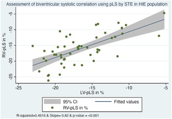
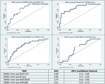
When evaluating the hypoxic-ischaemic encephalopathy population solely, right and left ventricular peak longitudinal strain were associated (R2 = 0.46, p < 0.01), indicating that some hypoxic-ischaemic encephalopathy newborns had various degrees of biventricular dysfunction (Fig 2). When taking into account the measurements of both groups combined (control and hypoxic-ischaemic encephalopathy newborns), tricuspid annular plane systolic excursion was associated with right ventricular peak longitudinal strain, and left-ventricular ejection fraction by Simpson’s was associated with left ventricular peak longitudinal strain, indicating that right and left ventricular conventional systolic markers of function corresponded to those estimated by speckle-tracking echocardiography (Supplementary Fig. S1-a). Also, the estimated stroke distance by velocity time integral of each ventricle was associated with the underlying systolic function by peak longitudinal strain rate (Supplementary Fig. S1-b). Interestingly, estimated systolic pulmonary arterial pressure and systolic pulmonary arterial pressure/systolic blood pressure ratio were not associated with tricuspid annular plane systolic excursion, right ventricular peak longitudinal strain, right ventricular peak longitudinal strain rate, or right ventricular fractional area change. Supra-systemic pulmonary pressure (defined as a systolic pulmonary arterial pressure/systolic systemic blood pressure > 110%) was not associated with abnormal tricuspid annular plane systolic excursion (<7mm, p = 0.96) or abnormal right ventricular peak longitudinal strain rate (>−14%, p = 0.64). Interestingly, a tricuspid annular plane systolic excursion <7mm was associated with pulmonary artery acceleration time to right ventricular ejection time <0.30 (p = 0.001 by Chi Reference Shankaran, Laptook and Ehrenkranz2 , sensitivity of pulmonary artery acceleration time to right ventricular ejection time was 59% and specificity 79%), while the receiver operating curve–area under the curve for pulmonary artery acceleration time to right ventricular ejection time for an abnormal tricuspid annular plane systolic excursion of <7mm was 0.74 (p = 0.002) – Figure 3. Furthermore, we found an association between tricuspid annular plane systolic excursion <7 mm and right ventricular outflow tract velocity time integral, as well as with right ventricular peak longitudinal strain by receiver operating characteristic curves analysis (Fig 3).
Association between right and left ventricular peak longitudinal strain in the hypoxic-ischaemic encephalopathy newborns. Right ventricular (RV) peak longitudinal strain (pLS) by speckle-tracking echocardiography (STE) was associated with left ventricular (LV) pLS within the hypoxic-ischaemic encephalopathy (HIE) population, indicating that some patients have some degree of biventricular dysfunction at the low end of systolic strain.

Receiver operating characteristics curves between markers right ventricular performance and pulmonary pressure. PAAT = pulmonary artery acceleration time; pLS = peak longitudinal strain; ROC = receiver operating characteristics curve; RV = right ventricle; RVET = right ventricular ejection time; RVOT = right ventricular outflow tract; sBP = systolic systemic blood pressure; sPAP = estimated systolic pulmonary arterial pressure; TAPSE = tricuspid annular plane systolic excursion; VTI = velocity time integral.

Discussion
In this retrospective case–control study, we compared the echocardiographic profile of newborns with moderate to severe hypoxic-ischaemic encephalopathy undergoing therapeutic hypothermia with a control population of newborns without perinatal distress. We were interested in outlining specific markers associated with an adverse cardiovascular profile that could be addressed and followed in the context of hypoxic-ischaemic encephalopathy management. We found that hypoxic-ischaemic encephalopathy infants had significantly altered left and right ventricular systolic and diastolic function compared to controls, as well as increased pulmonary pressures.
Pulmonary hypertension and right ventricular function
Persistent pulmonary hypertension of the newborn clinically presents as hypoxic respiratory failure, with deoxygenated blood entering the systemic circulation by the persistent fetal shunts. Reference Barrington, Finer, Pennaforte and Altit18 Persistent pulmonary hypertension of the newborn involves a failure of the natural decrease in resistances of the pulmonary vascular bed during the post-natal transition. Perinatal depression and concomitant neonatal hypoxic ischaemia are key mechanistic triggers of persistent pulmonary hypertension of the newborn. Reference Lakshminrusimha, Shankaran and Laptook3 Elevated pulmonary pressures and right ventricular dysfunction have been described in the neonatal hypoxic-ischaemic encephalopathy population and are associated with adverse outcomes. Reference Giesinger, El Shahed and Castaldo5 In our cohort, compared to controls, those with hypoxic-ischaemic encephalopathy had markers of increased right ventricular afterload (right to left or bidirectional shunts – Supplementary Table S1, systolic pulmonary arterial pressure, systolic pulmonary arterial pressure/systolic blood pressure ratio, left ventricular end-systolic eccentricity index, pulmonary artery acceleration time to right ventricular ejection time, main pulmonary artery dilation, and right to left ventricular diameter ratio). They also had evidence of right ventricular dysfunction (tricuspid annular plane systolic excursion, myocardial performance index, e’ velocity and E/e’ ratio, peak longitudinal strain and strain rate, as well as early diastolic strain rate). However, we did not find an association between the degree of right ventricular afterload and the functional alterations, possibly indicating an inherent injury or maladaptation of the right ventricular myocardium. These findings could be the result of the initial insult, the ongoing myocardial injury from acidosis and respiratory afterload, the adverse coronary perfusion, the iatrogenic effect of therapeutic hypothermia and antiseizure medications, as well as the underlying autonomic nervous system dysregulation. The impact of hypothermia alone on pulmonary vascular resistance is not well understood, although trials have not shown a statistical increase in persistent pulmonary hypertension of the newborn in the hypothermia groups. Reference Shah19 Being the standard of care, we did not have newborns unexposed to this treatment in order to further isolate its effect on pulmonary vascular resistance. As such, our study outlines the systolic and diastolic contributors to the right ventricular dysfunction in these patients and targeted management towards these functional properties may enhance their cardiovascular profile.
Left ventricle systolic contribution
Although we did not find depressed estimated left ventricular stroke distance by velocity time integral compared to controls, perhaps due to lower heart rate and increased filling time, we did report an altered left ventricular function by speckle-tracking echocardiography. Estimation of ejection fraction by conventional measurements remained similar between groups but was decreased when estimated by automated speckle-tracking echocardiography. As such, using automated tracking of the left ventricular myocardial wall, when assessing for subtle cardiac anomalies, may be of importance to detect underlying alteration in the left ventricular function. The left ventricular functional alterations may be secondary to the primary hypoxic-ischaemic injury, the ongoing post-natal homeostatic fluctuations, the inter-ventricular interactions, the therapeutic hypothermia, and/or the various degree of autonomic dysregulation. Recently, Yoon JH et al. described depressed estimated left ventricular output in their hypothermia population (n = 32) compared to controls, with preserved ejection fraction. Reference Yoon, Lee and Yum20 This group also reported that these patients had depressed systolic blood pressure, with maintained diastolic blood pressure, Reference Yoon, Lee and Yum20 a finding that we described here. Despite being lower, the average in our encephalopathy group was above the third centile for gestation, but with the caveat that some were already on cardiovascular medications. Depressed blood pressure may be secondary to various factors: abnormal cardiac function secondary to myocardial injury, depressed cardiac output, bradycardia, impaired filling, or increased biventricular afterload for increased systemic and pulmonary vascular resistances. Altered function by left ventricular myocardial performance index and decreased left ventricular output has also been described in newborns with hypoxic-ischaemic encephalopathy in the pre- and post-rewarming phases compared to controls, with improved myocardial performance index and output post-rewarming. Reference Hochwald, Jabr, Osiovich, Miller, McNamara and Lavoie21 While this may reflect the natural evolution of the disease, it is possible that therapeutic hypothermia may also influence myocardial recovery. In our cohort, we did not find altered ejection fraction by Simpson’s. However, a previous group has reported depressed ejection fraction by the same method in hypoxic-ischaemic encephalopathy newborns non-exposed to therapeutic hypothermia [59.7 (33.7)% versus 67.2 (1.8), p < 0.001]. Reference Bhasin and Kohli22 This discrepancy may be explained by the significant exposure to inotropic support in our cohort, variability of the measurements between the two studies, and/or some beneficial effect of therapeutic hypothermia on the left ventricular function. As such, vasoactive, inotropic, and hormonal (such as hydrocortisone) medications may further enhance ventricular function. Tailoring therapy as cardiac performance evolves during therapeutic hypothermia may be of benefit in a selected population presenting with an adverse left ventricular profile.
Biventricular performance and output
We found an association between ventricular performance by peak longitudinal strain rate and the respective ventricular outflow tract velocity time integral (a marker of stroke distance) in the combined populations (controls and hypoxic-ischaemic encephalopathy) – Supplementary Fig. S1. Thus, there is rational physiologic and echocardiographic evidence that more profoundly altered ventricular function is associated with altered output. Another group had previously described that most of their cohort undergoing therapeutic hypothermia for perinatal asphyxia had low biventricular output. Reference Sehgal, Wong and Mehta23 Also, right and left ventricular deformation by peak longitudinal strain was associated, indicating that those with decreased right ventricular deformation had a concomitant decrease in their left ventricular deformation in systole. This may be secondary to the right-left ventricular interactions, especially altered in the context of pulmonary hypertension, acidosis, ongoing adverse coronary perfusion, or due to more profound hypoxic stress affecting both ventricles. Indeed, coronary flow by echocardiography has been described as decrease in hypoxic-ischaemic encephalopathy newborns and may contribute to the altered ventricular filling and contraction. Reference Sehgal, Wong and Mehta23 Biventricular preload, contractility, and afterload may also be affected by acidosis, high lactate level and persistent hypoxia, nested in the context of altered vascular reactivity, exposure to catecholamines (endo- and exogenous), and hypothermia. Corroborating our findings, another group comparing newborns with hypoxic-ischaemic encephalopathy and healthy controls found that majority of their cohort was exposed to inotropic support (86%), with many (61%) requiring two or more agents. Reference Aggarwal and Natarajan24 In their cohort, they found a significantly decreased estimated right and left ventricular output, right and left ventricular myocardial performance index, as well as an altered end-systolic left ventricle eccentricity index. Our cohort also demonstrated evidence of biventricular dysfunction and of increased septal distortion.
Diastolic dysfunction
Our data indicate that newborns with hypoxic-ischaemic encephalopathy have signs of altered diastolic function compared to controls (biventricular e’ velocity and early diastolic strain rate by speckle-tracking echocardiography). This may be significant as these infants may be subjected to fluid resuscitation and blood products, particularly during initial post-natal transition and initial hypothermic therapy, as well as acute kidney injury, syndrome of inappropriate anti-diuretic hormone, and resultant fluid overload. Excessive volume repletion in the context of cardiac relaxation anomalies may further worsen tissue oedema and, as such, may impair adequate recovery. In another case–control study reporting on newborns with hypoxic-ischaemic encephalopathy, cases were found to have decreased left ventricular e’ and myocardial performance index by tissue Doppler imaging. Reference Sobeih, El-Baz, El-Shemy and Abu El-Hamed25 Although the authors did not report the E/e’ ratio of the left ventricle, the mitral E velocity was significantly lower in cases, indicating that, similar to our results, left ventricular diastolic function was impaired in their cohort of hypoxic-ischaemic encephalopathy newborns. Reference Sobeih, El-Baz, El-Shemy and Abu El-Hamed25 As such, considering the common practice of fluid repletion and transfusion to address haematological disturbances in this vulnerable population, future studies should consider the echocardiographic evaluation of such practice in those with altered markers of ventricular diastolic function.
Limitations
Our study was retrospective, single centre, and descriptive. Numerous newborns were identified during the study period with hypoxic-ischaemic encephalopathy undergoing therapeutic hypothermia, but echocardiography was not performed in all. There was no formalised echocardiography screening protocol for neonates with hypoxic-ischaemic encephalopathy undergoing therapeutic hypothermia, thus the population might be biased towards the sickest patients based on clinical concerns leading to obtaining an echocardiography. As such, these infants were likely to be exposed to inotropes during therapeutic hypothermia (and echocardiography), had high lactate on day 1 and day 2 of life, and four required extracorporeal membrane oxygenation during therapeutic hypothermia. However, when evaluating the excluded population (n = 113), 11 died (11%) compared to 10 deaths in the included cohort (18%), p = 0.22 (Fig 1). Due to the absence of such a formal protocol, the analysed echocardiography scans were not performed at a specific chronologic age for all the patients. Echocardiography was limited by the use of multiple scanning platforms, multiple professionals acquiring the images, and the absence of some markers or limited views in certain patients (tissue Doppler was not available in a third of the cohort). Many of the newborns were receiving inotropic support during echocardiographic assessment, without a standardised clinical approach, rendering it difficult to extrapolate the individual effect of certain medications and dosage regimens. Echocardiography measures were not analysed for intra-reader or inter-reader variability, although our methodology has been validated in previous reports. Reference Altit, Bhombal, Chock and Tacy8,Reference Altit, Bhombal, Van Meurs and Tacy9 Our methodology for velocity time integral reproducibility has been validated in a previous publication by our group, Reference Altit, Bhombal, Chock and Tacy8 and by other authors. Reference Chaiyakulsil, Chantra, Katanyuwong, Khositseth and Anantasit26–Reference Pees, Glagau, Hauser and Michel-Behnke28 Previous studies described strain assessment by speckle-tracking echocardiography as well as tricuspid annular plane systolic excursion to have high inter-reader and intra-reader agreement, Reference Mukherjee, Chung and Ton29,Reference Mercer-Rosa, Parnell, Forfia, Yang, Goldmuntz and Kawut30 with high reproducibility using different strain vendors Reference Costa, Beaver, Rollor, Vanichakarn, Magnus and Palac31 and different echocardiography machines. Reference Risum, Ali and Olsen32 Analysed echocardiography images were originally acquired at a lower frame rate of 30–60 Hz, when 2D speckle-tracking echocardiography recommendations advocate for 80–100 Hz. However, a report observed no difference between strain measurements at lower (below 60 Hz) and higher (≥ 60 Hz) frame rates. Reference Liu, Tacy, Chin, Obayashi and Punn33 Very few patients in our cohort had follow-up echocardiography which prevented evaluation of their longitudinal cardiovascular adaptation. Only time of birth was available for the hypoxic-ischaemic encephalopathy infants, precluding from calculating the hours of life at echocardiography for the control newborns. Hence, date of birth was used to calculate age in days of life at echocardiography for both groups. Data extractor was not masked to the hypoxic-ischaemic encephalopathy/control status at the time of echocardiography data extraction. Our control population was normothermic and without perinatal distress. In order to isolate the effect of hypothermia on cardiovascular function, we would need a population of neonates with moderate–severe hypoxic-ischaemic encephalopathy who did not undergo therapeutic hypothermia, but therapeutic hypothermia is the standard of care and thus it would be unethical to perform such a study. Finally, there were less control newborns than newborns with hypoxic-ischaemic encephalopathy. In most cases, the age-matched controls were referred to our echocardiography laboratory for a murmur evaluation and had sufficient medical information available in the chart for purpose of comparisons.
Conclusion
Newborns undergoing therapeutic hypothermia for hypoxic-ischaemic encephalopathy have concomitant cardiovascular disturbances. Compared to control newborns of the same age not exposed to therapeutic hypothermia and without perinatal asphyxia, many hypoxic-ischaemic encephalopathy newborns have signs of increased pulmonary pressures and altered biventricular systolic and diastolic performance. In our cohort, despite a significant exposure to inotropic support, these disturbances were quantifiable by echocardiography. As such, early echocardiography screening and targeted management may be avenues to explore as potential preventative measures to re-establish optimal cardiac function.
Supplementary material
To view supplementary material for this article, please visit https://doi.org/10.1017/S1047951122002839
Availability of data and material
Part of the data and material are shared in the Supplementary files. The rest of the derived data generated in this research will be shared on reasonable request to the corresponding author.
Code availability: Statistical analysis were done on STATA. Not applicable.
Acknowledgements
We want to acknowledge Ms Daniela Villegas Martinez for proof-reading the manuscript.
Authors’ contributions
Dr Altit conceptualised and designed the study, collected the data, extracted the data from echocardiography, analysed the data, drafted the manuscript, and adjusted the manuscript according to the comments of the co-authors.
Dr Bonifacio conceptualised and designed the study, collected the data, critically appraised the analysis of the data, and reviewed and revised the manuscript.
Dr Guimaraes collected the data, critically appraised the analysis, and revised the manuscript.
Mr Ganesh Sivakumar collected the data, critically appraised the analysis, and revised the manuscript.
Miss Beth Yan collected the data, critically appraised the analysis, and revised the manuscript.
Dr Chock conceptualised and designed the study, critically appraised the analysis of the data, and reviewed and revised the manuscript.
Dr Van Meurs conceptualised and designed the study, critically appraised the analysis of the data, and reviewed and revised the manuscript.
Dr Bhombal conceptualised and designed the study, supervised data collection, critically appraised the analysis of the data, wrote and critically reviewed the manuscript for important intellectual content.
All authors approved the final manuscript as submitted and agree to be accountable for all aspects of the work.
Financial support
This research received no specific grant from any funding agency, commercial, or not-for-profit sectors.
Conflicts of interest
None.
Ethics standards
This study was approved by the Institutional Review Board of Stanford University.
Consent to participate
Waiver of consent since retrospective study by IRB of Stanford.
Consent for publication
Not applicable.