Introduction
First branchial arch/cleft abnormalities are rare congenital abnormalities with an estimated incidence of approximately one case per one million people.Reference Arndal and Bonding1 First arch abnormalities account for 1–10 per cent of all branchial abnormalities.Reference El Omri, Naouar, Bellakhddher, Bergaoui, Kermani and Abdelkefi2, Reference Adams, Mankad, Offiah and Childs3 These involve the development of an abnormal cyst, sinus, cartilaginous remnant, or fistula between the skin just inferior to the mandible or in the pre-auricular region. These pass through the parotid region and opening in and around the external auditory meatus.Reference Adams, Mankad, Offiah and Childs3
Symptoms associated with first branchial arch fistulas may include recurrent infections, mucopurulent discharge from openings, pain or painless swelling.Reference Chen, Xu, Wang, Xu, Chen and Xu4 Treatment typically involves surgical excision of the tract to prevent recurrence with all literature reporting excision once diagnosis is made.Reference Chen, Xu, Wang, Xu, Chen and Xu4–Reference Li, Zhao, Xu and Li10 However, due to the serious potential for concomitant facial nerve paralysis, surgery can be challenging and requires meticulous planning. This can often be challenging given the difficulties in locating the extra-cranial facial nerve on magnetic resonance imaging (MRI) imaging in children.Reference Hwang, Yoon, Lee, Yoon, Jung and Cho11 Some patients may benefit from more conservative management when different surgical and patient circumstances are considered.
We present 10 years of experience managing first branchial arch abnormalities describing both successfully surgically managed and non-surgically managed paediatric patients in-order to highlight the different management strategies and when these may be used, in contrast with previously published literature.
Materials and methods
Clinical records were retrospectively reviewed from Alder Hey Children’s Hospital, a large UK paediatric tertiary ENT centre from January 2012 until September 2022. All patients with the ICD-10 code Q-180, Q181 or Q182 “Sinus, fistula and cyst of branchial cleft” were identified and included.
Patient records and imaging were reviewed, and only patients with confirmed first branchial arch abnormalities were included. Patient demographics, histories, examinations, investigations and intervention data were extracted and included. Management is discussed.
Results and Analysis
One hundred fifty-seven patients were identified meeting the search criteria, with a total of 4 patients meeting inclusion criteria. One hundred fifty-three patients were excluded as they had diagnoses other than first branchial arch abnormalities such as pre-auricular sinuses or other branchial arch abnormalities. All remaining patients had a first branchial arch fistula.
The average age of first presentation to ENT services was 2 years 4 months (10 months, 1 year 2 months, 5 years 5 months and 1 year 11 months). Three (75%) were female, all abnormalities were on the left side. Three patients underwent operative management at some point, one was managed conservatively. One patient who was operatively managed had an initial operative excision by another team and had recurrence, which was managed conservatively subsequently (Table 1).
Summary table of demographic details and general management principles of patients.

Patient A
A 1-year 11-month-old male patient presented to ENT outpatients with a left sided neck swelling since birth with oily discharge. There was no history of infection, purulent discharge or erythema. The child was otherwise healthy. Examination findings showed a 2-cm firm swelling at the angle of the mandible with discharge, but no infective signs.
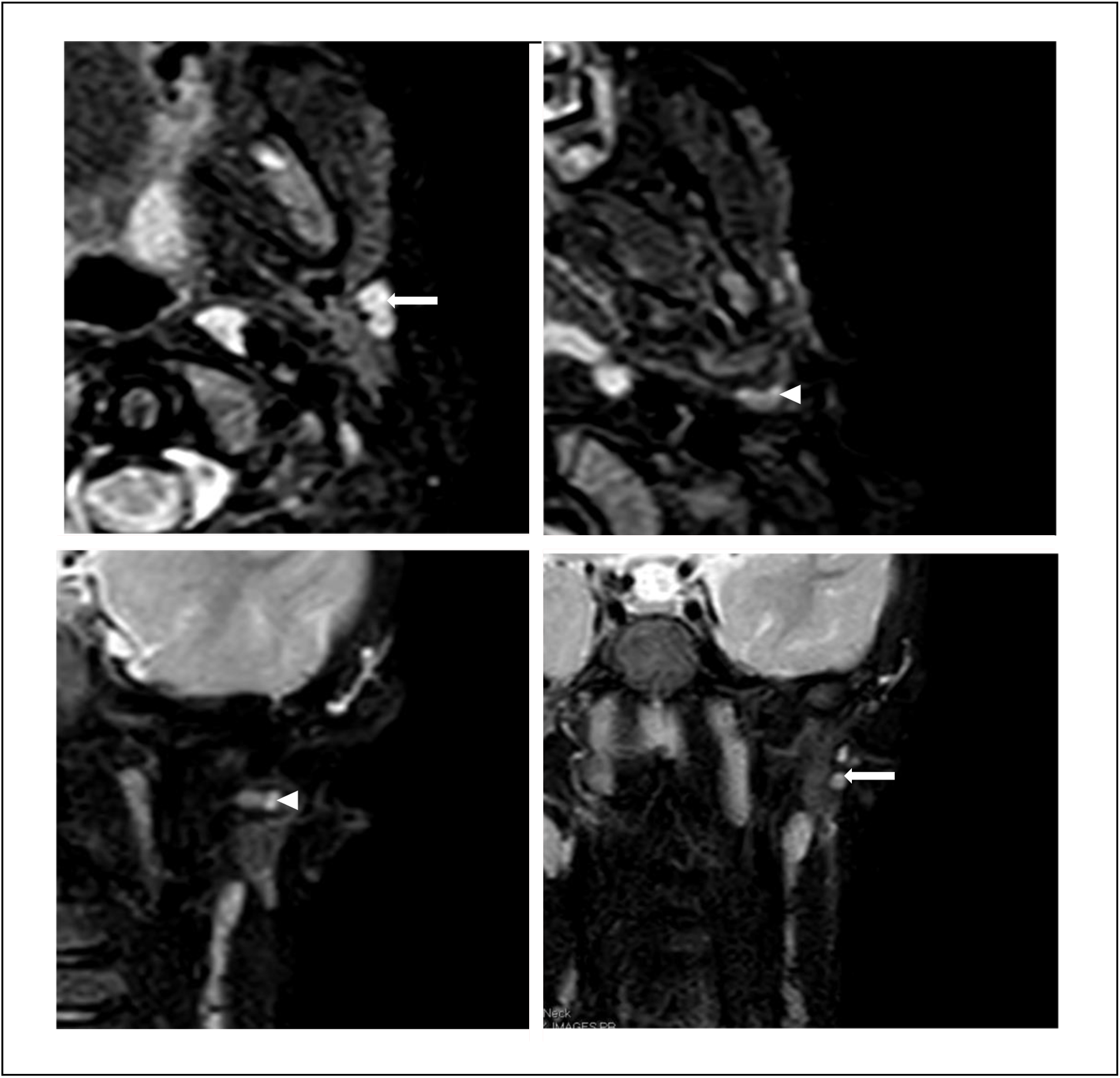
A sebaceous cyst was suspected. Ultrasound imaging of the lump showing the opening had a 4-mm tract leading to a complex cyst at the inferior lower pole of the parotid gland and extending anteriorly towards the skin. Subsequent MRI scanning showed the tract from the mandibular lump passing posterior-superiorly into the deep lobe of the parotid and terminated in the external auditory meatus (Figure 1).
Axial T2W TSE (top left), coronal T1W TSE (top right), coronal T1 post gadolinium (bottom left), axial T1W TSE (bottom right) MRI images of patient A. MRI displays a tract (white arrow) passing posterior-superiorly into the deep lobe of the parotid and terminating in the external auditory meatus.

The patient was discussed in the local ENT radiology meeting, and the child’s parents elected for excision of the tract which occurred 8 months after initial presentation. Post-operatively, the patient had no facial nerve deficits but had a wound infection which was treated with inpatient intra-venous antibiotics and analgesia. They had no further recurrence, and they were discharged just under 16 months following excision.
Patient B
A 5-year and 5-month-old female patient presented to ENT outpatients with left sided swelling in the pre-auricular region and discharge. The child had a prior history of excision of a first branchial arch sinus under the paediatric general surgeons at age 2 years and 5 months. The previous operation identified and excised a tract but was seen to taper out before the parotid gland. They developed swelling and discharge over the same left site 6 months later. The reason for delayed re-presentation was not known. Examination identified they had a thickened swelling under the angle of the left mandible. Recurrence of a first branchial arch abnormality was suspected.
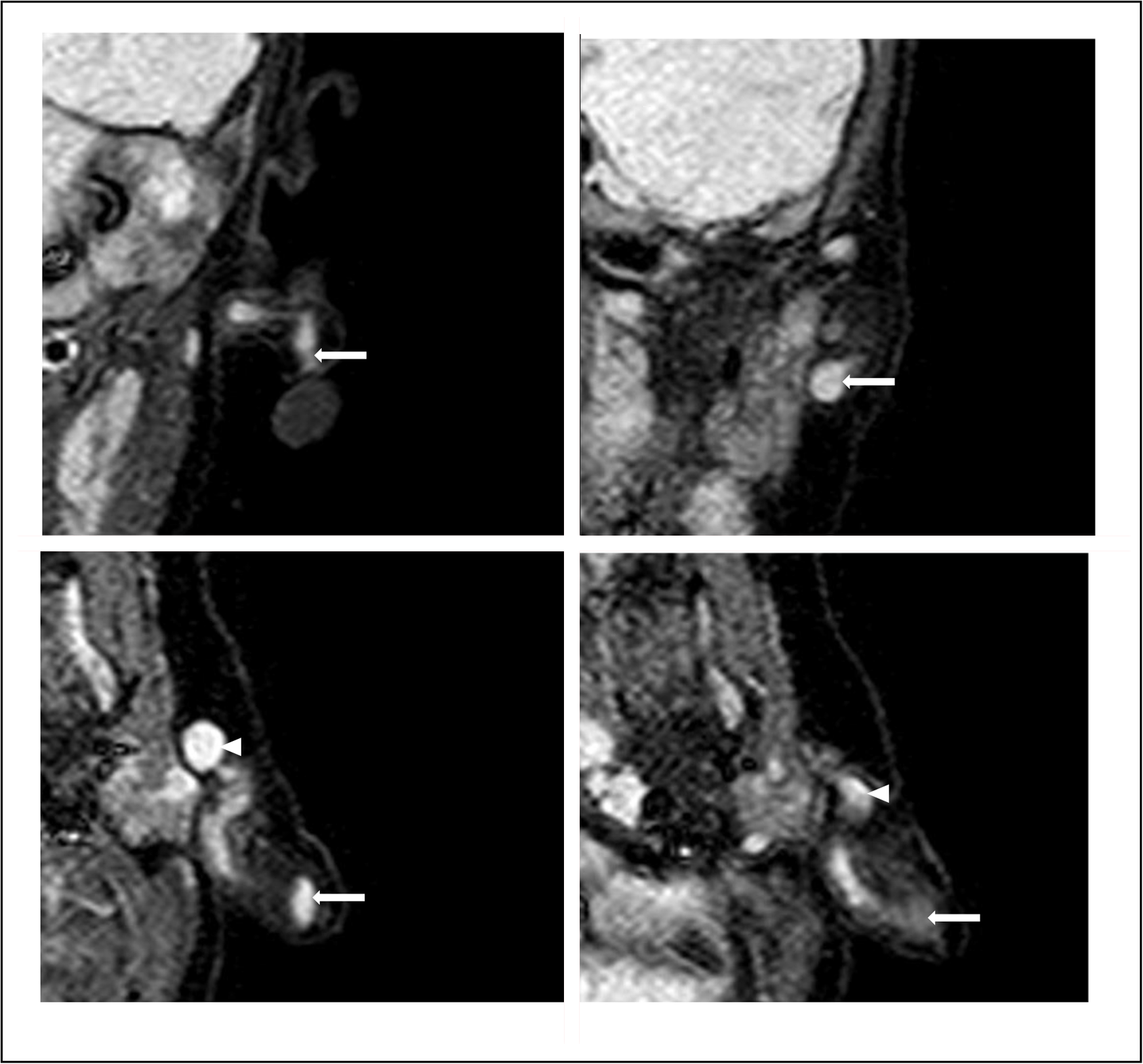
Ultrasound showed a 2-mm tract extending from the mandibular swelling passing superiorly across the parotid towards the external auditory canal. MRI showed a tract with 5 × 10-mm expansion lateral to the parotid extending across and deep to the parotid. The distal segment lay immediately inferior and parallel to the left external acoustic canal. The location of the abnormality was close to the usual tract of the facial nerve; however, its relationship to the facial nerve could not be determined due to limitations of the MRI imaging (Figure 2).
Axial STIR (top left and right) and coronal T2W TSE (bottom left and right) MRI images of patient B. MRI displays 5x10mm expansion (white arrow) lateral to the parotid extending across and deep to the parotid (white triangle). The distal segment lay immediately inferior and parallel to the left external acoustic canal.

Operative options were discussed with the parents, including the option of total parotidectomy for complete excision of the tract and significant subsequent risk of facial nerve damage. The parents elected for non-operative treatment.
The patient was discharged to local surveillance 3 years 5 months after initial excision.
Patient C
A 1-year and 2-month-old female patient presented to ENT outpatients with a history of two recurrently discharging left sided puncta. One punctum was located in the pre-auricular area and the other on the left ear lobe. Examination showed no signs of active infection.
Ultrasound showed a 3-mm tract between the two puncta passing inferior to the opening of the external auditory canal and which did not pass through the parotid parenchyma. The lobular element showed a 3 × 7-mm expansion. MRI scanning demonstrated a serpiginous cystic space extending from the left earlobe passing deep coursing beneath the external auditory meatus to pre-auricular region (Figure 3).
Coronal STIR (top left and right) and Axial STIR (bottom left and right) MRI images of patient C. MRI showed a serpiginous cystic space extending from the left earlobe (white arrow) passing deep coursing beneath the external auditory meatus to preauricular region (white triangle).

At this point, the patient had ongoing discharge; therefore, the decision was made to proceed with excision. This was completed 2 years and 2 months following initial presentation, and histology confirmed a first branchial cleft fistula. Post-operatively, the patient had no complications or recurrence and was discharged 8 months after excision.
Patient D
A 10-month-old female patient presented to ENT outpatients with a history of pit in the left mandibular region with thick discharge since birth. There was no prior history of infection. Examination showed no signs of infection, but thick discharge was expressed. No external auditory canal opening was found on otoscopic examination.
Ultrasound scanning demonstrated a tract leading from the punctum to a 1-cm cyst just deep to the ear lobe. MRI scanning demonstrated a tract from the posterior aspect of the cheek towards the parotid gland communicating with a 10 × 7 × 9-mm cyst. The tract was found to extend medially passing interior to the external acoustic canal and tapered in the left para-pharyngeal soft tissue (Figure 4).
Coronal MPR (top left and right), Axial MPR (bottom left and right) MRI images for patient D. MRI showed a tract from the posterior aspect of the cheek toward the parotid gland communicating with a 10 x 7 x 9mm cyst (white arrow). Tract extended medially (white triangle) passing interior to the external acoustic canal and tapered in the left para-pharyngeal soft tissue.

Management strategies were discussed with the parents and given the minimal symptom burden, risks of possible marginal mandibular nerve damage and potential need for parotidectomy, a watchful waiting strategywas used. They were last seen 4 years after initial presentation with no significant complications.
Discussion
We present a series of four patient with differing management strategies. Within the literature, the mainstay of treatment for first branchial arch anomalies is excision of the lesion.Reference Chen, Xu, Wang, Xu, Chen and Xu4–Reference Li, Zhao, Xu and Li10 In these studies, 414 patients are described with 0–11 per cent and 0–9 per cent facial nerve palsy and recurrence rates, respectively.Reference Chen, Xu, Wang, Xu, Chen and Xu4–Reference Li, Zhao, Xu and Li10 There are no previously published non-surgical management strategies for first branchial arch anomalies in children. We present the first case series to offer conservative alternative management strategies for these children to avoid high risk complications which can result in significant long-term morbidity. The children presented in this series demonstrate a range of clinical manifestations, aberrant anatomy and clinical journeys with a tailored management approach.
In this series, patient D had very little troublesome symptoms, whereas patient C suffered recurrent infections with concomitant morbidity. In the former patient, undertaking an operation with associated significant morbidity such as facial nerve paralysis would be difficult to justify with low symptom burden. In such cases, conservative measures, namely, close observation for changes in symptoms, antibiotic therapy with microbiology support, wound care including water precautions and analgesia will help optimise care. Counselling parents is key to aid them in recognising signs of infection and when to seek medical attention should complications arise.
Furthermore, patient B had more significant symptomology than patient D. However, from the result of the MRI scan, this patient presented greater operative challenges; therefore, a higher risk of complication.
Identification of relational anatomy pre-operatively is incredibly important in stratifying risk. Identifying the course of the extra-cranial facial nerve on imaging is challenging making surgical risk predication harder. Conventional MRI sequencing was used in the assessment of the patients in this series which has a limited ability to track the course of the extra-cranial facial nerve. Constructive interference in steady state (CISS) T2 MRI sequencing may offer superior facial nerve imaging. CISS MRI has been used to investigate and map the facial nerve in adults and has been described in the detection of paediatric facial nerve aplasia and Moebius syndrome.Reference Chalipat, Chavan, Mane, Taneja and Kumar12, Reference Decraene, Boudewyns, Venstermans and Ceulemans13 Its use in imaging the extra-cranial course is of particular interest in first branchial arch abnormalities given the often-close relationship of tracts with the facial nerve. In adults, CISS sequences show potential in fine visualisation of the extra-cranial course and are superior to double-echo steady state with water excitation sequences.Reference Guenette, Ben-Shlomo, Jayender, Seethamraju, Kimbrell and Tran14, Reference Fan, Ding, Zhao and Hou15 CISS MRI sequency for facial nerve identification has not been described in the literature for surgical planning of paediatric branchial arch abnormalities. This provides an opportunity for utilisation in the planning of excisions and in the risk stratification for patient and parents; however, this is yet to be investigated
Research covering larger patient groups needs to be collected to better understand the long-term outcomes of conservative management. Additionally, the role of CISS MRI in first branchial arch abnormalities should be explored with primary research and its impact on operative management.
Ultimately, clinicians need to support discussions with parents over the benefits and risks of each management strategy to allow tailored management decisions for each individualised case. This will provide superior outcomes whilst minimising potentially life-changing morbidity.
• First branchial arch abnormalities are rare causing pain, swelling, discharge and recurrently get infected
• Surgical excision is the only described technique in the literature which is not without complication including recurrence and facial nerve palsy
• This series adds that some patients may be more suitable for conservative measures when complication risks are higher
Conclusion
First branchial arch abnormalities are rare and result in aberrant tracts in and around the external auditory canal and angle of the mandible. These present with a range of symptomology. Excision is the mainstay of treatment and has dominated the research literature. This case series proposes a unique alternative use of conservative measures in selected patients. Patient and parent counselling supported by specialist input is vital to optimise outcomes and minimise morbidity.
Acknowledgements
The authors thank the patients and parents of the patients involved in this manuscript. We also thank the support staff in Alder Hey Children’s Hospital for their support.
Financial support
No financial support was provided for this study.
Competing interests
The authors declare none.