Anomalous origin of the left coronary artery from the pulmonary artery is a rare but well-known congenital coronary artery anomaly affecting approximately 1 in 300,000 births, and mainly expressing during early childhood.Reference Cowles and Berdon1 It is a common cause of myocardial ischaemia and infarction in children. If untreated, the mortality can reach 90% in the first year of life. Prenatal diagnosis is not possible because of equivalent pressures in the main pulmonary artery and in the aorta during foetal life, explaining that the clinical course is mostly represented by a severe cardiac failure in the first few months of life. Prognosis of patients with anomalous origin of the left coronary artery closely depends on early echocardiographic diagnosis.
Case report
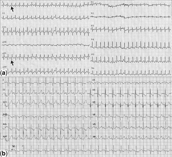
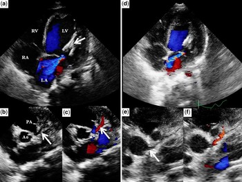
We describe the case of a 1-month-old boy referred to our emergency unit for excessive sleepiness and poor feeding. Pregnancy was normal. Parents described sweating during feeding and panting since 24 hours. On examination, the patient was tachypneic. He had tachycardia and hepatomegaly but was apyretic. Chest X-ray showed a cardiomegaly and a pulmonary oedema. Electrocardiogram revealed supraventricular tachycardia (heart rate of 260 beats per minute), which spontaneously reduced 10 minutes later (Fig 1). Lactate levels were 10.5 millimole per litre (pH 7.13), troponin level was 0.74 nanogram per millilitre, and brain natriuretic peptide level was initially 4500 picogram per millilitre. There was no renal function impairment. The first diagnosis was a rhythmic cardiomyopathy. Thereafter, the diagnosis of anomalous origin of the left coronary artery was affirmed in intensive care unit by two-dimensional echocardiography. The left ventricular ejection fraction was approximately 20% and the lateral wall of the dilated left ventricle was very hypokinetic. An eccentric moderate mitral regurgitation jet due to the restriction of the posterior leaflet of the mitral valve was seen and left ventricular papillary muscles were abnormally echogenic (Fig 2). There was no pericardial effusion. Filling pressures were high with dilated inferior caval vein, and systolic pulmonary pressure, estimated on tricuspid regurgitation, was 60 millimetres of mercury. Right coronary artery was dilated and left coronary artery arose from the main pulmonary artery (Fig 2). A bidirectional flow was seen into the left coronary artery on Doppler colour views. Electrocardiogram showed sinus rhythm with deep Q waves in derivations I and aVL, and diffuse ST-T wave changes consistent with an anterolateral infarction. Haemodynamic status was stabilised using diuretics, antiarrhythmic (amiodarone), and inotropic (dobutamine) drugs.
(a) A twelve-lead electrocardiogram (25 millimetres per second; 10 millimetres per millivolt) performed at admission showing supraventricular tachycardia. Deep Q waves are seen in the lateral leads I and aVL (arrow). Note the depression of the ST segment in the lateral leads. (b) A twelve-lead electrocardiogram performed 2 weeks after surgery showing normal sinus rhythm with the persistence of deep Q waves and ST segment depression in lateral leads I and aVL.

(a) Apical four-chamber echocardiographic view, before surgery, showing LV dilatation, “bright” papillary muscles (arrow), and eccentric mitral regurgitation; (b) parasternal short-axis echocardiographic view, before surgery, showing anomalous origin of the left coronary artery from the PA (arrow); and (c) in Doppler colour view the bi-directional flow into the coronary artery. (d) After surgery, a persistent hyperechogenicity of the papillary muscles was associated with mitral regurgitation despite the decrease of left ventricle diameter; (e) parasternal short-axis echocardiographic view, after coronary reimplantation, showing the new aortic implantation of the left coronary artery into the aortic root; and (f) the anterograde Doppler colour flow (RV = right ventricle; LV = left ventricle; RA = right atrium; LA = left atrium; PA = pulmonary artery; AO = aorta).

Surgical treatment, consisting of a direct left coronary reimplantation into the ascending aorta, was successfully performed. Because of an immediate poor haemodynamic tolerance, a temporary venoarterial mechanical circulatory support with extracorporeal membrane oxygenation was used for 5 days after surgery. The patient was discharged from the hospital on post-operative day 15 with a combination therapy of captopril and amiodarone. A month later, echocardiography showed a left ventricular ejection fraction of 60% with normal regional contraction, a trivial mitral regurgitation due to a persistent restricted motion of the posterior leaflet, and normal pulmonary pressures. Since then, the patient did not have any cardiac symptoms.
Discussion
Anomalous origin of the left coronary artery from the pulmonary artery is a rare malformation that usually leads to severe coronary hypoperfusion and left ventricular dysfunction when pulmonary vascular resistances decreased in the postnatal period. The physiological decrease of pulmonary arterial pressure is responsible for a coronary arterial steal due to a left-to-right shunt. Although complete heart block and ventricular tachycardia have been previously described in case of anomalous origin of the left coronary artery, no supraventricular tachycardia, to our knowledge, have been reported so far in this situation.Reference Math, Parakh, Sarin and Tyagi2–Reference Cohen and Berger4 It is unclear whether supraventricular arrhythmia was fortuitously associated with the coronary abnormality or whether it was an ischaemic consequence. The shunt into the left coronary artery was bidirectional, indicating that pulmonary vascular resistances were still relatively high. Furthermore, supraventricular tachycardia is not a common complication of myocardial ischaemia in children. Even if cardiac rhythm was reported as sinus during pregancy, supraventricular tachycardia was certainly prolonged, thus contributing to worsen cardiac failure. However, this arrhythmia allowed an early diagnosis of the coronary abnormality that led to rapid surgery before the patient developed severe ventricular impairment. Indeed, very early correction of an anomalous origin of the left coronary artery from the pulmonary artery is proved to improve post-operative management and prevent persistent mitral regurgitation.Reference Bonnemains, Lambert, Moulin-Zinch, Youssef and Serraf5 This report also confirms the importance of echocardiography for the diagnosis of abnormal coronary artery.Reference Yang, Nanda and Wang6, Reference Cohen, Herlong and Silverman7 Although signs of ischaemia were already present on the first electrocardiogram, they were “masked” by the rhythm disorder. Echocardiography corrected the diagnosis and allowed appropriate care.
Acknowledgements
The authors thank Loïc Macé, MD, for surgical assistance. The authors have no conflict of interest to declare.