INTRODUCTION
The genus Boccardia Carazzi, Reference Carazzi1893 currently includes 22 species, 14 of which are found boring into, or associated with molluscs, coralline algae, rock or foliose algae (see Ruellet, Reference Ruellet2004 for review). To date, two species have been recorded in the wild (i.e. dissociated from mariculture) in southern Africa (Day, Reference Day1955, Reference Day1961, Reference Day1967): Boccardia polybranchia (Haswell, Reference Haswell1885) and Boccardia pseudonatrix Day, Reference Day1961; a third, Boccardia cf. ligerica (Ferronnière, 1898) mentioned by Day (Reference Day1967), was transferred to Boccardiella sensu Blake & Kudenov (Reference Blake and Kudenov1978). Boccardia polybranchia was recorded widely along the south coast of South Africa and in Namibia; no indication was given of its habitat in South Africa, while it was recorded from shallow dredgings in Namibia (Augener, Reference Augener and Michaelsen1918; Day, Reference Day1967). By contrast B. pseudonatrix was found only once boring into rock in Knysna on the south coast of South Africa (Day, Reference Day1961). More recently the non-indigenous B. proboscidea Hartman, Reference Hartman1940 was detected on cultured abalone (Haliotis midae Linnaeus, 1758) at several on-shore aquaculture facilities (Simon et al., Reference Simon, Ludford and Wynne2006; Simon & Booth, Reference Simon and Booth2007; Simon et al., in review), stimulating renewed interest in shell-infesting Boccardia species in South Africa.
This paper provides a revision of the Boccardia species associated with both wild and cultured molluscs along the south coast of South Africa. The opportunity is also taken to describe B. polybranchia from South African material, provide comparisons with populations from other regions and discuss some of the taxonomic and nomenclatural issues associated with the species.
MATERIALS AND METHODS
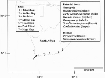
Molluscs were collected from the intertidal or shallow subtidal from five sites along the south and south-east coasts of South Africa in February and March 2005 and April 2006 (Figure 1). Worms were removed by immersing shells in a vermifuge, 0.05% phenol in seawater, for three hours to overnight. Once the worms abandoned their burrows, they were transferred to fresh seawater, relaxed with clove oil, preserved in 4% saline formaldehyde solution and stored in 70% ethanol. Material from abalone farms in Saldanha Bay on the south-west coast, Hermanus and Gansbaai on the south coast, and Haga Haga on the south-east coast was removed by dissolving the shells in which the worms had been fixed (in 4% saline formaldehyde solution and stored in 70% ethanol) in 5% HNO3 diluted in 70% ethanol (Simon et al., Reference Simon, Ludford and Wynne2006; Simon & Booth, Reference Simon and Booth2007).
Location of sampling sites and list of potential molluscan hosts collected.

For scanning electron microscopy (SEM), the specimens were dehydrated in a series of increasing concentrations (80–100%) of ethanol, critical point dried and sputter coated. Specimens were viewed on Vega Tescan and Leo 1430 VP scanning electron microscopes. Descriptions for each of the species were prepared, based on South African material. Partial synonymies were produced to include significant taxonomic works. Specimens are lodged at the Iziko South African Museum, Cape Town, South Africa.
RESULTS AND DISCUSSION
SYSTEMATICS
? Perialla claparedei Kinberg, 1866, p. 253; Reference Kinberg1910, p. 63, figure 9
? Polydora (Leucodore) polybranchia Haswell, 1885
Polydora (Boccardia) polybranchia Carazzi, Reference Carazzi1893, p. 16, pl. 2, figures 1–3
Polydora polybranchia Söderström, Reference Söderström1920, p. 256, figure 167
Polydora (Boccardia) polybranchia Fauvel, Reference Fauvel1927, pp. 58–59; Okuda, 1937
Boccardia polybranchia Imajima & Hartman, Reference Imajima and Hartman1964, p. 279; Day, Reference Day1967, pp. 463–464; Blake & Kudenov, Reference Blake and Kudenov1978, pp. 236–238; Blake, Reference Blake and Kornicker1983, pp. 248.
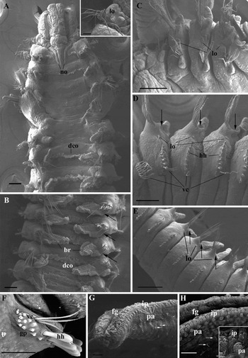
Boccardia polybranchia sensu Blake & Kudenov, Reference Blake and Kudenov1978, adult morphology. (A) Dorsal anterior showing ciliary bands of nuchal organs (no) and dorsal ciliary organs (dco). Inset shows lateral organ (lo) on first chaetiger; (B) dorsal mid-body showing branchiae (br) on chaetigers 10–14. Note attachment of the branchiae to the notopodial lobes (arrows); (C) lateral anterior showing lateral organs (lo) on chaetigers 2–4; (D) lateral view of chaetigers 6–10, showing lateral organs (lo) and connection of branchiae with notopodial lobes (arrows), hooded hooks (hh) with one or two ventral inferior capillary chaetae (vc); (E) ventro-lateral view of chaetigers posterior to chaetiger 16, showing lateral organs (lo) and change in the structure of the branchiae which are not here connected to notopodial lobes (arrows); (F) hooded hooks (hh) with external openings of glandular pouches (gp) and ventral inferior capillary chaetae (vc); (G & H) latero-frontal surface of palps showing the feeding groove (fg) large papillae with long cirri (pa) and inner row of papillae (ip), with tufts of cirri or cilia scattered on lateral and abfrontal surface of the palps (arrows). Scale bars: A–D, 100 µm; E, 200 µm; F, 25 µm; G, 20 µm; H, 10 µm. Insert to A, 30 µm; insert to H, 2.5 µm.

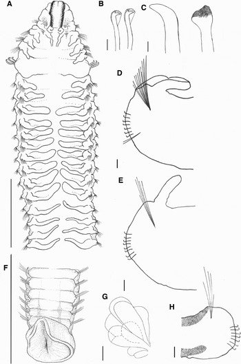
Boccardia polybranchia sensu Blake & Kudenov, Reference Blake and Kudenov1978. (A) Dorsal anterior showing the change in structure of branchiae after chaetiger 15; (B) hooded hooks; (C) falcate and brush-topped spines from chaetiger 5; (D) anterior view of left parapodium of chaetiger 9 showing the fold of skin connecting the branchia and notopodial lobe; (E) anterior view of left parapodium of a chaetiger posterior to chaetiger 17, without the fold of skin between the branchia and notopodial lobe; (F) dorsal view of pygidium, showing some pigmentation; (G) gross structure of the glandular pouches; note the relatively few, large pouches; (H) anterior view of right parapodium of posterior chaetiger showing the pigmentation on the dorsal and ventral surfaces. Scale bars: A, F, 1 mm; B, C, 20 µm; D, E, G, H, 50 µm.

MATERIAL EXAMINED
South Africa: Western Cape Province: Mossel Bay, 34°10′56″S 22°07′20″E, March 2005, associated with the turban shell Turbo sarmaticus Linnaeus, 1758 (SAMC, A21475 [7], A21476 [2, including slides]), mussel Perna perna Linnaeus, 1758, abalone Haliotis midae and limpet Scutellastra longicosta (Lamarck, 1819); A21477 (SEM); Eastern Cape Province: Grootbank (Tsitsikamma), 33°59′14″S 23°32′36″E, March 2005, associated with H. midae and T. sarmaticus A21515 & A21524 (SEM).
Brazil: Rio de Janeiro, Perialla claparadei (6 syntypes, SMNH 742). Australia: Coff's Harbour (2, AM, W13033), Jervis Bay (2, AM, W 24941). France: Biarritz (1, MNHNP UE 429).
MORPHOLOGY OF NEW MATERIAL
Medium-sized species, up to 20.3 mm long for 79 chaetigers. Prostomium bifid, caruncle extending to posterior margin of chaetiger 2, with lateral nuchal organs (Figures 2A & 3A); middle of caruncle swollen, no occipital antenna (Figures 2A & 3A). Up to two pairs of eyes. Preserved specimens tan with dark pigmentation along margin of prostomium, on dorsal surface of peristomium and chaetiger 1, on posterior chaetigers (Figure 3A, F & H), and sometimes on pygidium. Anterior end of body attenuated, widest at chaetiger 5 but from chaetiger 6 approximately the same width as chaetiger 4; posterior half of body narrows. Chaetiger 5 approximately three times as long as chaetigers 4 and 6.
Chaetiger 1 reduced, with small notopodia, lacking notochaetae (Figures 2A & 3A). Notochaetae on anterior chaetigers in three rows, first row with short winged chaetae, middle row chaetae slightly longer and spear-shaped; chaetae of last row longest, slender and lanceolate. No specialized notochaetae in posterior chaetigers. Hooded hooks from chaetiger 7, with 6–7 per ramus initially, then up to 11 hooks per ramus on later chaetigers (Figure 2D, E). No constriction on shaft; main fang 90° to shaft; secondary tooth about 45° to main tooth (Figure 3B). In all specimens examined, hooded hooks on chaetigers 7–9 accompanied by fascicles of up to three ventral inferior capillary chaetae (Figures 2D & 3D); a single ventral inferior capillary chaeta may be present with hooded hooks on more posterior chaetigers.
Chaetiger 5 has dorsal row of two or three falcate spines, ventral row of three or four bristle-topped spines (Figure 3C) and fascicle of ventral inferior capillary chaetae. Older falcate spines not strongly curved. Branchiae on chaetigers 2–4 and posteriorly from chaetiger 6 for 70–80% of body length. From chaetigers 6–16, branchiae short and broad, connected to notopodial lobe (Figures 2A, B, D & 3A & D). Succeeding branchiae filiform, longer than anterior branchiae, not connected to notopodia; branchiae never overlapping along midline of body (Figures 2E & 3A). Dorsal ciliary organs between and along inner surface of anterior branchiae (Figure 2A, B).
Paired glands composed of a few large sacs observed in chaetigers 7–9 (Figure 3G). External openings visible at base of hooded hooks on these chaetigers (Figure 2F).
Lateral organs observed on all chaetigers except the fifth, although not always visible on all specimens examined (Figure 2A, insert, C&E); largest on chaetigers 1 and 2 (Figure 2A, insert).
Row of large (5.5 µm diameter) papillae, 4.5 µm apart on outer latero-frontal edge of palps (Figure 2G, H). Each papilla bears a cirrus. Between the outer row of papillae and the feeding groove are rows of papillae (1.5 µm diameter), arranged in groups of three, and perpendicular to feeding groove, 1.75 µm apart, with shorter cilia or cirri (Figure 2H). Tufts of cilia or cirri scattered over lateral and abfrontal surface (Figure 2G).
Pygidium thick and fleshy, as wide as penultimate chaetiger, with dorsal notch (Figure 3F), sometimes also with ventral notch.
REMARKS
The specimens here assigned to Boccardia polybranchia sensu Blake & Kudenov (Reference Blake and Kudenov1978) were similar to each other (see more below & Table 1). Common to the B. polybranchia taxon as currently recognized are the bifid shape of the prostomium, length of the caruncle, absence of notochaetae on chaetiger 1, general pigmentation pattern (with some exceptions), distribution and number of hooded hooks (but see specimens from Jervis Bay, Table 1), shape and number of the modified spines on chaetiger 5 and distribution and general structure of the branchiae. The paired glands in chaetigers 7–9 of the specimens from South Africa and Jervis Bay, New South Wales, are composed of a few large sacs. The gross structure of these glands is not widely reported and it is not known how sensitive it is as a taxonomic character (but see Fauvel, Reference Fauvel1927; Hartman, Reference Hartman1940).
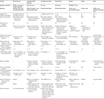
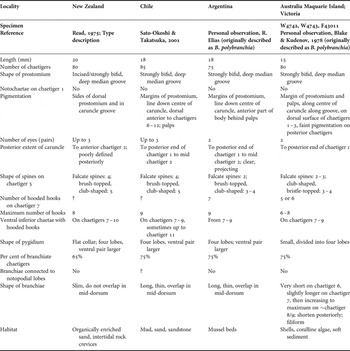
Morphological characteristics of the Boccardia polybranchia (Haswell, Reference Haswell1885) complex, sensu Blake & Kudenov, Reference Blake and Kudenov1978 from South Africa, South America, Australasia and Europe.

*, number in parentheses denotes the number of spines that had not yet extended beyond the surface of the chaetiger.
The South African specimens do, however, differ from other populations of B. polybranchia with respect to some of the characters. For example, the anterior branchiae of the specimens from South Africa and France are wider than those from Coff's Harbour and Jervis Bay. The only other description that mentions the attachment of the branchiae to the notopodial lobes is that of Imajima & Hartman (Reference Imajima and Hartman1964) for specimens from Japan, but no observations are made concerning a different structure to the branchiae after chaetigers 16 or 17. There is also considerable variation in the structure of the pygidium. In the specimens from South Africa, Biarritz and Jervis Bay, the pygidium is a small disc; it may be as wide as, or slightly wider than, the posterior chaetigers, notched dorsally, and sometimes divided into four lobes. The pygidia of the specimens from Coff's Harbour are much wider than the posterior chaetigers and split into two lateral halves. While the caruncles of most specimens (including those from South Africa) extend to the end of chaetiger 2, or the middle of chaetiger 3, those from Japan are longer, although this may be related to body length (Imajima & Hartman, Reference Imajima and Hartman1964; Sato-Okoshi & Takatsuka, Reference Sato-Okoshi and Takatsuka2001). The specimens compared also differ with respect to habitat; those from South Africa are associated with mollusc shells while the others are found in sandy or muddy tubes, associated with Lithothamnion and other coralline algae or Galeolaria (Serpulidae) tubes. The structure of the feeding palps of B. polybranchia in the current study differed considerably from that of specimens recorded as the same species, from Vancouver Island, Canada (Qian & Chia, Reference Qian and Chia1997). The larger, widely spaced, papillae on the latero-frontal edge of the palps, the regular arrangement of the smaller papillae and the tufts of cirri or cilia (these cannot be distinguished in preserved specimens) on the lateral and abfrontal surfaces described in the current study were not observed in the Canadian specimens. The specimens in the current study lack the large density of mucus glands described by Qian & Chia (Reference Qian and Chia1997).
No morphological characteristics were found that could distinguish between the specimens examined and assign them to separate species. Observed differences may be due to the size of the specimens (e.g. number of hooded hooks per ramus, number of branchiae attached to notopodial lobes, shape of pygidium) or preservation (pigmentation patterns and intensity). Although some species (e.g. Dipolydora armata (Langerhans, 1880)) do appear to be cosmopolitan (Radashevsky & Nogueria, 2003), the presence of the same species from such varied habitats and locations is usually indicative of either morphologically similar/identical sibling species (e.g. Marenzallaria spp: Bastrop & Blank, 2006) or of translocations mediated by human activity (e.g. Boccardia proboscidea: Simon et al., in review). Further work, including molecular studies and examination of material from more, different, localities will be necessary to elucidate this problem.
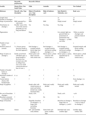
In addition to the specimens examined above, specimens lodged as Boccardia polybranchia from Macquarie Island and Kilcunda (AM W4742–W4743 and NMV G3011, respectively) were examined. These matched the description of B. wellingtonensis Read, 1975 (see Table 2).
Morphological characteristics of Boccardia wellingtonensis Read, 1975 and Boccardia polybranchia (Haswell, Reference Haswell1885) from Victoria and Maquarie Island, Australia and Argentina that have been re-identified as B. wellingtonensis.

NOMENCLATURAL CONSIDERATIONS
The original description of Boccardia polybranchia (Haswell, Reference Haswell1885), collected on oyster farms along the Hunter River, New South Wales, Australia, was limited, without figures and there are no type specimens (see also Blake & Kudenov, Reference Blake and Kudenov1978). More comprehensive descriptions, assigned to the same species, were subsequently produced by, amongst others, Carazzi (Reference Carazzi1893) and Fauvel (Reference Fauvel1927) for specimens collected in Naples and France, respectively. In 1978, Blake & Kudenov tried to provide a better description of B. polybranchia from the type locality. The only Boccardia species that they found at Hunter River was identified as B. chilensis Blake & Woodwick, 1971; they recorded specimens fitting the Carazzi (Reference Carazzi1893) description of B. polybranchia from Coff's Harbour, 325 km north of the Hunter River, and at Kilcunda, Victoria. These specimens, along with others from Macquarie Island, were used for their description but no neotypes were designated (presumably because the specimens came from localities too far from the type locality). Close examination of the above specimens during the current study showed that those from Kilcunda and Macquarie Island are in fact closer to Boccardia wellingtonensis (Table 2), while those from Coff's Harbour (and others from Jervis Bay, New South Wales) match the descriptions by Carazzi (Reference Carazzi1893) and Fauvel (Reference Fauvel1927) more closely than they do that of Haswell (Reference Haswell1885) (Tables 1 & 3).
Morphological characteristics of Boccardia polybranchia (Haswell, Reference Haswell1885) based on the type description, and Boccardia chilensis Blake & Woodwick, 1971.

The original description by Haswell (Reference Haswell1885) is very generic, and it is very difficult to be certain of what animal was really described. Blake & Kudenov (Reference Blake and Kudenov1978) suggested that ‘… B. chilensis may actually be what Haswell originally described, but because of inadequate descriptions, the lack of type material and alteration of the type locality, that can probably never be ascertained.’ There are certain similarities between Haswell's (1885) description and Australian material assigned to B. chilensis by Blake & Kudenov (Reference Blake and Kudenov1978). For example, chaetiger 5 is described as bearing a row of five falcate spines and another row of five that ‘end in a broad head having the form of an inverted cone with an oblique base; on the base of the cone are one or two small conical elevations’ (Haswell, Reference Haswell1885); the modified spines on chaetiger 5 are present in equal numbers. Also, while Haswell (Reference Haswell1885) does not specifically mention notochaetae on the first chaetiger, he does state that some anterior chaetigers bear long chaetae (Haswell, Reference Haswell1885; Blake & Woodwick, 1971; Table 3), a feature that is very clear in B. chilensis. Haswell's (1885) description is, however, also inadequate as a description of the species currently described as B. chilensis, in that it omits, for example, mention of the occipital antenna typical of that species.
Perialla claparedei Kinberg, 1866 described from Rio de Janeiro, was synonymized with B. polybranchia by Soderström (1921) and this was confirmed by Blake (Reference Blake and Kornicker1983). Perialla claparedei is very similar to the specimens of B. polybranchia from Biarritz, South Africa and New South Wales that were examined (see Table 1). However, none of the P. claparedei specimens from the type series are complete, and none are heavily pigmented. It is therefore difficult to confirm if it is the same as the B. polybranchia examined in this study. Furthermore, there is some doubt that all/many of the B. polybranchia in South America had been correctly identified. For example, Blake (Reference Blake and Kornicker1983) had synonymized B. wellingtonensis with B. polybranchia after examining many South American specimens. Sato-Okoshi & Takatsuka (Reference Sato-Okoshi and Takatsuka2001) reversed this synonymy with respect to some specimens collected in Chile. Preliminary observations of some specimens from Argentina suggest that these resemble B. wellingtonensis more closely than they do B. polybranchia (personal observation, see Table 2), and we were unable to secure ‘B. polybranchia’ from Brazil. It is, however, likely that there are several species currently included within B. polybranchia in South America; this situation is currently being reviewed (R. Elías, personal communication).
Given the confusion concerning the nomenclature and identity of B. polybranchia outlined above, its apparent cosmopolitan distribution, its importance as a potential pest on commercially important bivalves and as a pollution indicator species (Borja et al., Reference Borja, Franco and Pérez2000; Vallarino et al., Reference Vallarino, Rivero, Gravina and Elías2002; Ruellet, Reference Ruellet2004), it is important to clarify this problem. This would, however, require an extensive revision of the species, ideally including molecular comparisons from different localities, which is beyond the scope of the current study.
DISTRIBUTION
This species was previously recorded in Namibia, Saldanha Bay on the west coast of South Africa and from east of Cape Agulhas to north of East London (Augener, Reference Augener and Michaelsen1918; Day, Reference Day1967). Thus, the distribution of the species in the current study falls within its known range in South Africa. Boccardia polybranchia is considered cosmopolitan, and has, sensu Blake & Kudenov, Reference Blake and Kudenov1978, been recorded from Australia (New South Wales), South America (Brazil, Argentina, Straits of Magellan, Tierra del Fuego, Peru) and the Kerguelen Islands (Blake, Reference Blake and Kornicker1983; Elías et al., Reference Elías, Rivero, Palacios and Villarino2006). In the northern hemisphere it has been recorded in the Mediterranean (Naples) (Carazzi, Reference Carazzi1893), English Channel, France (Fauvel, Reference Fauvel1927; Ruellet, Reference Ruellet2004) and Japan (Imajima & Hartman, Reference Imajima and Hartman1964).
HABITAT
In the current study, the worms bored into the shells of a range of molluscs, including the abalone Haliotis midae, the turban shell Turbo sarmaticus, the limpets Scutellastra longicosta and Cymbula oculus and the bivalves Perna perna and Saccostrea cuccullata. Boccardia polybranchia sensu Blake & Kudenov, Reference Blake and Kudenov1978 has been recorded from amongst coralline algae, algae and Galeolaria tubes (Blake & Kudenov, Reference Blake and Kudenov1978) and from sand and rocks and in the intertidal algal zone (Blake, Reference Blake and Kornicker1983). In Italy and France they inhabit sandy and muddy tubes and are associated with oyster shells (Carazzi, Reference Carazzi1893; Fauvel, Reference Fauvel1927; Borja et al., Reference Borja, Franco and Pérez2000; Ruellet, Reference Ruellet2004).
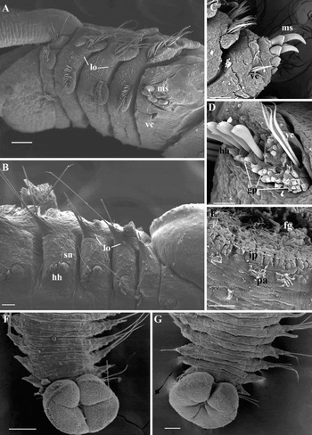
Boccardia proboscidea. (A) Lateral anterior showing lateral organs (lo) on chaetigers 2 and 3, and modified spines (ms) and ventral inferior capillary chaetae (vc) on chaetiger 5; (B) ventral posterior, showing lateral organs (lo) and single superior neuropodial capillary chaeta (sn) dorsal to the hooded hooks (hh) on each chaetiger; (C) ventral view of modified spines (ms) and ventral capillary chaetae (vc) of chaetiger 5; (D) hooded hooks (hh) with external openings of glandular pouches (gp) and ventral inferior chaetae (vc); (E) latero-frontal surface of the palps showing the feeding groove (fg) large papillae with long cirri (pa) and inner row of papillae (ip); (F & G) dorsal views of pygidia of different worms. Scale bars: A, C, F & G, 100 µm; B, 30 µm; D, 25 µm; E, 10 µm.

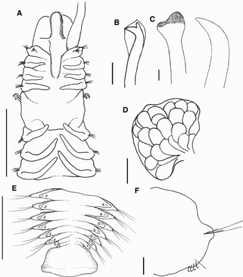
Boccardia proboscidea. (A) Dorsal anterior; (B) hooded hook; (C) brush-topped and falcate spines of chaetiger 5; (D) gross structure of glandular pouches; note the relatively large number of small pouches; (E) ventral view of pygidium and posterior chaetigers; (F) anterior views of posterior right chaetiger, showing position of ventral superior chaeta relative to hooded hooks. Scale bars: A, 1 mm; B, C, 20 µm; D, F, 100 µm; E, 500 µm.

Boccardia proboscidea Hartman, Reference Hartman1940, pp. 382–387; Woodwick, Reference Woodwick1963, p. 134, figure 2; Blake & Kudenov, Reference Blake and Kudenov1978, pp. 238–239, figure 33; Gibson et al., Reference Gibson, Paterson, Taylor and Woolridge1999, pp. 748–749, figures 3 & 4; Bailey-Brock, Reference Bailey-Brock2000, p. 28, figure 1.
MATERIAL EXAMINED
South Africa: Western Cape Province: Saldanha Bay, Jakobsbaai Sea Products (Pty) Ltd, January 2006, A21518 (17) & A21519 (22); Hermanus, Abagold (Pty) Ltd, August 2005, A21516 (14) and September 2005, A21517 (12), A21520 (SEM).
MORPHOLOGY OF NEW MATERIAL
Colour in life pale green, with bright red branchiae and black pigment along edge of prostomium; faint black markings on dorsal surface between chaetigers 1 and 2 and 2 and 3. Preserved specimens tan, dark line along the feeding groove of the palps and along margins of prostomium and caruncle.
Largest specimens examined 20 mm long for 99 chaetigers, but up to 33 mm long recorded in population dynamics study (Simon & Booth, Reference Simon and Booth2007). Prostomium rounded (Figure 5A), sometimes weakly indented, with 2–3 pairs of eyes; caruncle extending to middle or posterior margin of chaetiger 3 (Figure 5A), with nuchal organs consisting of paired ciliary bands along caruncle.
Notopodial lobe of chaetiger 1 with short notochaetae (Figures 4A & 5A). Anterior chaetigers with approximately 17 broad notochaetae with short tapering ends and few long needle-like chaetae. Posterior notochaetae long, needle-like, with two or three short chaetae with slightly curved ends that face posteriorly. Neurochaetae broad and blade-like with tapering ends, on chaetigers 2–4 and 6 a few capillary chaetae also present.
Hooded hooks start on chaetiger 7; bidentate, without constriction on shaft, angle between main fang and shaft >90°, angle between teeth 45° (Figure 5B). Seven or eight hooded hooks per ramus, decreasing to 2–4 on posterior chaetigers (Figure 4B). Chaetigers 7–9 with three long ventral inferior capillary chaetae (Figure 4D). Neuropodial lobe, associated with hooded hooks, up to chaetigers 10 or 11 (Figure 4D). Posterior chaetigers with single fine superior neuropodial chaeta (Figures 4B & 5E, F).
Chaetiger 5 with two heavy curved spines and three blunt, bristle-topped, spines on each side. Fascicle of short ventral inferior chaetae present (Figure 4C).
Branchiae filiform, separate from notopodial lobes, on chaetigers 2–4 and posteriorly from chaetiger 6 to near the posterior end of the body (Figure 4A&G) but absent from last 2–10 chaetigers. Longest branchiae (which may be as long as animal is wide) on chaetigers 8 or 9, decreasing in length towards end of the body (Figures 4G & 5A).
Large paired glands with many small glandular pouches observed in chaetigers 7–9 (Figure 5D). More than 50 openings visible at base of hooded hooks on these chaetigers (Figure 4D).
Lateral organs observed on chaetigers 2, 3, 6 and posteriorly (Figure 4A, B).
Row of papillae (3 µm diameter) each bearing ~8 cirri, 10 µm apart on latero-frontal surface of palp (Figure 4F). Towards the feeding groove, rows of papillae (2 µm diameter) arranged in pairs, about 1 µm apart, running perpendicular to length of palps, lacking cirri (Figure 4E).
Pygidium a fleshy cuff with dorsal notch; may be divided into four lobes or by folds in the pygidium (Figure 4F, G); often wider than posterior-most chaetigers.
REPRODUCTION
The reproduction of this species was examined at two farms: Jakobsbaai Sea Products (Pty) Ltd and Abagold (Pty) Ltd, on the west and south coasts, respectively. Worms reproduce throughout the year, but with increased numbers from the end of winter to early summer (Simon & Booth, Reference Simon and Booth2007). The worms are poecilogonous at both farms, with females producing capsules that contain: (a) adelphophagic and planktotrophic larvae; (b) planktotrophic larvae only; and (c) adelphophagic larvae only. At both farms, 50 to 80% of the brooding females brooded capsules containing both adelphophagic and planktotrophic larvae and brooding individuals were present throughout the year (Simon & Booth, Reference Simon and Booth2007). Within each category, larvae were of comparable size at the two farms (maximum length of planktotrophic larvae: 375 and 400 µm; maximum length of adelphophagic larvae: 1100 and 1000 µm at Abagold and Jakobsbaai, respectively). There was a tendency for worms at Jakobsbaai Sea Products (Pty) Ltd to brood more adelphophagic larvae per capsule (number of planktotrophic larvae per capsule: mean = 3.98, maximum = 7, number of adelphophagic per capsule: mean = 4.6, maximum = 12), while the opposite was true at Abagold (Pty) Ltd (number of planktotrophic larvae per capsule: mean = 5.5, maximum = 25, number of adelphophagic per capsule: mean = 3.19, maximum = 17).
REMARKS
Petch (Reference Petch1995) conducted an extensive morphological comparison of Boccardia proboscidea from the United States of America (Alaska, Washington State and California), Canada (British Columbia), Japan, Panama and Australia. He found that all specimens matched the original description of the species, with the exception of those from Alaska and Panama, which he suggested would represent separate species. Specimens from South Africa correspond well with other descriptions with just a few differences. South African specimens are similar in size to those from California (Hartman, Reference Hartman1940; Woodwick, Reference Woodwick1963), but larger than those described for Hawai'i (Bailey-Brock, Reference Bailey-Brock2000) and Japan (Sato-Okoshi, Reference Sato-Okoshi2000) and smaller than those from Barkley Sound, western Canada (Sato-Okoshi & Okoshi, Reference Sato-Okoshi and Okoshi1997). With respect to the shape of the pygidium, South African specimens differ from the type specimens (Hartman, Reference Hartman1940) but more closely resemble those described by Bailey-Brock (Reference Bailey-Brock2000). The branchiae of South African specimens are longer than those described by Woodwick (Reference Woodwick1963, figure 2) and Petch (Reference Petch1995). The fine ventral superior chaetae dorsal to the hooded hooks were also described by Gibson et al. (Reference Gibson, Paterson, Taylor and Woolridge1999) and Sato-Okoshi (Reference Sato-Okoshi2000).
HABITAT
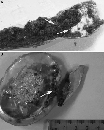
On cultured abalone in South Africa, Boccardia proboscidea is a secondary borer. It may form burrows on the surface of the shell (Figure 6A), in crevices on the shell surface as described for worms infesting the oysters Ostrea edulis and Crassostrea gigas, the barnacle Balanus cariosus and the abalone Haliotis roei (Sato-Okoshi & Okoshi, Reference Sato-Okoshi and Okoshi1997; Bailey-Brock, Reference Bailey-Brock2000; Sato-Okoshi, Reference Sato-Okoshi2000; Sato-Okoshi et al., Reference Sato-Okoshi, Okoshi and Shaw2008) or it may occur in the burrows and blisters of Polydora hoplura and Dipolydora capensis (see also Woodwick, Reference Woodwick1963). In extreme cases it forms ‘mudpacks’ which are covered with a thin layer of nacreous shell in the region of the respiratory pores. These packs usually contain several worms of different sizes and often cause the shell to break along the respiratory pores (Figure 6B). This pattern of infestation is similar to that of Polydora uncinata Sato-Okoshi, 1998 (Radashevsky & Olivares, Reference Radashevsky and Olivares2005).
Damage caused by Boccardia proboscidea. (A) Burrows formed on external surface of abalone shells, with B. proboscidia palps arrowed; (B) shell broken in the region of the respiratory pores, where it was weakened by infestation (arrow).

DISTRIBUTION
In South Africa, Boccardia proboscidea was found on cultured abalone at farms on the west, south and east coasts of South Africa. It is least abundant on the east coast. The presumed natural distribution range of B. proboscidea extends from Canada (British Columbia) to southern California, with unconfirmed reports from further south, and Japan (Hartman, Reference Hartman1940; Woodwick, Reference Woodwick1963; Fauchald, 1977; Petch, Reference Petch1995; Sato-Okoshi, Reference Sato-Okoshi2000; Oyarzun et al., in preparation). It has also been recorded from Australia (Blake & Kudenov, Reference Blake and Kudenov1978), Hawai'i (Bailey-Brock, Reference Bailey-Brock2000), New Zealand (Read, Reference Read2004) and Spain (Martínez et al., Reference Martínez, Adarraga and López2006) where it is considered non-native.
MATERIAL EXAMINED
Eastern Cape Province: Haga Haga, February 2005 A21521 (1) and April 2007, A21522 (7); A21523 (4, ethanol fixed).
MORPHOLOGY OF NEW MATERIAL
Specimens correspond well with the description by Day (Reference Day1961); largest specimen, 20 mm long for 83 chaetigers, was larger than described previously. Prostomium bilobed; caruncle extending to chaetiger 2; middle of caruncle is dark; with one pair of eyes (in one specimen, a second pair observed in the pigment of the caruncle). A mid-dorsal ridge from chaetiger 5 to the middle of chaetiger 8. Notochaetae present on chaetiger 1. No modified posterior notochaetae. Notopodial lobes small, inconspicuous in posterior chaetigers. Hooded hooks from chaetiger 7; eight and two hooks per ramus in anterior and posterior chaetigers, respectively; no accompanying chaetae. Chaetiger 5 with anterior row of spines with swollen tips, a raised central cone and raised ridge, and posterior row of falcate spines. Branchiae on chaetiger 2 longer than those on chaetigers 3 and 4; others short, never overlapping mid-dorsum; posterior third of body abranchiate. Pygidium reduced, forming pair of flattened cushions.
Female collected in April 2007 with ova in chaetigers 23–60.
HABITAT
Previously found in rock (Day, Reference Day1961) but associated with shells of cultured Haliotis midae and wild Saccostrea cuccullata in current study.
DISTRIBUTION
Previously found only in Knysna (Day, Reference Day1961). In the current study found only at Haga Haga.
ACKNOWLEDGEMENTS
Thanks are due to Andy Mackie (National Museum of Wales), Greg Rouse (Scripps Institution of Oceanography) and Frederik Pleijel (Göteberg University) for discussion of nomenclatural issues; the staff of South Africa National Parks and the Shell Museum and Aquarium, Mossel Bay, Anthony Bernard and Felicia Keulder for field assistance; abalone farmers who provided infested abalone; Pat Hutchings and Elin Sigvaldadóttir for finding references; Adrian Craig, Francesca Porri, Sophie Reinecke, Professor J.C. Zietsman and Mariaan Gaertner for translating manuscripts and Dylan McGarry for the drawings. Funding was provided by Marine and Coastal Management, the Abalone Farmers' Association of South Africa, a Rhodes University Joint Research Committee grant and a National Research Foundation post-doctoral grant.