Coronary artery fistulas are abnormal communications between a coronary artery and a cardiac cavity or nearby vessel. In general, they are asymptomatic and are discovered fortuitously in approximately 0.1% of adult patients undergoing routine coronary angiography.Reference Vavuranakis, Bush and Boudoulas1 Most coronary artery fistulas are repaired via surgical operation, while a few are filled with fistula by using coils via interventional surgery. This article reports how a successful closure for a case of coronary artery fistula is treated by using a patent ductus arteriosus occluder with a follow-up of 2.5 years.
Case report
An 8-year-old girl, weighing 22 kilograms, was hospitalised in July, 2007 due to coronary artery fistulas. She had no previous symptoms of chest tightness, panting, dyspnoea, cyanosis, crouch, dysplasia, etc. Cardiovascular ultrasound showed that the right coronary artery fistula was drained into the right ventricle and that the left ventricle was dilated.
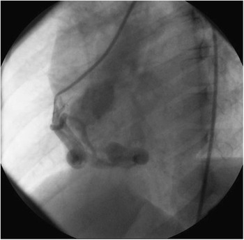
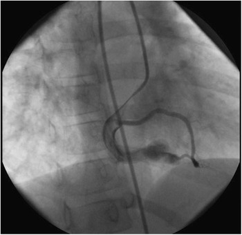
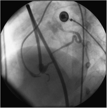
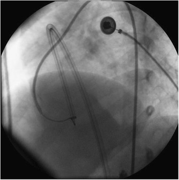
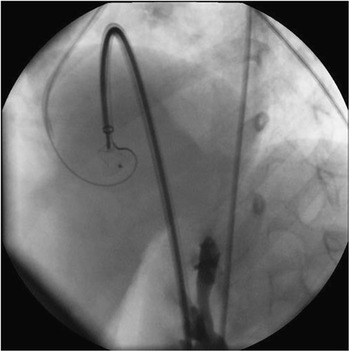
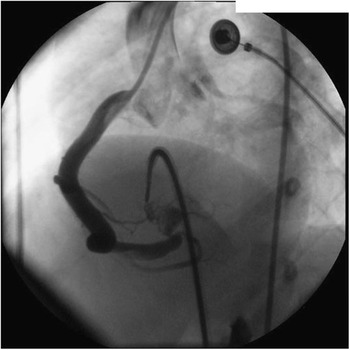
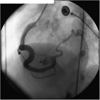
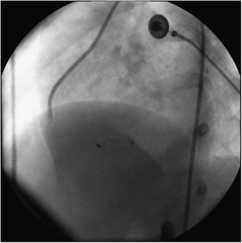
One day was selected for cardiac catheterisation and transcatheter interventional closure. After successful puncture of the right femoral artery and femoral vein, 2000 micron anticoagulant heparin solution was injected and a 6FJR3.5 Judkins Right duct was inserted into the right coronary artery. Radiography revealed the following (Fig 1): a big and tortuose fistula opening from the right coronary artery (Fig 2) and ending at the right ventricle. The maximum diameter of the fistula was 10 millimetres and the minimum diameter was 6 millimetres at the opening of the fistula. The right coronary artery was parallel to the fistula, whose ends were communicating with the ends of the left anterior descending coronary artery (Fig 3). An exchange guiding catheter 6FJR3.5 was placed at the opening of the fistula of the right coronary artery and a 2.60-centimetre long 0.032″ straight head super-slip exchange guide wire was threaded through the fistula into the right ventricle and pulmonary artery. A snare was then inserted into the pulmonary artery via the femoral vein to seize the straight head super-slip guide wire and pull it out of the body. Thus, a track of femoral artery – right coronary artery – fistula – right ventricle – femoral vein was established (Fig 4). A 7F delivery sheath was inserted from the end of the femoral vein and it could retrograde to reach the opening of the fistula (Fig 4). A patent ductus arteriosus occluder (by Shanghai Shape Memory Alloy Company Ltd, Shanghai, China) with a diameter of 9 millimetres was selected and pushed to the exit of the fistula by using a pushing bar with the guide wire track kept in place (Figs 5 and 6). With the pushing bar fixed, the long sheath was withdrawn to enable the waist of the occluder to be released in the narrow section. The waist was well shaped, which suggested that the occluder was sized to fit. The occluder was fixed well since resistance was felt by pushing gently. It was then opened and observed for 15 minutes to see whether the girl had chest pain or not. The electrocardiogram did not reveal ST segment elevation, and therefore myocardial infarction was excluded. As no residual shunt was found in a recheck by radiography, the pushing bar was rotated and the occluder was released (Figs 7 and 8). Radiography again showed that the right coronary artery was smooth without any coronary stenosis (Fig 7). The closure was successful.
By radiography of the right coronary artery, a big and tortuose fistula is seen to accompany the right coronary artery and to finally drain to the right ventricle.

By another body position of roentgenography, a coronary artery fistula opening in the right coronary artery is seen clearly.

By radiography of the sheath, ends of the right coronary artery are seen to communicate with the ends of the left anterior descending coronary artery.

A track of the femoral artery – right coronary artery – fistula – right ventricle – femoral vein is established successfully. A 7F sheath was used to retrograde to the opening of the fistula along the track.

With the method of guide wire track keeping, the patent ductus arteriosus occluder (by Shanghai Shape Memory Alloy Company Ltd) was sent to the exit of the fistula.

The performance of radiography of the right coronary artery is again an indication that the fistula exit is closed and the right coronary artery is clear.

The occluder is released after confirming that there is no myocardial infarction according to the clinical conditions and electrocardiogram.

An occluder with a diameter of 9 millimetres is released and is well shaped in the waist.

In the recheck 2.5 years after closure, the girl had no complaints and was generally in good condition. The size of each ventricle was in the normal range as indicated by cardiovascular ultrasonic diagnosis. A strong echo of the occluder could be seen at the outlet of the fistula in the right coronary artery. No displacement or residual shunt was found.
Discussion
Coronary artery fistula is an uncommon congenital malformation. It accounts for 0.27–0.4% in all congenital cardiac diseases. There are many types of coronary artery fistulas. This article discusses a case of the right coronary artery fistula connecting to the right ventricle. Cardiovascular ultrasonic diagnosis is an important method of determining coronary artery fistulas. At present, coronary angiography is the “golden standard” for determining coronary artery fistula.
There are currently three management options available for coronary artery fistula. Small and asymptomatic ones can be managed conservatively. Sometimes, they even close spontaneously.Reference Muhler, Keutel and Von2 The second therapeutic option is surgical closure, generally described as safe and effective even though myocardial infarction and recurrence have been reported.Reference Liberthson, Sagar, Berkoben, Weintraub and Levine3 In order to avoid median sternotomy and sometimes cardiopulmonary bypass, a percutaneous transcatheter closure can be proposed. There are many cases of closing relatively small coronary artery fistulas with a coil.Reference Luis, Diaz and Juan4, Reference Hoffer, Materne, Henroteaux, Markov and Boland5 It is, however, inappropriate to adopt the coil closure method for a relatively large and tortuose fistula. Individual cases have been reported where a patent ductus arteriosus occluder is used for a relatively large coronary artery fistula.Reference Sadiq, Wilkinson and Qureshi6, Reference Al-Ata, Amin, Galal, Kouatli and Hussain7 To achieve successful closure, the method of reserving the guide wire is very important, which not only could avoid the trouble of reestablishing the track due to the inappropriate size of the occluder, but also could reduce the damage of excessive intracardiac operation to the patient and damage of radiation to the medical staff.
A balloon inflation test is usually conducted while sealing the fistula with a coil to learn whether myocardial ischaemia occurs after coronary artery fistula closure.Reference Skimming, Gessner, Victorica and Mickle8 Along with the use of a controllable closure device, a patent ductus arteriosus occluder is used, however, which will not be released until closure is completed and until the possibility of myocardial ischaemia or infarct is excluded meaning that no change occurs with the electrocardiogram and other vital signs for at least 15 minutes. With this procedure, the balloon inflation test can be forgone, which will simplify the procedure and reduce the risks.
In the follow-up for 2.5 years, the girl had no complaints of discomfort and developed normally. The patient’s ventricle sizes are the same as for other children of the same age, as indicated by cardiovascular ultrasonic diagnosis. All of the above indicate that, through the application of a patent ductus arteriosus occluder, transcatheter closure of coronary artery fistulas proves to be a safe, feasible, and cost-effective alternative to surgery.