Balloon angioplastyReference Parsons, Ladusans and Qureshi1 and stentingReference Zahn, Chang, Aldousany and Burke2, Reference Gillespie and Rome3 of Blalock–Taussig shunts, to a diameter equal to the original nominal diameter, have been successfully performed in the past on clinical grounds, either as an emergency or as a semi-elective procedure. These procedures represent palliative interim interventions, performed until the patient is suitable for corrective surgery or the next stage of their staged palliation. Occasionally, however, patients are not clinically fit for the next stage of their surgery, and as they outgrow their shunt a second shunt needs to be inserted. This is applicable not only to infants and young children, but also to adults.Reference Bader, Somerville and Redington4 In such cases, the potential of transcatheter upsizing of existing patent aortopulmonary shunts could be a valuable alternative to surgery. We report the clinical course of an 8-month-old baby following Norwood I operation, who was deemed unsuitable to proceed to the next stage of Norwood palliation and had transcatheter upsizing of the Blalock–Taussig shunt with significant clinical improvement.
Clinical summary
A newborn was admitted to our unit with unbalanced atrioventricular septal defect and hypoplasia of the left ventricle and the aortic arch. Laryngeal cleft, bilateral choanal stenosis, severe tracheobronchomalacia, and bilateral optic disc colobomata were diagnosed postnatally. She underwent Norwood I operation with a 3.5-millimetre Blalock–Taussig shunt in the first week of life. The patient remained dependent on continuous positive pressure ventilation and desaturated with high carbon dioxide levels of approximately 10–13 kilopascals until the age of 7 months when she presented to our intensive care unit with drowsiness, increased secretions, and partial pressure of carbon dioxide of 19 kilopascals. Airway stenting was performed 3 weeks later and the patient was extubated soon after, although she remained desaturated at 65–70%, partly due to outgrowing the Blalock–Taussig shunt and also due to a fixed narrowing at the pulmonary arterial end as diagnosed on the magnetic resonance scan (Fig 1). After multidisciplinary assessment, she was deemed unsuitable for stage II Norwood operation due to chronically elevated carbon dioxide levels and the status of her comorbidities. High-risk interventional treatment was offered with Blalock–Taussig shunt stenting to a bigger diameter than the original one.
Magnetic resonance imaging showing the narrowing at the pulmonary end of the Blalock–Taussig shunt.

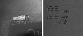
Bench testing was first performed to establish the stretch limits of the Gore-Tex shunt. A 6-millimetre diameter × 2-centimetre-long shunt was chosen and tested in vitro with implantation of a 10-millimetre diameter pre-mounted Genesis stent (Cordis, Johnson & Johnson, New Brunswick, New Jersey, United States of America). We implanted the stent along half of the shunt's length and further expanded it using a 10-millimetre Powerflex balloon that reached up to 14 Atmospheres of pressure. No tears or foreshortening of the Gore-Tex tube were noted (Fig 2). The part of the stent covered by the Gore-Tex increased from 6 to 8 millimetres, whereas the uncovered part increased to the nominal balloon diameter of 10 millimetres (Fig 2).
The 15-millimetre-long pre-mounted stent is implanted along half of the 20-millimetre-long Blalock–Taussig shunt with obvious macroscopic enlargement of the shunt diameter, which was quantified under fluoroscopy. The part of the stent covered by the Gore-Tex increased from 6 (X2) to 8 millimetres (X3), whereas the uncovered part increased to the nominal balloon diameter of 10 millimetres (X4).

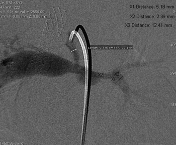
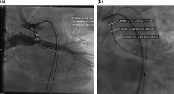
During catheterisation, shunt angiogram showed a narrowed Blalock–Taussig shunt, which measured 3.1 millimetres, and diffusely narrow left pulmonary artery (Fig 3). The left pulmonary artery was initially stented and over the same 0.018-inch platinum plus wire (Boston Scientific, Natick, Massachusetts, United States of America) a 5 × 15-millimetre pre-mounted Palmaz Genesis stent was advanced into the shunt and inflated slowly to 15 Atmospheres of pressure. The stent was dilated with its own balloon and no adjunctional balloons were needed. The Blalock–Taussig shunt diameter enlarged to 4.7 millimetres at its aortic and pulmonary end and 4.4 millimetres at its mid-portion (Fig 4a and b). There was no evidence of dissection or extravasation on the repeat angiogram. Although there was no improvement to the patient's saturations following left pulmonary artery stenting, there was an immediate increase in the saturations from 70–75% to 85% following shunt stenting, indicating the restrictive nature of the relatively small shunt and the fixed obstruction at its distal end. The patient remained self-ventilating in air and was discharged home with saturations of 85%.
Angiographic Blalock–Taussig shunt measurements before intervention (note the tapering of the distal end of the shunt and the two other stents, previously implanted in the trachea and the right main bronchus).

(a) Blalock–Taussig shunt measurement after stent implantation and (b) measurement of the final stent diameter.

Discussion
Blalock–Taussig shunts (GORE-TEX Vascular Graft – Paediatric Shunt, W.L. Gore & Associates Inc., Flagstaff, Arizona, United States of America) are made of e-polytetrafluoroethylene material. They are produced in different diameters ranging from 3 to 5 millimetres and varying lengths of 5, 10, 15, and 20 millimetres. Gore-Tex stretch paediatric shunts are known to have a degree of longitudinal extensibility to allow for easier tailoring and sizing and potential of 50% elongation of the bevelled end; however, there is no solid evidence on the degree of their safe radial expansion. Brown et alReference Brown, Boshoff and Heying5 recently published their experience with stent expansion of stretch Gore-Tex grafts in children with congenital cardiac lesions. Our bench testing and clinical outcome are in complete agreement with the findings of the only other publication in the literature referenced above.
The mean time interval between a modified Blalock–Taussig shunt and a second procedure – reshunting/open repair – has been found to be approximately 12.4 months.Reference Gold, Violaris and Engle6 The effectiveness of the 4-millimetre diameter conduit has been doubted due to the occurrence of conduit failure at 1 year after implantation, whereas other sized conduits remained widely patent.Reference Karpawich, Bush and Antillon7 Bove et alReference Bove, Kohman and Sereika8 have reported 56 modified Blalock–Taussig shunts in 50 patients with different pathologies, stating that the majority of the patients had satisfactory palliation for at least 3 years after the procedure. Overall, patient age at operation, weight less than 3.0 kilograms, and graft size have been found to be risk factors for shunt patency.Reference Karpawich, Bush and Antillon7–Reference Tsai, Chang and Lin9
The potential of upgrading existing patent aortopulmonary shunts by transcatheter techniques could be a valuable alternative to surgery. In our patient's case, no extravasation was noted at a nearly 30% shunt diameter increase, while the patient's saturations improved by 10%. Although the Blalock–Taussig shunt was taken up only by 1 millimetre, the degree of flow improvement achieved from such a small increase in diameter proved to be very significant. This is in accordance with the cubic law, stating that the flow rate is proportional to the cube of the vessel's diameter.Reference Zamir10 In our patient's case, an increase in the shunt diameter by 1 millimetre would translate into more than doubling the flow through it. Improvement of the patient's saturations only following Blalock–Taussig shunt stenting was indicative of the fact that there was limitation to the pulmonary flow by the shunt size and the additional fixed narrowing of its distal end.
We suggest that the procedure of percutaneous upsizing of surgically placed Blalock–Taussig shunts by transcatheter techniques may become a useful and safe alternative to either surgical upsizing or placing of further shunts. Further studies and clinical experience are required before this becomes more routine practice on a select number of patients.
Acknowledgements
We are very grateful to Drs Philipp Beerbaum and Kuberan Pushparajah from the Evelina Children's Hospital for their valuable input in this case.