Percutanous closure of a patent foramen ovale in patients having suffered from an ischaemic stroke is a treatment option. We report on successful closure of a pulmonary arteriovenous malformation in a young adult after an ischaemic stroke with suspected patent foramen ovale.
Case report
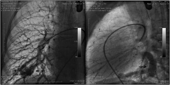
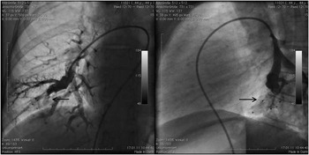
A 44-year-old woman with an ischaemic stroke was referred for catheter-interventional patent foramen ovale closure. The medical history revealed recurrent epistaxis and an iron-deficiency anaemia. On cerebral magnetic resonance imaging, additional older ischaemic lesions were seen. Her transcutaneous oxygen saturation was slightly decreased (94%), her haemoglobin was 10.1 g/dl, and her thrombocyte count was in the normal range. On trans-oesophageal echocardiography, the interatrial septum was intact. However, during an echocontrast study a significant amount of air bubbles were seen in the left atrium after three cardiac cycles. A right-to-left shunt of an extracardiac origin was suspected. Angiography revealed an arteriovenous malformation in the lower lobe of the right lung (Fig 1). Through a 6 F coronary guide catheter, a total of three Amplatzer vascular plugs I (6, 8, and 12 mm; AGA/St. Jude Medical, St. Paul, Minnesota, United States of America), a single vascular plug II (6 mm), and a detachable coil (8/5; Cook Ireland Limited, Limerick, Ireland) were deliverd into the feeding vessels. This led to complete occlusion of the arteriovenous malformation (Fig 2). The transcutaneous oxygen saturation improved to 99% after the closure of the pulmonary arteriovenous malformation, and the patient was discharged home on the following day.
The angiogram shows a large pulmonary arteriovenous malformation in the right lower lobe with a significant right-to-left shunt. Left side – anteroposterior projection; right side – lateral projection

The angiogram after successful closure shows complete occlusion of the pulmonary arteriovenous malformation with no right-to-left shunt. Left side – anteroposterior projection; right side – lateral projection.

Discussion
This case report shows that in a patient with ischaemic stroke, intact interatrial septum and positive bubble test for a right-to-left shunt, the underlying disease could be a pulmonary arteriovenous malformation.Reference Morier, Goncalves-Matoso and Michel 1 Retrospectively, the pulmonary arteriovenous malformation is the most probable cause for the ischaemic cerebral incidents in our patient. The combination of epistaxis, iron deficiency, anaemia, and pulmonary arteriovenous malformation led to the diagnosis of hereditary haemorrhagic teleangiectasia, validated by the mutation in the endoglin gene – hereditary haemorrhagic teleangiectasia type 1.
Catheter-interventional treatment of pulmonary arteriovenous malformations has proven to be safe and effectiveReference Letourneau-Guillon, Faughnan and Soulez 2 – Reference Gupta, Faughnan and Bayoumi 5 and is recommended for all adults with pulmonary arteriovenous malformations and children with symptomatic pulmonary arteriovenous malformations.Reference Faughnan, Palda and Garcia-Tsao 6
Hereditary haemorrhagic telangiectasia is a vascular disorder due to gene mutations with autosomal-dominant inheritance. So far, mutations in the endoglin gene and activin receptor-like kinase gene have been discovered, resulting in hereditary haemorrhagic telangiectasia type 1 and 2 with slightly differing phenotypes.Reference Govani and Shovlin 7 Moreover, hereditary haemorrhagic telangiectasia in association with juvenile polyposis is linked to the mutation of Smad4. Further genes are under investigation, probably resulting in more subtypes.Reference Govani and Shovlin 7 Hereditary haemorrhagic telangiectasia is being diagnosed clinically via the Curaçao Criteria where four criteria – epistaxis, telangiectasia, visceral lesions, and positive family history for hereditary haemorrhagic telangiectasia – are being taken into consideration; three criteria make the diagnosis definite, two possible, and one unlikely.Reference Shovlin, Guttmacher and Buscarini 8 If the clinical diagnosis of hereditary haemorrhagic telangiectasia is confirmed, then genetic testing is recommended to identify the causative mutation and help establish the diagnosis in other – especially asymptomatic – family members.Reference Faughnan, Palda and Garcia-Tsao 6
Owing to the fact that there is a strong association between pulmonary arteriovenous malformations and hereditary haemorrhagic telangiectasia, the detection of the former should always lead to a search for the diagnosis of the latter.Reference Kurshid and Downie 9
Through molecular analysis, a mutation in the endoglin gene was detected in our patient, thus placing her in the subtype hereditary haemorrhagic telangiectasia type 1. Studies have shown that endoglin mutation is highly associated with pulmonary arteriovenous malformations, as well as with cerebral arteriovenous malformations.Reference Govani and Shovlin 7 Current international guidelines for hereditary haemorrhagic telangiectasia recommend routine screening for pulmonary arteriovenous malformations in all patients with suspected hereditary haemorrhagic telangiectasia,Reference Faughnan, Palda and Garcia-Tsao 6 as they bear the risk of causing severe neurological problems such as ischaemic stroke or brain abscess.Reference Letourneau-Guillon, Faughnan and Soulez 2 Therefore, contrast echocardiography should be performed to unveil these vascular abnormalities and refer the patient to the recommended management, which is embolotherapy.Reference Faughnan, Palda and Garcia-Tsao 6 It has been shown that even small pulmonary arteriovenous malformations can be the cause for ischaemic stroke.Reference Shovlin, Jackson and Bamford 10 This makes screening for pulmonary arteriovenous malformations and closure even more important. In addition, a considerable number of patients with hereditary haemorrhagic telangiectasia and pulmonary arteriovenous malformations are undiagnosed at the time of the ischaemic stroke as was the case in our patient. This emphasises that the event of an ischaemic attack/stroke in a young adult should give rise to a diagnostic work-up covering congenital malformations of the heart, for example patent foramen ovale, acquired cardiac lesions, haematologic abnormalities, as well as vascular lesions such as pulmonary arteriovenous malformations – highly suspicious for hereditary haemorrhagic telangiectasia.