

Introduction
In the literature, detailed anatomic descriptions of normal and abnormal laryngeal and pharyngeal structures as observed through nasopharyngeal fibre-optic endoscopy are widely availableReference Byars and Slovis1. In contrast, the caudo ventral oropharynx has not been as well described. This region cannot be observed during a routine (standing) nasopharyngeal endoscopic examination; the best way to visualize the caudal oropharynx is through oropharyngeal endoscopy. This is problematic in the conscious horse as it requires valuable endoscopic equipment to be placed in the oral cavity. Additionally, movement of the tongue in a conscious horse can make a thorough examination of the caudal oropharynx difficult.
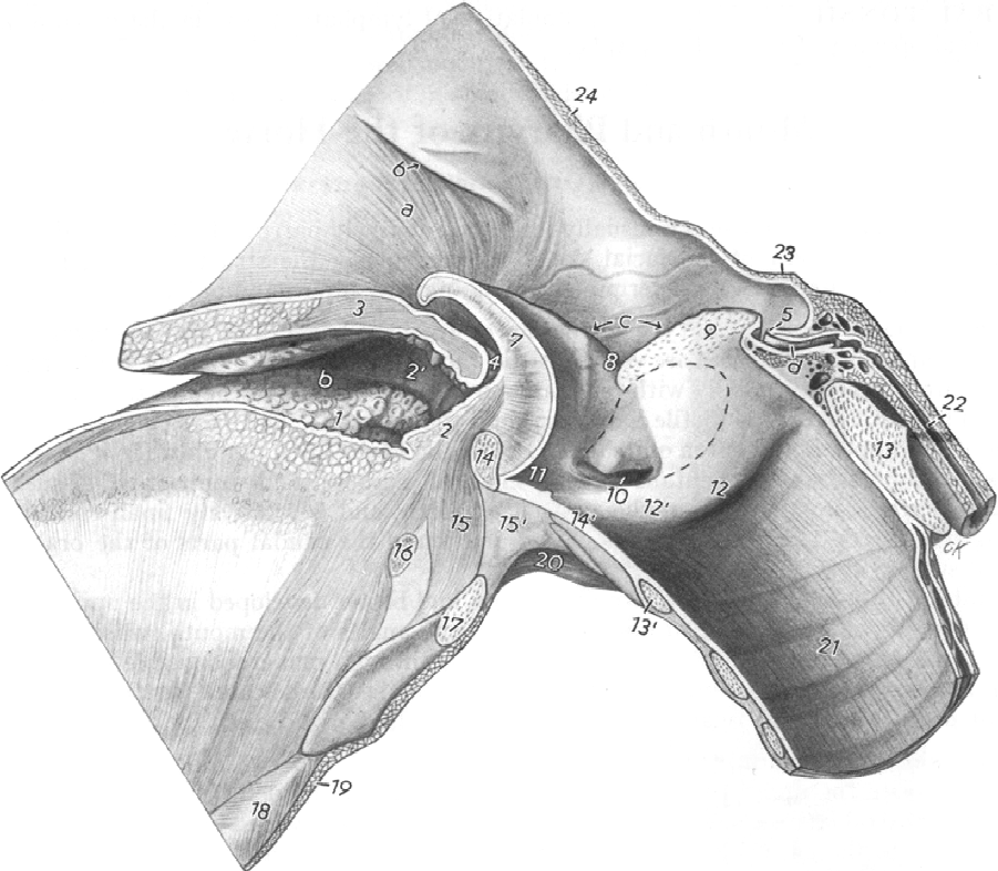
One structure believed by some authors to be of clinical significance in the oropharynx is the glossoepiglottic fold (GEF)Reference Noden, de Lahunta, Noden and de Lahunta2. A congenital abnormality of this structure, described as a frenulum of the epiglottis, has been reported previously in four foals and was implicated in the abnormal epiglottic function and dorsal displacement of the soft palate (DDSP) observed in those animalsReference Yarbrough, Voss, Herrgesell and Shaw3. More recently, a report of persistent DDSP in an adult horse has been attributed to a frenulum of the epiglottisReference Moorman, Marshall and Jann4. The epiglottis and tongue originate from swellings on the floor of the pharynx in the embryo. These swellings are in contact embryologically, and incomplete separation of the swellings could result in an abnormal connection, i.e. post-epiglottic frenulumReference Schummer, Schummer, Nickel and Wolfgang5. To date, this structure has been poorly described in the literature. Anatomical texts identify this fold as having contact with the base of the tongue rostrally, the ventral aspect of the base of the epiglottis caudo-dorsally and to the hyoid bone rostro ventrally through its associations with the hyoepiglottic ligament and the hyoepiglotticus muscle, respectively (Fig. 1)Reference Noden, de Lahunta, Noden and de Lahunta2.
Sagittal section of the equine pharynx and larynx demonstrating the relationship between the tongue (1), glossoepiglottic fold (2), soft palate (3), boundary of intrapharyngeal opening (4), epiglottis (7), median laryngeal ventricle (11), thyroid cartilage (14), hyoepiglotticus muscle (15), hyoideus transversus (16) and basihyoid with lingual process (17). From SchummerReference Schummer, Schummer, Nickel and Wolfgang5 with kind permission of Springer Science and Business Media

The purpose of this study was to document the endoscopic appearance of the GEF in horses with normal upper airway function, so that variations of normal anatomy may be differentiated from abnormalities that may contribute to upper respiratory tract dysfunction.
Materials and methods
Six clinically normal adult Quarter Horses between 5 and 7 years of age and ranging from 480 to 520 kg bodyweight were obtained for the purposes of this study. All horses were currently being used for respiratory physiology studies at Oklahoma State University (OSU). Documentation of normal upper airway function was predicated upon a lack of exercise intolerance and abnormal airway noise during numerous high-speed treadmill examinations prior to the commencement of this study. Horses performed a single high-speed treadmill examination, starting with a warm-up of 5 min at 2 m s− 1 and 1.5° incline, followed by a 5 min at 3.8 m s− 1 and 2.5° incline. The horses then cantered (depending upon the size and subjective gait of the horse) on a 3.5° incline until they were fatigued and unable to maintain position on the treadmill. Duration was variable, but all horses were capable of cantering for at least 5 min.
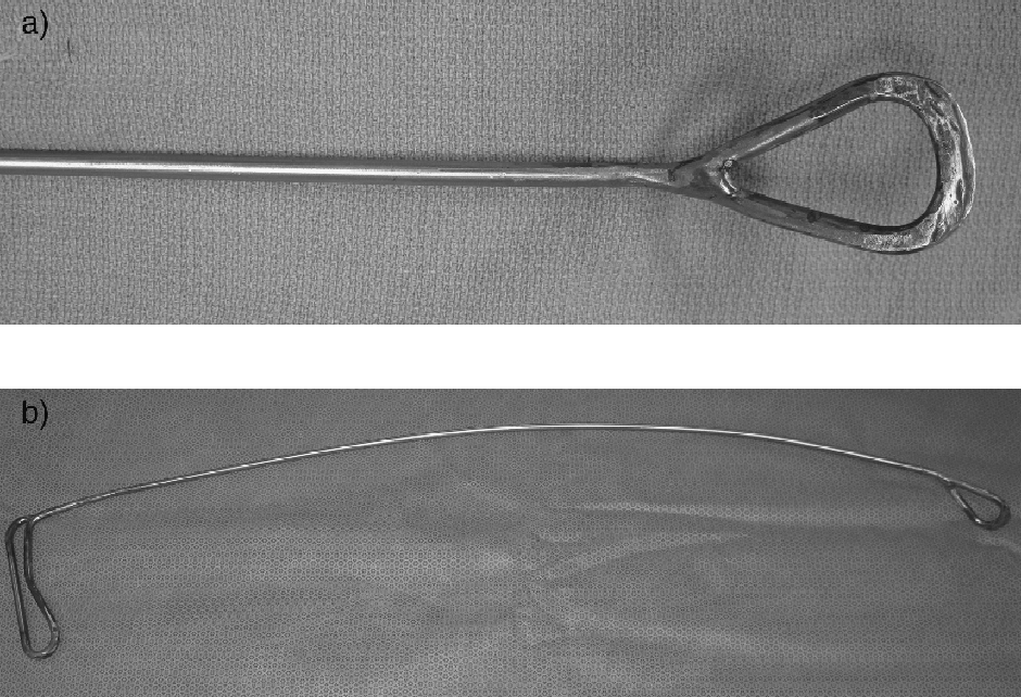
To facilitate examination of the ventral aspect of the epiglottis, a specialized epiglottic elevation instrument was designed. A 4-feet (105 cm) length of stainless steel rod of 3/16th in. (4.5 mm) diameter was forged to give a spatulated triangular tip 2 in. (48 mm) long and 1 in. (24 mm) wide. The other end of the steel rod was folded back on itself for a length of 5 in. (120 mm) and bent through 90° to provide a handle. A gentle downward curve was incorporated into the design of the instrument. Metal inert gas welds were used to seal the rod ends at the handle and retractor tip. Any rough edges were then removed with a bench-top angle grinder (Fig. 2a and 2b).
Epiglottic elevation instrument

Approval from the Institutional Animal Care and Use Committee of OSU was obtained. All horses were placed under general anaesthesia for the purpose of safe and complete endoscopic examination of the oropharynx. General anaesthesia was induced using an intravenous protocol of xylazine hydrochloride (1.1 mg kg− 1), diazepam (0.05 mg kg− 1) and ketamine hydrochloride (2.2 mg kg− 1). A PVC pipe speculum/gag provided oral access of instrumentation between the maxillary and mandibular incisors. The ventral aspect of the epiglottis in the oropharynx was examined by dorsally displacing the soft palate with the epiglottis elevator. A 1 m flexible endoscopeFootnote a was then passed through the mouth gag for oropharyngeal examination. The epiglottic elevator was then used to manipulate the epiglottis dorsal and caudal, so that the nature of the GEF and its relationship to the ventral aspect of the epiglottis could be assessed. The endoscopic appearance of the oropharynx was documented using digital image capture technology.
Conclusion
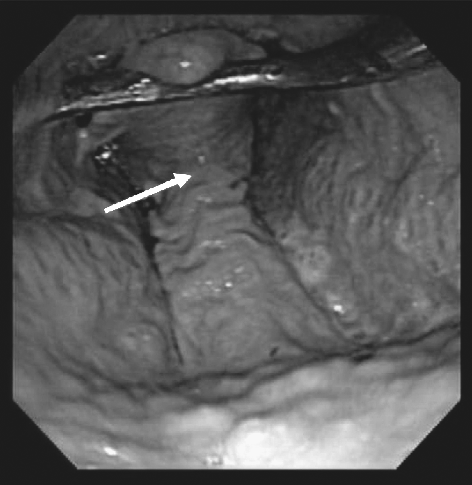
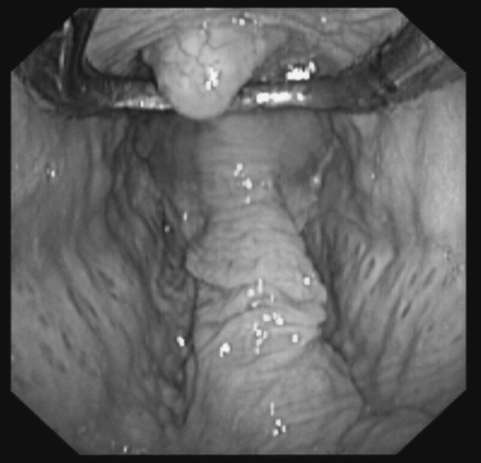
The endoscopic appearance of the GEF and its attachment to the epiglottis was consistent in all horses. The fold ended at the caudal aspect of the ventral epiglottis with no redundant mucosa reflected rostrally or plicated sagitally along the midline of the ventral epiglottis (Figs 3 and 4).
Horse #3. Normal appearance of the glossoepiglottic fold, which shows attachment to the base of the epiglottis and plication oriented in the transverse plane (arrow)

Horse #5. Normal appearance of the glossoepiglottic fold

Discussion
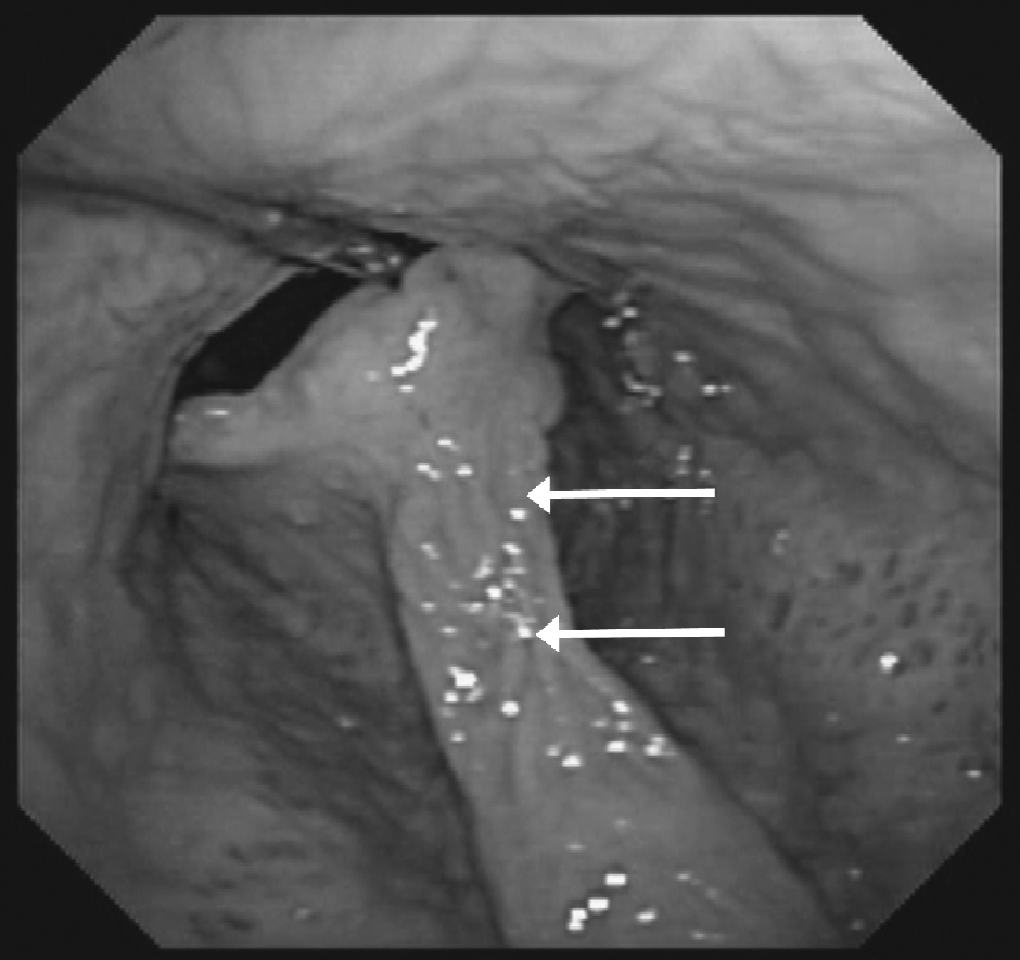
This study served to document the normal endoscopic appearance of the GEF and its relationship to the ventral aspect of the epiglottis. In all cases, the GEF was plicated in the transverse plain at the junction of the fold with the epiglottis (Fig. 3). In none of the horses was there evidence of subepiglottic mucosa plicated in the sagittal plain and extending rostrally along the ventral aspect of the epiglottis towards the apex as reported previously (Fig. 5).
A previously unpublished image of the 507 kg, 7-year-old Thoroughbred stallion with a frenulum of the epiglottis described by Moorman et al. Reference Moorman, Marshall and Jann4. There is plication in the sagittal plane from the tip of the epiglottis to the glossoepiglottic fold (arrows)

The appearance of the GEF in the horses examined in this study contrasts with the anomalies described by Yarbrough et al. Reference Yarbrough, Voss, Herrgesell and Shaw3 and Moorman et al. Reference Moorman, Marshall and Jann4, where an epiglottic frenulum was identified as a cause of persistent DDSP. During the act of swallowing, the epiglottis moves caudally to cover the opening of the larynx as the swallowed material moves dorsally from the oropharynx to the opening of the oesophagus. A substantial frenulum could impair this movement, resulting in dysphagia. This hypothesis is supported by the fact that in both reports, affected horses had dysphagia that resolved with transection of a subepiglottic frenulum.
Whether an epiglottic frenulum is a cause of DDSP (and subsequent exercise intolerance) is less clear. The response of some clinicians and researchers in this field to the published case report by Moorman et al. would indicate that the significance of a subepiglottic frenulum and its role (if any) in the etiopathogenesis of DDSP are controversialReference Madison and Robertson6. The horse is unique in domestic athletes in being an obligate nasal breather due to the normal orientation of the epiglottis relative to the caudal edge of the soft palate, and DDSP is a well-recognized cause of exercise intolerance. Whether the epiglottis has a clear role in preventing DDSP is less clear. Various treatments have been described based on the premise that the epiglottis restrains the soft palate in the ventral position, but these treatments are not universally successful. In the case described by Moorman et al., persistent DDSP did not resolve with transection of the subepiglottic frenulum, while in the case series described by Yarborough et al., no abnormal upper respiratory function was reported by the owners of the three foals surviving to adulthood. Upon review of the clinical evidence available in the literature, it may be more appropriate to make an association between the presence of an epiglottic frenulum and dysphagia rather than DDSP, as follow-up treadmill endoscopic examinations of the upper respiratory tract are lacking in all cases of subepiglottic frenula published to date.
In this study, six horses with normal upper airway function demonstrated considerable uniformity in the appearance of the GEF as visualized through oropharyngeal endoscopy. The subepiglottic structure similar to that identified by Moorman et al. was not found in any of the horses examined (Fig. 5). It is possible that the sample size utilized in this study was too small to document all variations of normal anatomy. The results of this study may facilitate the differentiation of normal oropharyngeal anatomy from malformations believed by some authors to contribute to upper airway dysfunction.
Acknowledgements
The authors would like to thank John Bennett for his technical assistance.