Introduction
Throughout folliculogenesis, Bufo arenarum oocytes, like those of other amphibian and vertebrates species, remain arrested at the prophase of the first meiotic division (Masui, 1985). At the end of follicle growth, which occurs during the preovulatory period, the oocytes exhibit a maximum degree of development (fully grown oocytes) and are meiotically competent, that is, gametes are capable of meiotic resumption and progression of the cell cycle from prophase I to metaphase II (Maller, Reference Maller1985; Jalabert et al., Reference Jalabert, Fostier, Breton, Weil, Pang and Schreibman1991). This process, known as nuclear or meiotic maturation, is necessary to insure that once ovulation takes place the gamete can undergo fertilization and normal embryonic development.
Physiologically, maturation is a hormone-dependent process. It has been demonstrated that during the preovulatory period the increase in circulating gonadotropins induces follicular cells to secrete ovarian steroids, among them progesterone (Polzonetti-Magni et al., Reference Polzonetti-Magni, Mosconi, Carnevali, Yamamoto, Hanaoka and Kikuyama1998). This hormone, whose plasma concentration reaches a maximum before ovulation (Fortune, Reference Fortune1983; Medina et al., Reference Medina, Ramos, Crespo, González and Fernández2004), is considered as the natural inducer, both in vivo and in vitro, of amphibian oocyte nuclear maturation (Sadler & Maller, Reference Sadler and Maller1982; Liu & Patiño, Reference Liu and Patiño1993; de Romero et al., Reference de Romero, de Atenor and Legname1998).
During maturation, besides biochemical changes (Fernández & Ramos, Reference Fernández, Ramos and 2003), oocytes undergo a series of morphological modifications such as those involving the plasma membrane, the cytoplasm and the nucleus or germinal vesicle (GV) (Bement & Capco, Reference Bement and Capco1990; Ramos et al., Reference Ramos, Cisint, Alcaide and Casal1998, Reference Ramos, Winik, Cisint, Crespo, Medina and Fernández1999). Coordinated progression and completion of all these changes make the oocytes ready for fertilization and subsequent development (Yamashita et al., Reference Yamashita, Mita, Yoshida and Kondo2000).
Reorganization and redistribution of several structures such as the endomembrane system, some of whose components are related to sequestration and release of intracellular calcium, take place at the cytoplasmic level (Campanella et al., Reference Campanella, Andreuccetti, Taddei and Talevi1984; Mehlmann et al., Reference Mehlman, Terasaki, Jaffe and Kline1995; Kume et al., Reference Kume, Yamamoto, Inoue, Muto, Okano and Mikoshiba1997; Ramos et al., Reference Ramos, Winik, Cisint, Crespo, Medina and Fernández1999).
Several results indicate that an intracellular calcium increase is required for this process, as reported for amphibian (Wasserman et al., Reference Wasserman, Pinto, O'Connor and Smith1980; Duesbery & Masui, Reference Duesbery and Masui1996) and mammalian species (Fujiwara et al., Reference Fujiwara, Nakada, Shirakawa and Miyazaki1993; Homa, Reference Homa1995; Boni et al., Reference Boni, Cuomo and Tosti2002). On the other hand, results from other authors suggest that calcium is not important for the development of this process (Cork et al., Reference Cork, Cicirelli and Robinson1987). Thus, the role played by Ca2+ in nuclear maturation is still controversial.
Taking these data into account and considering that intracellular calcium can increase as a result of the release of intracellular stores or of calcium movement across the plasma membrane from the extracellular medium, the aim of the present study was to determine the ultracytochemical distribution of Ca2+ deposits, the presence of Ca-ATPase activity in Bufo arenarum fully grown oocytes during progesterone-induced nuclear maturation in vitro, and the influence of extracellular calcium in this process.
Materials and Methods
Animals
Sexually mature Bufo arenarum females were collected in the neighbourhood of San Miguel de Tucumán, Argentina, during the breeding period of the species (August–November). Animals were used within 24 h after capture.
Follicle treatment
Fully grown oocytes (1.5–1.7 mm in diameter) surrounded by one layer of follicle cells were manually isolated from the ovary using watchmaker forceps.
The analysis of in vitro oocyte maturation was performed in lots of 100 follicles that were randomly selected from a single animal incubated with 15 ml of amphibian Ringer's solution (112.92 mM NaCl; 2.01 mM KCl; 1.35 mM CaCl2 plus 30 mg penicillin G sodium and 50 mg streptomycin sulphate per litre, pH 7.4) containing progesterone 1 μg/ml, which had been previously determined as the optimal concentration to induce nuclear maturation (de Romero et al., Reference de Romero, de Atenor and Legname1998). A stock solution of progesterone (supplied by Sigma Chemical Co.) was obtained by dissolving the steroid in ethanol at a ratio of 1 mg/ml. Control follicles were maintained in the same incubation medium without progesterone.
In order to asses the influence of extracellular calcium in maturation, follicles were incubated with Ringer's solution containing different CaCl2 concentrations.
All incubations were carried out at 25 ± 1°C. The germinal vesicle breakdown (GVBD) was used as the indicator of oocyte meiotic maturation.
Ultrastructural studies
Subcellular calcium localization
Control and progesterone-treated fully grown follicle oocytes were removed at random at different times of incubation ranging between 0–8 h, taking into account that during the breeding period Bufo arenarum progesterone-induced nuclear maturation in vitro is completed around 8 h after follicles are treated with the steroid (de Romero et al., Reference de Romero, de Atenor and Legname1998).
Subcellular location of ionic Ca2+ was examined according to the method of Spicer et al. (Reference Spicer, Hardin and Greene1968) re-evaluated by Klein et al. (Reference Klein, Yen and Thureson-Klein1972).
Follicles were fixed in 2% potassium pyroantimonate with 1% osmium tetroxide adjusted to pH 7.8 with 0.05 N acetic acid, for 2 h at 4°C. Parallel samples were fixed in 1% osmium tetroxide alone as controls. Then all samples were dehydrated in a graded ethanol series, exchanged through acetone and embedded in Spurr resin. Ultrathin sections stained with uranyl acetate and lead citrate were examined with a Zeiss EM 109 transmission electron microscope.
The calcium ion precipitation formed by the treatment of K+ pyroantimonate was observed as electron-dense opaque deposits. The nature of the antimony deposits was evaluated by treating oocyte samples with 3 mM EGTA.
Ultracytochemical Ca-ATPase determination
Ca-ATPase detection was studied following the method of Ando et al. (Reference Ando, Fujimoto, Mayahara, Miyajima and Ogawa1981). Samples were fixed in 1% glutaraldehyde–paraformaldehyde mixture in 0.1 M cacodylate buffer, pH 7.4, at 4°C for 2 h and then washed in the same buffer for 3 h at 4°C. Samples were then immersed in 0.1 M cacodylate buffer (pH 7.4) containing 10 mM ethylenediaminetetraacetic (EDTA) to remove any calcium that might be present. After rinsing in the same buffer without EDTA, samples were incubated in a medium consisting of 250 mM glycine–KOH buffer (pH 9.0), 3 mM ATP, 10 mM CaCl2 as an activator, 2 mM lead citrate and 2.5 mM l-tetramisole as an inhibitor of alkaline phosphatase. Controls were incubated in a similar medium without CaCl2. Incubation was carried out for 30 min at room temperature. After rinsing in 0.1 M cacodylate buffer (pH 7.4), the samples were post-fixed in 1% osmium tetroxide buffered with 0.1 M cacodylate buffer, pH 7.4, for 1 h at 4°C and then dehydrated in a graded alcohol and acetone series and embedded in Spurr resin (Pelco Inc.). Ca-ATPase activity was observed as electron dense deposits.
Data analysis
Statistical analysis was undertaken by Student'st-test, with p < 0.05 considered as statistically significant. Data are presented as means ± standard error of the mean (S.E.M.).
Results
Ultrastructural detection of calcium and Ca-ATPase
In the present study the observations were made at the level of the animal pole in both control and progesterone-treated oocytes, considering that during maturation the most important structural changes take place in this region.
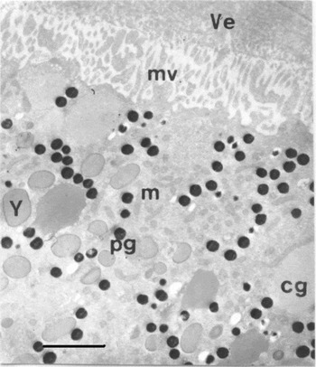
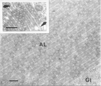
At the cortical level, in both control oocytes and oocytes treated with progesterone for 1–2 h, all of them with the germinal vesicle (GV) present, the most relevant cytoplasm structures observed were mitochondria, pigment granules, yolk platelets and amorphous cortical granules. The oocyte membrane was characterized by the presence of numerous microvilli arranged perpendicularly to the surface (Fig. 1). In a deeper region scattered stacks of annulate lamellae (AL) were found (Fig. 2), sites of continuity between each lamella and cisternal elements of the endoplasmic reticulum (ER) being observed (Fig. 2 inset). At this time, electron-dense Ca2+ deposits could not be observed in any of the control or progesterone-treated samples analysed (Figs. 1 and 2).
Bufo arenarum oocytes incubated for 1–2 h with progesterone. The image at the cortical level reveals the absence calcium deposits. cg, cortical granules; Y, yolk platelets; pg, pigment granules; m, mitochondria; mv, microvilli and Ve, vitelline envelope. Scale bar, 2 μm.

Bufo arenarum oocyte treated for 1–2 h with progesterone. At the subcortical region no calcium deposits could be detected. AL, annulate lamellae; Gl, glycogen granules. Scale bar, 0.25 μm. Inset: Detail indicating site of continuity between AL, annulate lamellae and endoplasmic reticulum cisternae (arrows head). Scale bar, 0.25 μm.

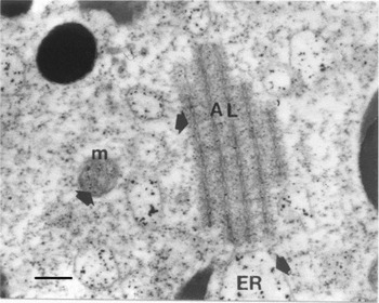
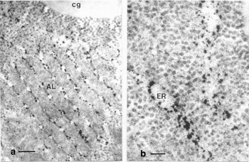
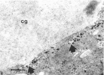
After 3 h of progesterone treatment and concurrently with the beginning of nuclear migration, conspicuous electron dense Ca2+ deposits were visualized within the spaces limited by the AL membranes, in the cisternae of ER and in mitochondria (Fig. 3). The presence of Ca-ATPase activity, revealed as a punctuate linear distribution of dense precipitates, was also observed in the membrane of the above structures (Fig. 4a, b).
Localization of calcium at the subcortical region of Bufo arenarum oocyte after 3 h of treatment with progesterone. Arrows indicate Ca2+ deposits in the stacks of annulate lamellae (AL), endoplasmic reticulum vesicles (ER) and in the mitochondria (m). Scale bar, 0.25 μm.

Ca-ATPase activity in Bufo arenarum oocyte after 3 h of incubation with progesterone. Conspicuous electron dense deposits indicate the presence of Ca-ATPase activity in the membranes of: (a) AL, annulate lamellae. cg, cortical granules. Scale bar, 2 μm. (b) ER, endoplasmic reticulum vesicles. Scale bar, 0.10 μm.

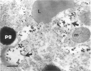
Between 4–5 h of incubation with progesterone, at the same time as nuclear membrane dissolution, a rearrangement of cytomembrane structures was observed. At this time the AL stacks had practically disappeared because of vesiculation, giving rise to a large number of vesicles and cisternae that migrated to the oocyte cortex. At this level important electron dense calcium deposits accumulated in ER vesicles of irregular sizes and shapes (Fig. 5).
Localization of calcium in Bufo arenarum oocyte treated for 4–5 h with progesterone. Oocyte cortex exhibiting a large number of vesicles and cisternae containing conspicuous calcium deposits (arrows). L, lipid droplet; m, mitochondria; pg, pigment granules. Scale bar, 0.25 μm.

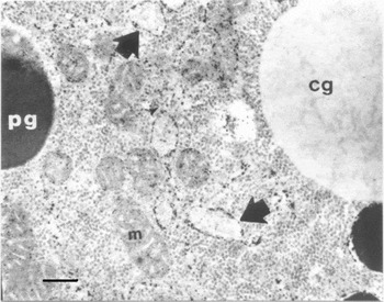
Six hours after progesterone treatment, at the time when the GV reached the surface of the oocyte and the nuclear envelope was undergoing the dissolution process, the presence of a considerable number of Ca2+ deposits could be observed in individual cisternae that form an interconnected network of ER. Calcium precipitates were also detected in some vesicles distributed very close to the cortical granules which were now placed in the proximity of the oocyte membrane. On the membrane of these vesicles an evident reactivity indicated the presence of active Ca-ATPase (Fig. 6).
Ca-ATPase activity in Bufo arenarum oocyte after 6 h incubation with progesterone. Note the prominent Ca-ATPase pump activity in the membrane of vesicles located in the cortex cytoplasm (arrows). cg, cortical granules; m, mitochondria; pg, pigment granules. Scale bar, 0.25 μm.

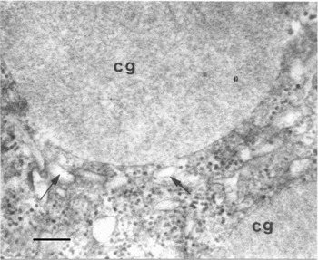
Up to 8 h after progesterone treatment, the time during which the oocytes underwent GVBD and the expulsion of the first polar body took place, most of the Ca2+ precipitates were detected within numerous flat vesicles. These vesicles were found surrounding the cortical granules aligned in close contact with the plasma membrane and also between them. Concurrently, the membranes of these vesicles showed a positive reaction for Ca-ATPase (Fig. 7).
Ca-ATPase activity in Bufo arenarum oocyte after 8 h of incubation with progesterone. Positive Ca-ATPase activity was detected (arrows) in the membrane of flat vesicles placed very close to and around the cortical granules. cg, cortical granules. Scale bar, 0.25 μm.

The use of EGTA allowed us to show that calcium was the major component of the precipitate in our material. The specificity of the Ca-ATPase reaction was carefully checked using appropriate controls. When the substrate CaCl2 was omitted from the procedure, no reaction products were detected (Fig. 8).
Bufo arenarum oocyte samples after 8 h of treatment with progesterone for Ca-ATPase control detection. The reaction products disappear when CaCl2 was removed (arrows). cg, cortical granules. Scale bar, 0.25 μm.

Effect of extracellular calcium on nuclear maturation
Taking into account that the increase in intracellular calcium can originate from either the ion release from intracellular stores or a calcium influx from the extracellular medium, in this work we also analysed the influence of extracellular Ca2+ in progesterone-induced meiotic resumption. For this purpose, fully grown follicle oocytes were incubated in a medium containing different calcium concentrations in the presence of progesterone 1 μg/ml as a nuclear maturation inducer.
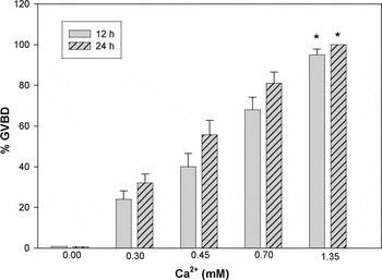
The results obtained, expressed as % GVBD, showed that no effect of progesterone was observed in the absence of calcium in the incubation medium (Fig. 9). However, a progressive increase in the percentage of nuclear maturation in response to progesterone was obtained together with the increase in the calcium concentration added to the incubation medium. The maximum GVBD percentage was observed at the normal concentration (1.35 mM) of the cation present in amphibian Ringer's solution (p < 0.001 with respect to the lowest calcium dose assayed).
Effect of extracellular calcium on Bufo arenarum oocyte maturation induced by progesterone. Fully grown follicle oocytes were incubated in amphibian Ringer's solution containing different concentration of calcium plus progesterone 1 μg/ml. After 12 and 24 h of incubation, follicles were fixed in Ancel and Vintemberger and then examined to detect the presence of the germinal vesicle. In calcium free conditions, CaCl2 was absent in Ringer's solution and 0.1 mM EGTA was also added to the medium to chelate residual calcium. Data obtained from duplicate incubations (n = 6) are expressed as % GVBD mean ± S.E.M.*, asterisk indicates a significant difference when compared with the lowest calcium concentration assayed (*p < 0.001).

Discussion
The results of the present study indicate that both in control oocytes with no progesterone treatment and in oocytes treated with progesterone for the first 2 h, no Ca2+ deposits were present in the cytoplasm of the cortical region. These data suggest that, at the stage when the germinal vesicle is intact, calcium would not be necessary for any event to occur at the gamete level. These results are in agreement with the ones reported for oocytes from mammals (Mehlman et al., Reference Mehlman, Mikoshiba and Kline1996). However, in Xenopus immature follicle oocytes, Bertout et al. (Reference Bertout, Flament, Browaeys-Poly and Vilain1997) reported the presence of Ca2+ deposits at two different sites, the follicle cells and the pigment granules, although the functional significance of this Ca2+ store is unknown.
The electron-dense Ca2+ deposits were visualized in the oocytes 3 h after the beginning of the progesterone treatment. The presence of Ca2+ deposits between the membrane stacks in AL and in ER cisternae indicates that these endomembrane structures would function as storage sites as reported for sea urchin eggs (Terasaki & Sardet, Reference Terasaki and Sardet1991). We also detected Ca-ATPase activity in the membrane of both ER and AL; these data would suggest that Ca2+ sequestration at this level is an ATP-dependent process.
The cortical detection of calcium at this time would be related to the already known first steps in the mechanism of progesterone action that include a transient increase in intracellular calcium leading to microtubule depolymerization (Riabova, Reference Riabova1990; Duesbery & Masui, Reference Duesbery and Masui1996) necessary for GV migration. In agreement with this, previous results showed that GV migration in Bufo arenarum started around 3 h after progesterone treatment (de Romero et al., Reference de Romero, de Atenor and Legname1998).
On the other hand, progesterone is known to promote an increase in the levels of the maturation-promoting factor (MPF), one of whose functions is the induction of changes in ER structure temporally correlated with calcium oscillations as reported for marine protostome worms (Stricker & Smythe, Reference Stricker and Smythe2003).
As the maturation process progressed (4–5 h after progesterone treatment), the Ca2+ deposits could be observed on the surface of the oocyte cortex. At this level, the presence of the calcium inside the vesicles originated either by the disruption of AL or from the ER together with the detection of Ca-ATPase at the membrane of these structures suggests the participation of this enzyme in the maturation process by controlling cytosolic calcium and/or intracellular Ca2+ stores (Kobrinsky & Kirchberger, Reference Kobrinsky and Kirchberger2001).
When GVBD occurred, most Ca2+ deposits were already placed in the cortical ER cisternae organized into a network. The functional significance of this disposition becomes important at the time of fertilization. In fact, it was suggested that the large ER clusters are correlated with an increase in the releasing capacity of calcium mediated by inositol triphosphate (IP3), which determines open Ca2+ channels. In agreement with the above, immunofluorescent techniques have shown the presence of IP3 receptors in the ER clusters (Terasaki et al., Reference Terasaki, Runft and Hand2001).
On the other hand, it has been suggested that the presences of several vesicles containing Ca2+ deposits placed in the cortex and close to or around the cortical granules would be essential not only for the propagation of the activating signal (Nucciteli et al., Reference Nuccitelli, Yim and Smart1993) at the time of fertilization but also for the cortical granules exocytosis that causes the vitelline envelope modification during egg activation (Stricker, Reference Stricker1999; Jaffe et al., Reference Jaffe, Giusti, Carrol and Foltz2001).
Our results showed a close dependence between the calcium concentration in the incubation medium and the progesterone response, that is, meiotic resumption. In agreement with the above, Tosti et al. (Reference Tosti, Boni and Cuomo2000) reported for bovine oocytes that a calcium influx from the extracellular medium was required for in vitro germinal vesicle breakdown and that the cation movement was effected through voltage-dependent calcium channels. These results were supported by those of Morrison et al. (Reference Morrison, Waggoner, Whitworth-Langley and Stith2000), who found that lowering the calcium concentration in the incubation medium inhibited progesterone effect. Contrasting results were reported for Xenopus oocytes (Sun & Machaca, Reference Sun and Machaca2004) where low calcium in the medium failed to affect the rate and extent of germinal vesicle breakdown, indicating that calcium influx is not required for meiotic resumption.
Our results indicate that during in vitro Bufo arenarum meiotic maturation endomembrane reorganization and distribution are established, and generate numerous vesicles located at the oocyte cortex. These vesicles, which show intracellular Ca2+ stores, are placed close to the site of sperm–egg fusion where they would serve as a calcium source necessary for cortical granules exocytosis to form the fertilization barrier that blocks polyspermy during oocyte activation (Stricker, Reference Stricker1999). On the other hand, our results suggest that the plasma membrane and the calcium from the extracellular medium would play an important functional role during oocyte maturation.
Acknowledgements
This work was supported by a grant (026/D347) from CIUNT, Consejo de Investigaciones de la Universidad Nacional de Tucumán, Argentina.