Introduction
Thyroid carcinoma is a relatively uncommon entity, representing only 1 per cent of all new malignancies.Reference Sherman1 Ninety per cent of thyroid tumours are differentiated thyroid carcinomas, including the papillary and follicular subtypes.Reference Yilmazlar, Kocaeli and Cordan2 Differentiated thyroid carcinomas tend to be low-grade and slowly progressive; the overall survival rate of papillary thyroid carcinoma is 98 per cent.Reference Gilliland, Hunt, Morris and Key3
Metastasis of differentiated thyroid carcinoma to the skull base (including the temporal bone) is extremely uncommon: only six cases involving papillary thyroid carcinoma and 17 cases involving follicular thyroid carcinoma have been reported. In four of these 17 latter cases, histopathological investigation was unable to identify a definitive primary thyroid gland lesion, raising the question of whether these cases were due to hyperplastic ectopic thyroid tissue rather than skull base metastasis. Two cases of follicular thyroid carcinoma metastasis to the petrous bone have been reported.Reference Kachhara, Nair, Radhakrishnan, Pandey, Ahmed and Kumar4, Reference Song, Chan, Tey and Choi5 However, to our best knowledge there have been no previous reports of papillary thyroid carcinoma metastasis to the petrous portion of the temporal bone.
Due to its rarity and anatomical location, a papillary thyroid carcinoma metastasis in the skull base may be mistaken for other skull base tumours. The differential diagnosis includes meningioma, schwannoma and, more rarely, chordoma, chondrosarcoma, paraganglioma and endolymphatic sac tumour. An incorrect diagnosis (which may lead to inappropriate clinical management) is more likely to occur when the diagnosis is made based on clinical presentation and radiological findings, without a tumour biopsy.
We present a case of a temporal bone tumour centred in the jugular fossa, which was initially thought to be a benign skull base tumour, based on radiological evaluation and an indeterminate initial biopsy, but which was later found to be a metastatic follicular-variant papillary thyroid carcinoma, based on further histopathological investigation.
Case report
A 64-year-old woman was referred to our tertiary-care clinic for investigatation of a 2.1 × 2.9 × 3.9 cm, left-sided, lateral skull base mass seen on magnetic resonance imaging (MRI) and computed tomography (CT). The patient had a six-month history of fatigue, and was being followed for a left-sided thyroid nodule which had been investigated with fine needle aspiration (FNA), with non-diagnostic results.
On examination, the patient had no evidence of any lower cranial nerve palsies. The remainder of her physical examination was unremarkable.
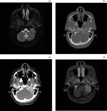
A repeated MRI showed significant tumour growth in the five-month interval since the patient's initial MRI. The tumour now measured 2.9 × 3.8 × 4.1 cm. The diffusely enhancing mass was seen to occupy much of the left petrous bone, extending into the low left cerebellopontine angle and down into the infratemporal fossa, and was centred on the left jugular fossa (Figure 1). Based on this radiological appearance, the mass was believed to be a paraganglioma; however, the absence of commonly seen flow voids within the mass led to the addition of schwannoma to the differential diagnosis.
Axial imaging scans showing a large, enhancing mass located in the posterior left petrous bone, with bone destruction, seen on (a) T2-weighted, fat-saturated magnetic resonance imaging (MRI), (b) bone window computed tomography (CT), (c) contrast-enhanced CT, and (d) contrast-enhanced, T1-weighted, fat-saturated MRI.

To exclude possible metastasis from a distant site, the patient was sent for a mammogram and chest X-ray; the results of both were unremarkable.
She was otherwise healthy, and functional inquiry was benign. Abdominal and pelvic imaging were not indicated.
A repeated CT with contrast demonstrated irregularity of the edges of the bone defect around the left-sided lesion, which was best seen at the level of the occipital condyle, occipital bone lateral to the foramen magnum, and two mastoid air cells. The enhancing mass was seen to extend into the mastoid, the left cerebellopontine angle, the infratemporal fossa and posteriorly into the upper neck muscles. Vessels were visualised in the centre of the mass.
Following the repeated MRI and CT, the patient was reviewed in the clinic. On otoscopy, there was evidence of a reddish mass in the left inferior mesotympanum. Audiological testing indicated that the patient's hearing was within normal limits.
After neurosurgical consultation, radiation therapy was offered and a trans-tympanic biopsy of the mass was arranged to determine pathology (to aid radiotherapy planning). Histological examination of the biopsy demonstrated closely packed glands, and the specimen was positive for pan-keratin and S100 protein. Features were consistent with a middle-ear adenoma; however, given the extent of bony invasion of the lesion and its location both inside and outside the tympanic cavity, endolymphatic sac tumour was added to the differential diagnosis (although the specimen lacked the papillary features expected for such pathology).
Repeated FNA of the left thyroid nodule was performed; histopathological examination showed a follicular lesion of undetermined significance.
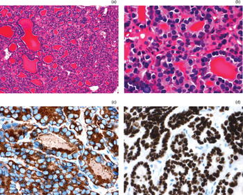
Given that the results of radiological and pathological analysis remained inconclusive as to the aetiology of the skull base lesion, a repeated biopsy was performed via a trans-mastoid approach, and a third FNA of the left thyroid nodule was taken. Fine needle aspiration cytology results were non-diagnostic. However, histopathological examination of the skull base mass demonstrated cellular thyroid tissue with a predominantly microfollicular pattern admixed with occasional macrofollicles. Follicular cells showed mild to moderate nuclear enlargement, with smooth to slightly irregular nuclear contours and occasional nuclear grooves. On immunohistochemical analysis, the tissue stained for cytokeratin-7 (CK7), thyroid transcription factor-1 and thyroglobulin (Figure 2).
Photomicrographs of skull base mass showing hypercellular thyroid tissue with microfollicular architecture, seen at (a) ×100 and (b) ×400 (H&E). Immunoperoxidase staining shows positivity for (c) thyroglobulin and (d) thyroid transcription factor-1, indicating a thyroid origin (original magnification ×400).

As a result, the differential diagnosis was revised to include metastatic follicular or follicular-variant papillary thyroid carcinoma and hyperplastic ectopic thyroid tissue.
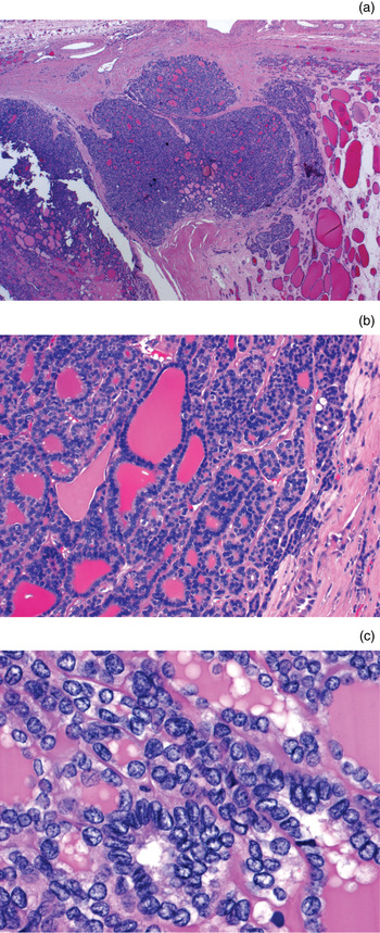
The patient underwent a total thyroidectomy and neck dissection, with 24 lymph nodes removed. The final surgical pathology showed a unifocal, right-sided, 0.7 cm thyroid mass; histological examination showed papillary thyroid carcinoma of the follicular variant. The appearance of the tumour was similar to that of the repeated biopsy (Figure 3). There was no lymph node involvement. The final pathological tumour-node-metastasis (TNM) staging was T1 N0 M1.
Photomicrographs of the primary thyroid tumour, showing: (a) an area of invasive growth into the adjacent parenchyma; (b) follicular tumour architecture identical to that of the skull base metastasis; and (c) focal areas of nuclear clearing and nuclear grooves (H&E; original magnifications ×25, ×100 and ×400, respectively). The tumour was diagnosed as a follicular-variant papillary thyroid carcinoma.

Following surgery, the patient underwent external beam radiation to treat her skull base metastasis.
Discussion
Metastasis of papillary thyroid carcinoma to the skull base is an uncommon clinical entity, and only six cases have been reported. Of these, papillary thyroid carcinoma metastases have been reported to arise in the following skull base sites: sella turcica and pituitary gland,Reference Bell, Kovacs, Horvath, Smythe and Asa6–Reference Sziklas, Mathews, Spencer, Rosenberg, Ergin and Bower8 cavernous sinusReference Masiukiewicz, Nakchbandi, Stewart and Inzucchi7, Reference Takami, Ohata, Tsuyuguchi, Mao, Inoue and Wakasa9 and sphenoid sinus.Reference Freeman, Gershon, Liavaag and Walfish10 To our best knowledge, our patient represents the first reported case of a papillary thyroid carcinoma metastasis arising in the petrous bone. A review of the English language literature of the Medline database, using the medical subject heading key words ‘thyroid neoplasms’, ‘neoplasm metastasis’ and ‘carcinoma, papillary’ yielded no relevant results. We recognise that there is some controversy surrounding the differentiation of follicular thyroid carcinoma and follicular-variant papillary thyroid carcinoma. However, our patient's diagnosis of follicular-variant papillary thyroid carcinoma was determined by consensus by the experienced pathologists at our institution.
Local and regional spread of papillary thyroid carcinoma tends to occur via the lymphatic system, with distant metastases developing later in the course of the disease.Reference Sherman1 Papillary thyroid carcinoma generally metastasises through local and regional spread. In cases of cranial papillary thyroid carcinoma metastasis, brain parenchyma is more frequently involved than bony structures. This is in contrast to follicular thyroid carcinoma, the cranial metastases of which tend to be found in the skull, skull base and paranasal sinuses.
A review of the literature revealed that papillary thyroid carcinoma metastases to the skull base present with symptoms of a local mass effect. In the previously described six cases, the skull base metastases were found after diagnosis of papillary thyroid carcinoma, and caused symptoms of visual disturbance, ptosis, hypopituitarism and facial pain. In our case, while the patient had a history of a left-sided solitary thyroid nodule, it had been repeatedly investigated and found unremarkable, and was unrelated to the temporal bone lesion. The patient's right-sided thyroid carcinoma was not found until histopathological analysis following thyroid resection, after the skull base lesion had been completely investigated.
The behaviour of our patient's primary papillary thyroid carcinoma was unusual in that it lacked nodal involvement and the only extra-thyroidal manifestation was a metastasis to a previously unreported site. However, follicular-variant papillary thyroid carcinoma has been reported (in a case series of five patients) to metastasise directly to a bony site, without nodal involvement. In each of these cases, the primary thyroid lesion was 1.5 cm or smaller.Reference Baloch and LiVolsi11
While metastatic papillary thyroid carcinoma arising from the petrous bone has not previously been described, two cases of follicular thyroid carcinoma metastasis to the petrous bone have been described.Reference Kachhara, Nair, Radhakrishnan, Pandey, Ahmed and Kumar4, Reference Song, Chan, Tey and Choi5 In both of these cases, as in ours, the patient had no history of thyroid pathology, and presented with symptoms related to what were later found to be skull base follicular thyroid carcinoma metastases. In contrast to the symptomatology described in the two previous cases of differentiated thyroid carcinoma metastasis to the petrous bone, our patient was asymptomatic, and continued to experience no symptoms and to remain neurologically intact despite the large size and obvious rapid growth of the tumour.
The location and invasive behaviour of our patient's lesion mimicked other skull base lesions, including paraganglioma, schwannoma and endolymphatic sac tumour. Imaging studies also demonstrated characteristics consistent with the various pathologies in our differential diagnosis. The correct diagnosis of a skull base tumour has major implications for tumour management, since tumour types respond differently to different treatment modalities. The treatment of metastatic differentiated thyroid carcinoma is surgical, comprising total thyroidectomy together with neck dissection in the case of cervical lymphadenopathy. Post-operative ablation by radioactive iodine (131I) and external beam radiation should be considered for control of residual disease and metastases.Reference Nar Demirer, Ayturk, Tutuncu, Gursoy, Pak and Demirag12, Reference Rosahl, Erpenbeck, Vorkapic and Samii13
• Differentiated thyroid carcinoma has an overall survival of 90 per cent; subtypes include papillary and follicular
• Only 23 cases of papillary or follicular thyroid carcinoma skull base metastasis have been reported
• Skull base metastasis is more common for follicular than papillary thyroid carcinoma
• The first case report of papillary thyroid carcinoma metastasis to the petrous bone is presented
Papillary thyroid carcinoma metastasis to the skull base is extremely rare. However, because of the uniqueness of its treatment plan in comparison with other skull base tumours, clinicians should retain a high index of suspicion given a skull base tumour with an unusual presentation or radiological features. Biopsy is not the conventional management in such situations; however, if the tumour growth rate, location, spread and radiological features are atypical, then pathological investigation can help to raise suspicion of an unusual neoplastic entity such as metastatic thyroid cancer. Although unexpected, the diagnosis of such an unusual tumour will have grave consequences on the choice of clinical management and, indeed, on patient outcomes.