Implications
Myostatin, a member of the transforming growth factor-β superfamily, is a negative regulator of skeletal muscle growth. Myostatin plays an important role in myoblast proliferation and differentiation. In this study, a miR-27a binding site in the 3′-untranslated region of porcine myostatin was identified. There was an inverse correlation between miR-27a and myostatin expression in fat and heart of pigs as well as in proliferating porcine myoblasts. We identified that miR-27a could positively regulate porcine myoblast proliferation via targeting myostatin. This work may lead to new strategies for blocking porcine myostatin activity and improving lean meat percentage of pigs.
Introduction
Myostatin, a member of the transforming growth factor-β (TGF-β) superfamily, is a critical autocrine/paracrine inhibitor of skeletal muscle growth, and is expressed in developing muscle of embryos and muscle of adult animal (McPherron et al., Reference McPherron, Lawler and Lee1997; Lee, Reference Lee2004). Myostatin plays a crucial role in myoblast proliferation and differentiation (Huang et al., Reference Huang, Chen and Chen2011). However, the regulatory mechanisms of myostatin during myoblast proliferation and differentiation remain largely unknown.
MicroRNAs (miRNAs) are endogenous ~22nt RNAs that negatively regulate gene expression at the posttranscriptional level via binding to the 3'-untranslated region (3'UTR) of target mRNAs (Gladka et al., Reference Gladka, da Costa Martins and De Windt2012). MicroRNAs are able to suppress translation and/or induce mRNA degradation (Tanzer and Stadler, Reference Tanzer and Stadler2004). Translational control via the 3'UTR can affect formation of the closed loop translation initiation complex, ribosome binding or a post-initiation step (de Moor et al., Reference de Moor, Meijer and Lissenden2005). The sequence of mature miRNAs are well conserved, whereas the sequence and length of 3'UTR of mRNAs are different among mammalian species (Bartel, Reference Bartel2004). Therefore, it seems especially essential to explore miRNAs regulating target gene expression starting from the animal itself.
MiR-27a, a member of miR-27 family, which has negative impacts on kinds of cells through different pathways, has been demonstrated to promote murine myoblast proliferation and differentiation through targeting myostatin in our previous studies (Huang et al., Reference Huang, Chen, Yu, He and Chen2012; Chen et al., Reference Chen, Huang, Chen, Yang and Liu2014). Nevertheless, we have no idea whether miR-27a could also promote porcine myoblast proliferation via downregulation of myostatin expression. In this study, we cloned the full-length 3'UTR of porcine myostatin by rapid amplification of 3'-cDNA ends (3'-RACE), demonstrated that the 3'UTR of porcine myostatin is targeted by miR-27a through luciferase reporter assay, and further confirmed that miR-27a suppresses myostatin expression, thereby accelerating porcine myoblast proliferation.
Material and methods
Cloning of porcine myostatin 3'UTR
To obtain the full-length of porcine myostatin 3'UTR, 3'-RACE experiment was performed using a 3'-Full RACE Core Set Ver.2.0 Kit (TaKaRa, Dalian, China) according to the manufacturer’s directions. The gene-specific primer used in 3'-RACE was 5'-GTAGATCGCTGTGGGTGCTCATGA-3', which was designed according to the porcine myostatin coding sequence (CDS) (GenBank accession no. NM_214435). The full-length 3'UTR of porcine myostatin was PCR amplified from the total RNA isolated from the longissimus lumborum muscle of a 28-day-old, 7.6 kg female Duroc×Landrace×Yorkshire (DLY) pig using the gene-specific primer (forward) and the 3'RACE inner primer (5'-CGCGGATCCTCCACTAGTGATTTCACTATAGG-3') (reverse). The purified PCR product was cloned into pMD19-T vector (TaKaRa) and sequenced, resulting in pMD19-T- pMyostatin3'UTR.
Plasmid construction
The wild type 3'UTR of porcine myostatin was PCR-amplified using the specific primers (forward primer 5'-CCGCTCGAGGATTTATATTTGGTTCATTAC-3' and reverse primer 5'-CCTGCGGCCGCTTTTTTTTTTCCTTTACTG-3') and the recombinant plasmid pMD19-T-pMyostatin3'UTR as a template. The forward and reverse primers introduced Xho I and Not I sites (underlined), respectively. After digestion with Xho I and Not I, the PCR product was cloned into psiCHECK-2 dual-luciferase reporter vector (Promega, Madison, WI, USA) to generate psiCHECK-2-pMyostatin3'UTR-wt. The mutant porcine myostatin 3'UTR reporter, designated as psiCHECK-2-pMyostatin3'UTR-mu, was created by mutating the seed region of the predicted ssc-miR-27a site (ACUGUGA to UGACACU) by nested PCR.
Luciferase reporter assay
The human embryonic kidney (HEK) 293T cell line (CRL-11268) was from the American Type Culture Collection (Rockville, MD, USA). Luciferase reporter assay was performed as previously described (Huang et al., Reference Huang, Chen, Yu, He and Chen2012). Briefly, the HEK 293T cells were plated into a 24-well plate and maintained in Dulbecco modified Eagle medium (DMEM) (Invitrogen, Carlsbad, CA, USA) containing 10% fetal bovine serum (FBS) (Invitrogen) at 37°C, 5% CO2 in a humidified atmosphere. After 24 h, cells were cotransfected with 0.5 μg of either the psiCHECK-2-pMyostatin3’UTR-wt, psiCHECK-2-pMyostatin3’UTR-mu or empty plasmid psiCHECK-2, and 50 nM of either miRNA mimics Negative Control or miR-27a mimics (RiboBio, Guangzhou, China) using Lipofectamine 2000 (Invitrogen) according to the manufacturer’s protocols. Forty-eight hours after the transfection, cells were lysed in Passive Lysis Buffer (Promega) and Firefly and Renilla luciferase activities were measured with a GloMax 20/20 Luminometer (Promega) using Dual-Luciferase Reporter Assay System (Promega) according to the manufacturer’s instructions.
Animals and tissue sample collection
Three 10-week-old female DLY pigs (BW of 31~31.6 kg) were slaughtered in a humane manner according to protocols approved by the Animal Care Advisory Committee of Sichuan Agricultural University. The heart, fat, psoas major (PM) muscle, longissimus lumborum (LL) muscle, extensor digitorum longus (EDL) muscle and Soleus (SOL) muscle were removed and immediately snap frozen in liquid nitrogen before being stored at −80°C for RNA isolation.
Cell isolation and culture
Porcine myoblasts were isolated from longissimus lumborum muscle of 3-day-old male DLY pigs. The pig obtained from the farm of Sichuan Agriculture University was slaughtered according to protocols approved by the Animal Care Advisory Committee of the University. Briefly, muscle was removed and washed twice in cold PBS (pH 7.4) under sterile conditions, and then cut into small pieces with scissors. The dissected muscle was digested with 0.2% collagenase type I (Sigma, St. Louis, MO, USA) for 2 h at 37°C in a water bath. A single cell suspension was obtained after filtering the samples through 37-µM mesh, and then collected cells were purified by differential adhesion method. The cell suspension were plated on rat-tail collagen coated-flask and cultured in DMEM/Ham’s F-12 (1 : 1) medium (Invitrogen) supplemented with 20% FBS (Invitrogen), 5 nM FGF-basic (Invitrogen) and antibiotics (100 U/ml penicillin and 100 μg/l streptomycin) (Invitrogen). Cells were maintained at 37°C in a saturated humidly atmosphere containing 95% air and 5% CO2, with the medium being renewed every 2 days. The cells were identified by immunofluorescence with anti-myosin heavy chain antibody (Santa Cruz Biotechnology, Santa Cruz, CA, USA).
RNA isolation and reverse transcription
Total RNA was extracted from the collected tissue samples and adherent cultured porcine myoblasts using TRIzol reagent (Invitrogen) according to the manufacturer’s directions. The concentrations of total RNA were determined spectrophotometrically using a Beckman Coulter DU800 (Beckman Coulter, Fullerton, CA, USA). Reverse transcription was done with 500 ng total RNA from each sample using a PrimeScript RT reagent Kit with gDNA Eraser (TaKaRa) according to the manufacturer’s instructions.
miR-27a overexpression
Porcine myoblasts were cultured in 24-well plates (Corning) and transfected with 50 nM of miR-27a mimics or 50 nM of miRNA mimics Negative Control (RiboBio) using Lipofectamine 2000 (Invitrogen) following the manufacturer’s instructions.
Real-time quantitative PCR
Real-time quantitative PCR was performed in a 7900HT real-time PCR system (384-cell standard block) (Applied Biosystems, Foster, CA, USA) using the iTaq SYBR Green Supermix with ROX (Bio-Rad, Hercules, CA, USA) in a final volume of 10 μl. The primers for quantification of mRNA expression as follows: porcine myostatin (forward) 5'-TCTGAATGAGAACAGCGAGCAA-3' and (reverse) 5'-TCGTTTCCGTCGTAGCGTGA-3'; porcine β-actin (forward) 5'-CATCGTCCACCGCAAAT-3' and (reverse) 5'-TGTCACCTTCACCGTTCC-3'. The Bulge-Loop™ miRNA qPCR Primer Sets specific for miR-27a and U6 were purchased from Ribobio. The PCR conditions were as follows: predenaturation at 95°C for 10 min, followed by 45 cycles of 95°C for 15 s and 60°C for 30 s. A single sharp peak was detected in the melting curve. Relative expression of the transcript/miRNA was calculated using comparative C t method (Livak and Schmittgen, Reference Livak and Schmittgen2001) and was normalized by β-actin or U6 expression, respectively.
EdU proliferation assay
EdU (5-ethynyl-2'-deoxyuridine) is a nucleoside analog of thymidine that is incorporated into DNA during active DNA synthesis only by proliferating cells. Proliferating porcine myoblasts were determined by using the Click-iT EdU Alexa Fluor 594 Imaging Kit (Invitrogen). EdU proliferation assay was performed as previously described (Huang et al., Reference Huang, Chen, Yu, He and Chen2012), with minor modification. Briefly, porcine myoblasts were incubated with 10 μM EdU for 6 h before fixation, permeabilization, and EdU staining. Cell nuclei were stained with Hoechst 33342 (Invitrogen) at a concentration of 5 μg/ml for 30 min.
Statistical analysis
Data expressed as mean±s.e. were compared by one-way ANOVA followed by Tukey’s tests (SPSS Inc., Chicago, IL, USA). P<0.05 was considered to be statistical significant.
Results
Cloning and sequence analysis of porcine myostatin 3'UTR
DNA sequencing showed that the length of amplified fragment is 638 bp. The nucleotide sequence of porcine myostatin 3'UTR has been deposited in GenBank database under accession number KC299915. Sequence analysis showed that porcine myostatin 3'UTR cloned in this study shares 99.36% homology with the known porcine myostatin 3'UTR (GenBank accession no. AF033855). A polyA tail was observed at 14 nucleotides downstream from typical polyadenylation site (AATAAA). A potential binding site for miR-27a was found at its N-terminus (ACTGTGA) (63–70 bp), indicating that porcine myostatin might be a miR-27a target.
The 3'UTR of porcine myostatin is targeted by miR-27a
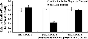
To examine whether porcine myostatin 3'UTR can be directly targeted by miR-27a, the entire wild-type 3'UTR of porcine myostatin or the mutant 3'UTR with 7 bp mutation in seed region was cloned downstream of the luciferase gene and assayed in HEK 293T cells. As shown in Figure 1, a highly significant decrease (56%) was observed in luciferase activity when miR-27a mimics and the wild-type porcine myostatin 3'UTR reporter were cotransfected into HEK 293T cells. By contrast, cotransfection of miR-27a mimics with the mutant porcine myostatin 3'UTR reporter didn't result in decrease in luciferase activity (Figure 1), indicating that the predicted site is a direct target of miR-27a.
The 3'UTR of porcine myostatin is targeted by miR-27a. Porcine myostatin 3'UTR reporter, its miR-27a site mutant, or empty vector was cotransfected with either miR-27a mimics or miRNA mimics Negative Control into HEK 293T cells, followed by cell lysis and luciferase assays 48 h later. Data were presented as mean±s.e. (n=3). ***P<0.001 as compared with empty vector.

miR-27a level inverse correlates with porcine myostatin mRNA expression in vivo and in vitro
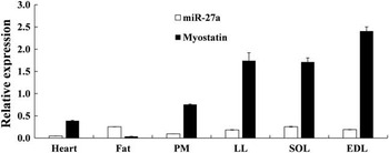
The expression profiles of porcine myostatin mRNA and miR-27a were determined by real-time quantitative PCR in various tissues. As shown in Figure 2, porcine myostatin mRNA was abundant in the EDL muscle, LL muscle, SOL muscle and PM muscle, followed by the heart and fat. MiR-27a level was most abundant in the fat and was least in the heart in examined tissues (Figure 2). There was an inverse correlation between myostatin and miR-27a expression in fat and heart of pigs (Figure 2).
Relative expressions of porcine myostatin mRNA and miR-27a in different tissues. Total RNA from different tissues of three healthy Duroc×Landrace×Yorkshire (DLY) pigs was used to perform the real-time quantitative PCR. Samples were performed in duplicate. The amount of porcine myostatin mRNA and miR-27a was normalized to the amount of porcine β-actin mRNA or U6 snRNA, respectively. Data were the mean and standard errors (n=3). PM=psoas major muscle; LL=longissimus lumborum muscle; EDL=extensor digitorum longus muscle; SOL=Soleus muscle.

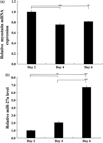
To determine the correlation with expressions of myostatin mRNA and miR-27a in vitro, porcine myoblasts were seeded in a 24-well plate at 2×10⁴ cells per well. After 8 days, the cells reached ~100% confluence (Supplementary Figure S1). Then, we detected the expressions of myostatin mRNA and miR-27a at day 2, 4 and 6. As show in Figure 3, the expression of mature miR-27a was up-regulated and myostatin was downregulated during porcine myoblasts proliferation.
Relative expressions of endogenous porcine myostatin and miR-27a during porcine myoblast proliferation. Porcine myoblasts were seeded in a 24-well plate at 2×10⁴ cells per well. The cells were harvested at 2, 4, 6 days after initial plating and RNA was extracted. Porcine myostatin mRNA (a) and miR-27a level (b) were determined using real-time quantitative PCR. The amount of porcine myostatin mRNA and miR-27a were normalized to the amount of porcine β-actin mRNA and U6 snRNA, respectively. Data were the mean and standard errors (n=3). **P<0.01 and ***P<0.001 as compared with Negative Control.

Overexpression of miR-27a promotes proliferation of porcine myoblasts
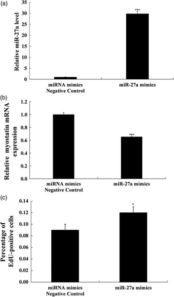
A miR-27a binding site in the porcine myostatin 3'UTR and negative correlation between mature miR-27a and porcine myostatin transcription gave us the motion to examine whether miR-27a could modulate proliferation of porcine myoblasts. In order to verify this point, porcine myoblasts were transfected with miR-27a mimics or miRNA mimics Negative Control and EdU incorporation experiments were performed to assess its proliferation. As shown in Figure 4a, compared to the cells transfected with miRNA mimics Negative Control, miR-27a mimics transfected cells had a significance increased in miR-27a level. By contrary, level of porcine myostatin transcript reduced 35% (Figure 4b). In EdU proliferation assay, miR-27a mimics transfected cells had higher proliferation (Figure 4c). These data suggest that the miR-27a targeted 3'UTR of porcine myostatin and suppressed porcine myostatin expression, then promoted proliferation of porcine myoblasts.
Overexpression of miR-27a promotes proliferation of porcine myoblasts. Porcine myoblasts were seeded in a 24-well plate at 2×10⁴ cells per well. After 96 h, the cells were transfected with 50 nM of either miR-27a mimics or miRNA mimics Negative Control. Transfection mix was removed 6 h later and cells were grown for another 18 h in DMEM/20%FBS medium. (a) The amount of porcine myostatin mRNA normalized to the amount of porcine β-actin mRNA was measured by real-time quantitative PCR. Data were presented as mean±s.e. (n=3). (b) The amount of miR-27a normalized to the amount of U6 snRNA was measured by real-time quantitative PCR. Data were presented as mean±s.e. (n=3). (c) Quantification of EdU labeling experiments. The percentage of EdU-positive cells was calculated as the number of EdU-positive cells out of total number of cells indicated by Hoechst 33342-positive staining for each microscopic field. Data were presented as mean±s.e. from two independent experiments. *P<0.05 and ***P<0.001 as compared with Negative Control.

Discussion
Myostatin belongs to the TGF-β superfamily and is known to play an essential role in the regulation of skeletal muscle mass (Joulia-Ekaza and Cabello, Reference Joulia-Ekaza and Cabello2006). The complete CDS of myostatin has been cloned from swine (Ji et al., Reference Ji, Losinski, Cornelius, Frank, Willis, Gerrard, Depreux and Spurlock1998), cattle (Grobet et al., Reference Grobet, Martin, Poncelet, Pirottin, Brouwers, Riquet, Schoeberlein, Dunner, Ménissier, Massabanda, Fries, Hanset and Georges1997), horse (Hosoyama et al., Reference Hosoyama, Kawada, Oshiumi, Yoneda, Soeta, Yamanouchi, Hasegawa, Ishida, Mukoyama, Ishii and Tachi2002) and sheep (Sharma et al., Reference Sharma, Kambadur, Matthews, Somers, Devlin, Conaglen, Fowke and Bass1999). MiRNAs negatively regulate gene expression at the posttranscriptional level via binding to the 3'UTR of target mRNAs (Bartel, Reference Bartel2004; Tanzer and Stadler, Reference Tanzer and Stadler2004; de Moor et al., Reference de Moor, Meijer and Lissenden2005; Gladka et al., Reference Gladka, da Costa Martins and De Windt2012). The myostatin 3'UTRs of some species have been known, such as 1552 bp (1262–2814 bp) in goats, 1506 bp (1261–2767 bp) in cattle, 498 bp (1234–1732 bp) in rat (data from GenBank database) and so on. In this study, we cloned the full-length porcine myostatin 3'UTR (638 bp) by 3'-RACE. These results suggested that the complete CDS of myostatin is well conserved among mammalian species, but the length of myostatin 3'UTR is different among different species.
Myostatin has been reported to be regulated by several miRNAs, such as miR-208a (Callis et al., Reference Callis, Pandya, Seok, Tang, Tatsuguchi, Huang, Chen, Deng, Gunn, Shumate, Willis, Selzman and Wang2009), miR-27b (Allen and Loh, Reference Allen and Loh2011) and miR-499 (Zhang et al., Reference Zhang, Liu, Liu, Liu, Wang, Zhu and Yu2012). In Texel sheep, the myostatin allele characterized by a G to A mutation in myostatin 3'UTR creates a target site for miR-1 and miR-206 (Clop et al., Reference Clop, Marcq, Takeda, Pirottin, Tordoir, Bibe, Bouix, Caiment, Elsen, Eychenne, Larzul and Laville2006). High expression of miR-1/206 in muscle inhibits transcript level of myostatin, which contributes to muscular hypertrophy (Clop et al., Reference Clop, Marcq, Takeda, Pirottin, Tordoir, Bibe, Bouix, Caiment, Elsen, Eychenne, Larzul and Laville2006). In our previous work, we demonstrated that miR-27a suppresses transcript level of mouse myostatin (Huang et al., Reference Huang, Chen, Yu, He and Chen2012). It should be pointed out that mature miR-27a sequence (5'-UUCACAGUGGCUAAGUUCCGC-3') is well conserved among mammalian species. In the known myostatin 3'UTR of rat, dog, horse and cattle, the potential miR-27a binding site (5'-ACUGUGA-3') is found, suggesting that miR-27a might regulate myostatin expression of these species at the posttranscriptional level. In this study, we found a potential miR-27a target site in 3'UTR of porcine myostatin, and we demonstrated that the porcine myostatin is a bona fide target of miR-27a, by luciferase reporter assay. Besides, we also observed an inverse correlation between expressions of myostatin mRNA and miR-27a in fat and heart of pigs and in proliferating porcine myoblasts. The reciprocal expression of miR-27a and myostatin has also been reported in other studies (Allen and Loh, Reference Allen and Loh2011; Huang et al., Reference Huang, Chen, Yu, He and Chen2012; Chen et al., Reference Chen, Huang, Chen, Yang and Liu2014).
The miR-27 family consists of two members (miR-27a and miR-27b) located at different chromosomes. It has been reported that miR-27 enhances differentiation of osteoblasts and strongly inhibits adipogenesis (Lin et al., Reference Lin, Gao, Alarcon, Ye and Yun2009; Wang and Xu, Reference Wang and Xu2010). MiR-27b is a regulatory hub in lipid metabolism through repressing critical regulators of lipid homeostasis, such as PPARα, HMGCR (Kida et al., Reference Kida, Nakajima, Mohri, Oda, Takagi, Fukami and Yokoi2011, Vickers et al., Reference Vickers, Shoucri, Levin, Wu, Pearson, Osei‐Hwedieh, Collins, Remaley and Sethupathy2013). In a recent study, miR-27b was reported to specifically target to bovine myostatin (Miretti et al., Reference Miretti, Martignani, Accornero and Baratta2013). MiR-27a shares the same seed region with miR-27b, but with a C to U mutation at C-terminal. MiR-27a modulates the growth of breast and liver cancer cells by targeting FOXO1 and FZD7/β-catenin, respectively (Guttilla and White, Reference Guttilla and White2009; Chen et al., Reference Chen, Ma, Huang, Zhang, Lv, Xu, Hu and Li2013). Besides, miR-27a also suppresses adipocyte differentiation through targeting PPARγ (Kim et al., Reference Kim, Kim, Lee, Son, Lee, Lee, Lee and Kim2010). We previously demonstrated that miR-27a inhibits myostatin expression then promotes proliferation of C2C12 myoblasts (Huang et al., Reference Huang, Chen, Yu, He and Chen2012). In this study, we showed that miR-27a enhances proliferation of porcine myoblasts through downregulation of myostatin expression.
In summary, we cloned the full-length of porcine myostatin 3'UTR and confirmed that miR-27a positively regulates porcine myoblast proliferation via targeting myostatin. In the future, it will be interesting to determine whether miR-27a could modulate the differentiation of porcine myoblasts.
Acknowledgments
This work was supported by the National Natural Science Foundation of China (No. 31272459), the National Basic Research Program of China (No. 2012CB124701), the Sichuan Youth Science and Technology Foundation (No. 2012JQ0049) and the Specific Research Supporting Program for Academic Sustentation Research Team in Sichuan Agricultural University.
Conflicts of Interest
None.