Recent regulatory guidance requires that medical device manufacturers validate the cleaning instructions as well as the disinfection or sterilization instructions. 1 In addition, the American National Standards Institute/Association for the Advancement of Medical Instrumentation (ANSI/AMMI) guidance document 2 emphasizes the need for adequate endoscope cleaning. However, recent outbreaks of infections transmitted through contaminated duodenoscopesReference Epstein, Hunter and Arwady 3 , 4 have raised questions about the adequacy of the manufacturer’s cleaning and disinfection instructions for duodenoscopes. Some sites have indicated that contamination of duodenoscopes with bacteria of concern persists despite following the manufacturer’s instructions for use (MIFU).Reference Ross, Baliga and Verma 5 – Reference Naryzhny, Silas and Chi 7 In particular, concern has arisen regarding the ability to adequately clean the complex elevator lever mechanism.Reference Alrabaa, Nguyen and Sanderson 8 This focus has resulted in newly validated cleaning instructions for duodenoscopes from Olympus. 9
The study by Brandabur et alReference Brandabur, Leggett and Wang 6 of surveillance cultures from 21 different facilities offering procedures utilizing endoscopes with lever mechanisms documented that of 2, 238 endoscope encounters evaluated, 8.4% were culture positive for any organism and 0.9% were culture positive for organisms of concern. Furthermore, they reported variability in culture rates, with pathogenic organisms detected in 9 of 21 healthcare facilities included. They identified the elevator lever as the most commonly culture-positive site. In addition, their data support the role of biofilm within endoscope channels as the basis for persistent culture positivity.Reference Brandabur, Leggett and Wang 6 Although endoscopes should not develop biofilm if they are adequately disinfected and stored totally dry, recent evidence indicates that up to 95% of patient-used endoscopes still have visible water inside the instrument channel after overnight storage despite being processed in an automated endoscope reprocessor (AER) with a “dry cycle.”Reference Ofstead, Wetzler, Heymann, Johnson, Eiland and Shaw 10 The 1-minute default dry cycle in the AER was inadequate, and even an increase in dry time to 3 minutes was insufficient to adequately dry the endoscopes evaluated after overnight storage.Reference Ofstead, Wetzler, Heymann, Johnson, Eiland and Shaw 10 Moisture in the channel during storage indicates a strong likelihood that biofilm may form. A key consideration in the root-cause analysis of how pathogens survive within flexible duodenoscopes is determining whether, under “worst-case” conditions where each night there is biofilm formation, full cleaning and disinfection following the MIFU each day can prevent accumulation of biofilm residuals.
The objectives of this study were to assess (1) the impact of 5 repeated rounds of overnight biofilm formation, each followed by full reprocessing on the accumulation of organic and microbial residuals and (2) the effect of enzymatic versus nonenzymatic detergent combined with bristle or pull-through channel cleaners on this accumulation.
MATERIALS AND METHODS
Biofilm Formation
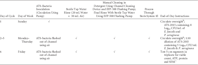
New 3.7-mm × 1,800-mm polytetrafluorethylene (PTFE) channels (Endoscopy Development Company, Maryland Heights, MO) were used in simulated-use testing of the impact of repeated cycles of cleaning and disinfection on the accumulation of biofilm residuals within the channel. The PTFE channels used in this research study were identical to those used as replacements for damaged instrument channels for Olympus endoscopes. Each PTFE channel was processed through the Steris System 1E (Steris, Mentor, OH) prior to use (active ingredient was peracetic acid). Artificial test soil ATS-2015 (Healthmark Industries, Fraser, MI) was rehydrated and supplemented with 20% defibrinated sheep blood (Cedarlane, Burlington, Ontario) according to the manufacturer’s instructions. ATS-2015 is an appropriate test soil because it has been shown to mimic the composition and viscosity of patient secretions that gastrointestinal flexible endoscopes are exposed to.Reference Alfa and Olson 11 The ATS-2015 was supplemented with 108 CFU/mL of both Enterococcus faecalis (ATCC 29212) and Pseudomonas aeruginosa (ATCC 15442) and circulated through the PTFE channel on the first day of biofilm formation. On subsequent days, a 1:10 dilution of ATS-2015 containing 105 CFU/mL of both bacteria was circulated overnight for biofilm formation. (See Table 1 for details of this protocol.)
Protocol for Repeated Rounds of Biofilm Formation in PTFE ChannelsFootnote a and Reprocessing

NOTE. PTFE, polytetrafluorethylene; ATS, artificial test soil; EFP, endoscope flushing pump; CFU, colony-forming units.
a 5 separate 40-cm PTFE channels were used to form the biofilm needed for this experiment.
b A peristaltic pump was used to circulate the fluid at 72 mL/h through each PTFE channel.Reference Alfa, Ribeiro, da Costa Luciano, Franc, Olson, DeGagne and Singh 25
c Positive controls had repeated biofilm formation and rinsing but did not have cleaning or disinfection.
Channel Cleaning and Disinfection Protocol
Subsequent to overnight biofilm development, the PTFE tubing was fully reprocessed, including manual cleaning per the Olympus MIFU using a channel-cleaning brush (or pull-through channel cleaner), the EFP-500 endoscope flushing pump for all flushing and rinsing steps and disinfection using the Steris System 1E (SS1E) per the manufacturer’s instructions. The detergents evaluated included (1) a neutral pH enzymatic detergent, Renuzyme Plus (Getinge, Rochester, NY), containing proteases (dual enzyme), D-glucitol, sodium polyacrylate, 1,2 propylene glycol) and (2) a neutral-pH nonenzymatic detergent, Intercept (Medivators, Minneapolis, MN), containing benzylkonium chloride, diethyl glycol monoethylester, lactic acid, and alkyl polyglycoside. The source of friction for channel cleaning was either an appropriately sized bristle brush (ConMed, Utica, NY) or the pull-through channel cleaner (Ruhof, Mineola, NY). Thus, the 4 cleaning interventions were set up as follows:
-
1. Enzymatic detergent + bristle brush
-
2. Enzymatic detergent + pull-through channel cleaner
-
3. Non-enzymatic detergent + bristle brush
-
4. Non-enzymatic detergent + pull-through channel cleaner
Notably, this experimental approach represents a “worst-case” situation where, after a single round of full reprocessing, biofilm was actively stimulated overnight using organic material and bacteria. In clinical practice, multiple rounds of use and reprocessing would likely occur each day, and only after prolonged use could there be potential biofilm formation overnight (ie, our experimental protocol expedited the biofilm challenge).
The positive control consisted of PTFE channel with biofilm formation over 5 days without cleaning or disinfection; the positive control was only rinsed each day with 20 mL sterile tap water.
Testing Residuals
Viable bacteria
The exterior of each 40-cm length of PTFE was wiped with alcohol and then aseptically cut into 5-cm lengths using a sterile scalpel. Each 5-cm PTFE segment (in triplicate) was further aseptically cut lengthwise and then crosswise into 10 small pieces (each approximately 1 cm × 0.5 cm), and all pieces were totally immersed in 5 mL neutralizer containing Tween 80 (Sigma, St Louis, MO) 3% (v/v), lecithin (Sigma) 0.3% (w/v), L-histidine (Sigma) 0.1% (w/v), and sodium thiosulfate (Sigma) 0.5% (w/v).Reference Pineau and De Philippe 12 Each sample was sonicated at 50/60 Hertz using a Bransonic 1200 Ultrasonic clear (Branson Canada, Pickering, Ontario) for 5 minutes and then mixed for 1 minute. For the positive controls (in triplicate), the extracted sample was serially diluted 1:10, then 0.1 mL of the direct sample and each dilution were plated onto a blood agar plate. For the test samples, the PTFE segments were aseptically removed and the sample was spun at 3,500 rpm. The supernatant was then removed, leaving 0.5 mL to suspend the pellet, and the entire sample was then inoculated onto a blood agar plate. All cultures were incubated aerobically for 72 hours. The colony morphology is distinct, so E. faecalis and P. aeruginosa colony types were enumerated and colony-forming units per square centimeter was determined using the following equation:
Log10 (total CFU per filter/internal surface area of a 5-cm segment of PTFE)=Log10 CFU/cm2
where the internal surface area of a 5-cm segment=5.81 cm2
Protein and adenosine triphosphate (ATP) determinations
From the same 40-cm PTFE channel, a separate set of (triplicate) 5-cm PTFE lengths were aseptically cut into 10 small pieces and immersed in 5 mL sterile reverse-osmosis water. Each sample was sonicated for 5 minutes and then mixed for 1 minute as previously described. A portion of the sample was used for protein determination using the QuantiPro BCA assay kit (Sigma, St Louis, MO), which included a bovine serum albumin protein standard and is a quantitative assay based on bicinchoninic acid. Protein results were converted to micrograms per square centimeter. The inner-channel surface area of each 5-cm segment was 5.81 cm2.
A separate portion of the extracted sample was tested using the 3M Clean Trace ATP water test (3M, St Paul, MN). Results were recorded as relative light units (RLUs) and reported as Log10 RLUs per 5-cm segment.
Benchmarks for adequate cleaning and disinfection
For pump-assisted manual cleaning, the expected cutoff for protein residuals is <2 μg/cm2, and for ATP residuals, the cutoff is <200 RLUs, which is Log10 2.30 RLU.Reference Alfa, Olson and Murray 13 The expectation was that there would be no detectable viable bacteria after disinfection using the SS1E.
Statistical Analysis
Data are displayed using descriptive statistics. Kruskal–Wallis and Wilcoxon rank-sum tests were used to assess the differences of bacterial counts and protein and ATP residues after the cleaning methods described above.
RESULTS
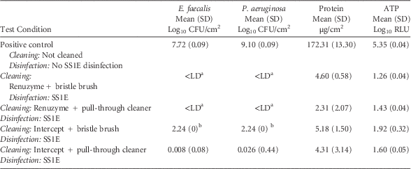
Biofilm was allowed to form overnight, then the PTFE-biofilm channel was fully manually cleaned (pump assisted) and disinfected using the SS1 AER. This process was repeated for 5 consecutive days. The viable count, protein, and ATP residuals for various cleaning conditions are shown in Table 2. The positive control consisted of no cleaning and no disinfection using SS1E and showed very high levels of bacteria, protein, and ATP (Table 2). Significantly less E. faecalis (P=.019), P. aeruginosa (P=.016), protein (P=.024), and ATP (P=.024) were detected for all 4 of the cleaning conditions tested compared to the positive control. Differences were detected in E. faecalis (P=.022), P. aeruginosa (P=.023), and ATP (P=.022), among the 4 different cleaning interventions. Furthermore, in comparison of all Renuzyme versus Intercept data (regardless of bristle brush or pull-through device), significantly less E. faecalis (P=.024), P. aeruginosa (P=.049), ATP (P=.017), and protein residuals (P=.049) were detected when cleaning used Renuzyme as the detergent compared to Intercept.
Impact of 5 Repeated Rounds of Biofilm Formation Followed by Cleaning and Disinfection on Biofilm Accumulation in PTFE Channel

NOTE. PTFE, polytetrafluorethylene; RLU, relative light units; SS1E; Steris System 1E; SD, standard deviation.
a <LD indicates less than the limit of detection of 1 CFU/5.81 cm2 = 0.357 CFU/cm2 = 0.047 Log10CFU/cm2.
b The samples tested after high-level disinfection were expected to have very low viable bacteria, so the sample was concentrated by centrifugation and the entire sample was inoculated onto the culture plate. When the number of colonies on the culture plate was too numerous to count (ie, >1,000 CFU), it was set at 1,000 CFU for presentation purposes, which converts to 2.24 Log10 CFU/cm2.
For both detergents tested and both channel cleaning methods, the ATP residuals were within the expected cutoff (ie, ATP <200 RLUs), but the protein was not below the expected cutoff (ie, <2 μg/cm2). Only the Renuzyme detergent provided residual bacterial levels that were within the expected cutoff (ie, viable count < limit of detection [LD]) regardless of the channel-cleaning friction method used. Although, there was no statistical significant difference overall between the 2 channel-cleaning friction methods (P>.200), the counts were lower with Intercept cleaning with the pull-through channel cleaner than the Intercept cleaning with the bristle brush channel cleaner. Statistical testing could not be performed for stratified testing of friction channel cleaner by detergent due to the small number of the experiments in each stratified subgroup.
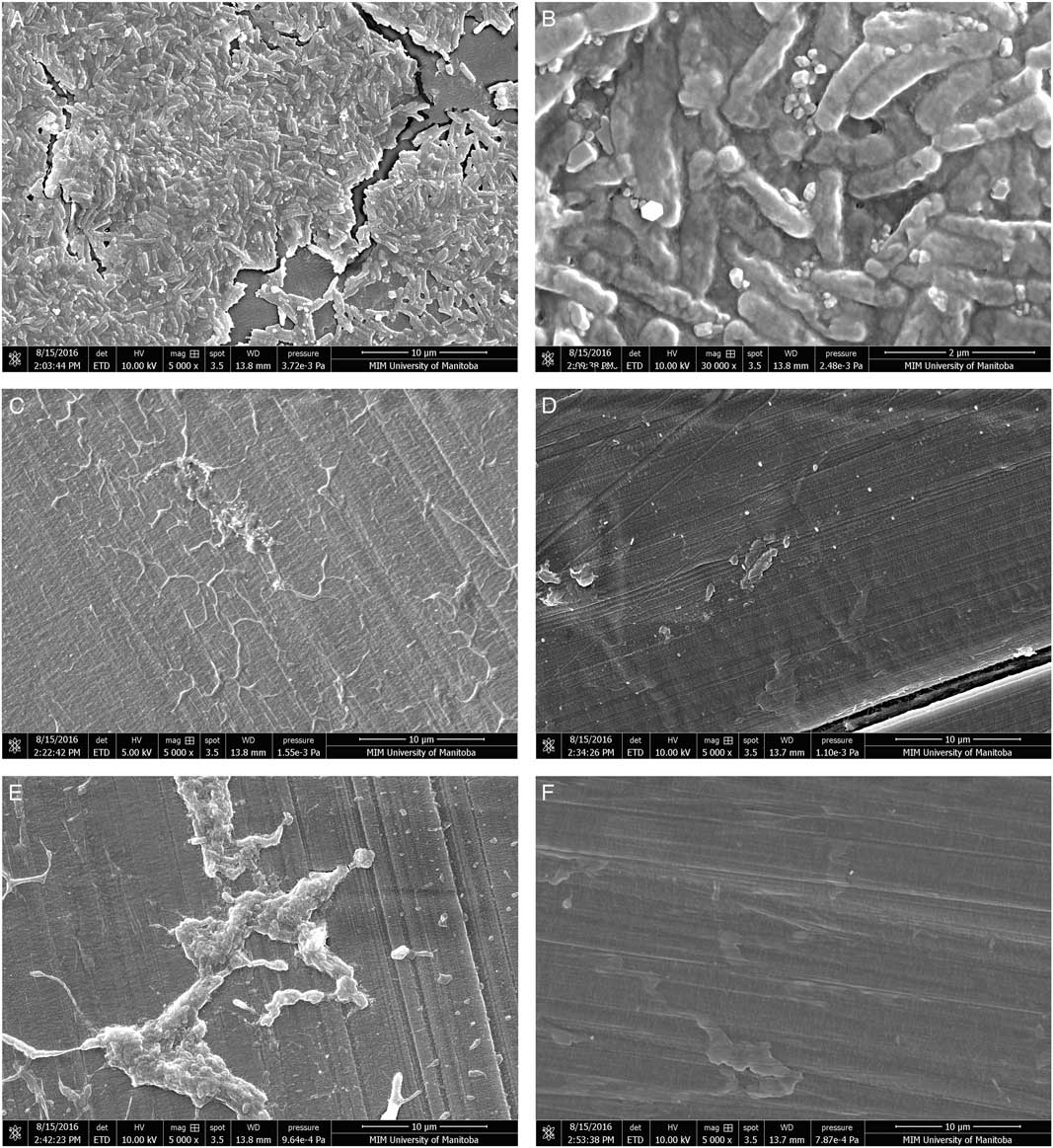
The SEM images (Figure 1) support the efficacy of Renuzyme detergent with either bristle or pull-through channel cleaners, whereas Intercept detergent showed residual bacteria by SEM for the bristle brush but not the pull-through channel cleaner. Residual organic debris was apparent for all cleaning methods assessed but was lowest for the pull-through cleaner regardless of detergent.
Impact of detergent and channel cleaning brush on removal of biofilm in polytetrafluorethylene (PTFE). Biofilm was developed in PTFE channels overnight and then subjected to full cleaning and disinfection. This process was repeated 5 times. The positive control (A, B) had only daily rinsing, whereas full cleaning each day was performed as follows: (C) Renuzyme detergent and a bristle brush, (D) Renuzyme detergent and a pull-through cleaner, (E) Intercept detergent and a bristle brush, and (F) Intercept detergent and a pull-through cleaner. The PTFE channels in C, D, E, and F all had SS1E sterilization after each round of full cleaning. (See Materials and Methods and Results for full details.)

DISCUSSION
A key objective of this study was to determine whether adequate cleaning and disinfection could eliminate repeated rounds of biofilm that formed overnight (ie, to mimic the worst-case conditions, contaminated endoscopes were stored under wet conditions overnight to allow biofilm formation). Our model is an adaptation of the France model for flexible endoscopes. 14 The level of protein in the positive control in our study was similar to that reported as the worst-case level (116 µg/cm2) in patient-used colonoscopes prior to cleaning.Reference Alfa, DeGagne and Olson 15 Our data showed that protein accumulation occurred after 5 rounds of reprocessing regardless of the detergent used or the channel-cleaning method used. Although the mean level of residual protein was low (<6.4 µg/cm2), it still exceeded the benchmark level of 2 µg/cm2, which should be achievable with pump-assisted manual cleaning.Reference Alfa, Olson and Murray 13 Our findings support those of others who have also reported that regardless of cleaning, there is a slow but gradual accumulation of protein (or organic material seen on SEM) with repeated exposure of a medical device to organic material followed by cleaning and high-level disinfection.Reference Verran and Boyd 16 – Reference Vickery, Ngo, Zou and Cossart 20
Verran et alReference Verran and Boyd 16 reviewed how rapid formation of a conditioning film occurs on a pristine surface, which acts as a “linker layer” between the surface and the accumulation of organic material; this determines the stability of the attached organic material. Furthermore, surface defects enhance this gradual buildup of organic debris.Reference Verran and Boyd 16 This accumulation of visible debris over time has been substantiated for flexible endoscope channels in recent reports by Ofstead et alReference Ofstead, Wetzler, Heymann, Johnson, Eiland and Shaw 10 , Reference Ofstead, Wetzler and Doyle 21 on patient-used endoscopes. Our data showed that for PTFE channels after only 5 repeated rounds of biofilm formation followed by full pump-assisted cleaning using channel friction (bristle brush or pull-through) and disinfection, there was an accumulation of organic material (shown by both protein quantitation and SEM observation). Our study results confirm that even when no viable bacteria were found, some organic debris, observed by SEM, remained. Our findings are similar to those of Vickery et al,Reference Vickery, Ngo, Zou and Cossart 20 who reported visible exopolysaccharide and bacterial cells despite no detectable viable bacteria. However, our data extend those of Vickery et al because neutralizer and concentration of the sample were used in our study to ensure optimal culture results for viable but nonculturable bacteria.Reference Li, Mendis, Trigui, Oliver and Faucher 22
Our data demonstrate that after 5 rounds of biofilm formation and full reprocessing, the quantity of residuals observed on SEM and the level of residual viable bacteria differed depending on the detergent used for the cleaning step. There were no detectable viable bacteria when enzymatic detergent (Renuzyme) was used, regardless of the use of a bristle-brush or a pull-through device. When the nonenzymatic cleaner (Intercept) was used, between 0.008 and 2 Log10/cm2 CFU remained regardless of the channel friction method used. The residuals after cleaning with these 2 detergents were different for bacterial residuals and ATP independent of the type of friction used for cleaning (Table 2). These data suggest that the stability of the “linker layer”Reference Verran and Boyd 16 left after Renuzyme cleaning was less stable compared to that left after Intercept cleaning (regardless of the friction method used in the cleaning process) and that this difference in cleanability could be the basis for the differences observed after 5 repeated rounds of reprocessing using the 2 detergents. Notably, this difference in cleaning translated to a difference in the ability of bacteria to survive the disinfection stage. There were no detectable bacteria after disinfection associated with 5 rounds of Renuzyme cleaning, but detectable bacteria were associated with 5 rounds of Intercept cleaning. Clearly, our data show that ATP is not sensitive enough to detect low levels of viable bacteria in biofilm residuals in PTFE channels after disinfection, supporting the conclusion of Visrodia et alReference Visrodia, Hanada, Pennington, Tosh, Topazian and Petersen 23 that ATP cannot substitute for culture to detect contamination of patient-used duodenoscopes after disinfection.
The SS1E AER has recently been revalidated as an effective liquid chemical sterilization method for flexible endoscopes providing that adequate cleaning has been achieved.Reference McDonnell, Ehrman and Kiess 24 Our data do not negate this revalidation; rather, we demonstrate how residual biofilm can prevent effective action of an AER, and we support the AER manufacturer’s statement that adequate cleaning must be done prior to disinfection in an AER. Further studies are needed to fully understand this phenomenon. However, the SEM results showed a higher level of accumulated debris after the 5 rounds of Intercept cleaning, and this may have been the basis for bacterial survival of the disinfection process. The rate of organic accumulation after cleaning appears be a key determinant in the survival of microorganisms and may be the basis of sustained biofilm formation in patient-used flexible endoscopes. Biofilm formation during storage is an underappreciated phenomenon, but the recent data of Ofstead et alReference Ofstead, Wetzler, Heymann, Johnson, Eiland and Shaw 10 from stored patient-used endoscopes showed that up to 95% of endoscopes had residual fluid in the suction channel after overnight storage, highlighting the extent of fully processed endoscopes that could potentially develop biofilm because they were not dry.
Our data also support the role of friction in the cleaning process because the use of the pull-through channel cleaner left less organic debris than bristle brush, when evaluated by SEM. The flexible “discs” of the pull-through device would have better surface contact with the inner surface of the PTFE channel compared to the bristles of the traditional bristle brush used for cleaning. This may explain why the SEM images showed far less residual debris and bacterial forms when the pull-through device was used, regardless of which detergent was used. Indeed, for the nonenzymatic detergent (Intercept) the pull-through device left much lower bacterial levels than when the bristle brush was used. The recent report by Alfa et alReference Alfa, Ribeiro, da Costa Luciano, Franc, Olson, DeGagne and Singh 25 also supports the value of adequate friction during cleaning and that flushing fluid alone is not sufficient to remove build-up biofilm from PTFE channels. There is an urgent need for new developments in endoscope reprocessing to improve the friction aspect of channel cleaning, especially for those channels that are too small for traditional brushes or pull-through devices.
A limitation of our study is that only 1 enzymatic and 1 nonenzymatic detergent were evaluated. The efficacy of each detergent formulation in this type of biofilm challenge is unique and should not be generalized. Despite this limitation, our data support the concept that the efficacy of manual cleaning (ie, type of detergent and method of friction used) on biofilm formed within a PTFE channel does impact microbial survival post disinfection.
In conclusion, this study used simulated-use testing to show that the efficacy of the cleaning step affects the adequacy of biofilm removal. Furthermore, our data demonstrate that even after just 5 repeated rounds of biofilm formation, in which each round was followed by full reprocessing, both organic debris and viable bacteria accumulated depending on the cleaning method used.
ACKNOWLEDGMENTS
The funds for this research project were provided by the American Society of Gastroenterology (ASGE). The pull-through cleaning devices were provided by Ruhof; the bristle brushes were supplied by Olympus; and the ATS-2015 was provided by Healthmark. M.J.A. is on the advisory board for 3M and has provided consulting services for 3M, Karl Storz, Novaflux, Sealed Air Diversey, Olympus, and Ofstead Associates. M.J.A. received royalties from the University of Manitoba for a patent license to Healthmark. H.S. is on the advisory boards of Pendopharm and Ferring Canada, and he has received research funding from Merck Canada. D.D. is on the advisory board of Shire. All authors report no conflicts of interest relevant to this article.