INTRODUCTION
Kinetoplastid protozoa, including the genera Trypanosoma and Leishmania, are responsible for many parasitic diseases of humans and animals worldwide (McGhee and Cosgrove, 1980). The peptidases present in different protozoa appear to be relevant for several aspects of host-parasite interactions, quite apart from their obvious participation in the nutrition of the parasite at the expense of the host (McKerrow et al. 1993; Yao, Donelson and Wilson, 2003). Metallopeptidases have been described in a number of trypanosomatids, but only those present in Leishmania spp. have been thoroughly characterized (reviewed by Yao et al. 2003). Parasites that belong to the Leishmania genus are digenetic trypanosomatids that cycle between the gut of the sand fly vector (promastigote forms) and the phagolysosome of a mammalian host macrophage (amastigote forms) (McGhee and Cosgrove, 1980). Several Leishmania surface molecules facilitate survival in these diverse host environments. For instance, Leishmania spp. express a major glycosylphosphatidylinositol (GPI)-anchored glycoprotein of 63 kDa named gp63 or leishmanolysin (Etges, Bouvier and Bordier, 1986; Medina-Acosta, Beverley and Russell, 1993). Leishmanolysin is a Zn+2-dependent HEXXH metallopeptidase with a broad range of substrate specificity and optimum pH activity (Yao et al. 2003). Structural and biochemical similarities exist between gp63 and members of the metzincin class of matrix metallopeptidases (Stöcker and Bode, 1995). The latter are important for enhancing the migration of some tumor cells through the extracellular matrix and basement membrane, aiding in their metastasis (Stöcker and Bode, 1995). In this context, Leishmania species engineered to express high levels of the surface metallopeptidase gp63 had enhanced capacity of migration through extracellular matrix, since type IV collagen and fibronectin were extensively degraded in vitro (McGwire, Chang and Engman, 2003).
The amount of gp63 protein expressed by Leishmania spp. is modulated in different parasite growth-phase and life-cycle stages (Yao et al. 2003). In this sense, gp63 is abundantly expressed on the surface of the promastigote forms, up-regulated in infectious metacyclic promastigotes, and has a low but detectable expression level in the intracellular amastigote stage (Medina-Acosta et al. 1989). Leishmanolysin plays different roles in the host-parasite interactions and has been postulated as a virulence factor (Joshi et al. 2002; Yao et al. 2003). The finding of gp63 gene homologues in the insect trypanosomatids Crithidia fasciculata and Herpetomonas samuelpessoai (Inverso et al. 1993; Yao et al. 2003) suggests that leishmanolysin may be important for the interaction with the invertebrate host. However, gp63 is apparently not essential for insect stages of the life-cycle, since L. major promastigote-specific genes can be knocked out with no deleterious effects on the growth and development of the parasite in 3 Old World Phlebotomus species (Joshi et al. 2002). In contrast, Hajmová and co-workers (2004) reported that the down-regulation of gp63 in a L. amazonensis clone adversely affects its early development in the neotropical Lutzomyia longipalpis sand fly. The possibility exists that gp63 may function differently for these two distinct Leishmania species in their interactions with different invertebrate vector species.
The identification, characterization and cloning of parasite cellular antigens have been the focus of extensive investigations. Previous studies of our group and others, showed the production of a similar cell-surface metallopeptidase activity of 60–66 kDa in some monoxenous trypanosomatids, including C. fasciculata (Etges, 1992), C. deanei, C. desouzai and C. oncopelti (d'Avila-Levy et al. 2001), C. guilhermei (Melo et al. 2002), C. luciliae (Jaffe and Dwyer, 2003), H. samuelpessoai (Etges, 1992; Santos et al. 2003), H. roitmani and H. anglusteri (Santos et al. 1999), Leptomonas seymouri (Jaffe and Dwyer, 2003) and Blastocrithidia culicis (Santos et al. 2001a; d'Avila-Levy et al. 2005). On the other hand, less is known about the identity and functions of molecules secreted or released during the growth of these parasites into their environment. In the present study, we showed that H. samuelpessoai cells extracellularly released a metallopeptidase of 66 kDa, which shared biochemical and immunological similarities with the leishmanolysin of the human pathogen L. amazonensis.
MATERIALS AND METHODS
Chemicals
Media constituents, reagents used in electrophoresis, buffer components, nitrocellulose membrane and reagents for chemiluminescence detection were purchased from Amersham Life Science (Little Chalfont, England). The proteolytic inhibitors (phenylmethylsulfonyl fluoride [PMSF], trans-epoxysuccinyl L-leucylamido-(4-guanidino) butane [E-64], ethylene glycol-bis(β-aminoethyl ether) [EGTA], ethylenediaminetretraacetic acid [EDTA] and 1,10-phenanthroline), the protein substrates (gelatin, bovine serum albumin [BSA], haemoglobin and casein), p-chloromercuriphenylsulfonic acid (p-CMPS), Bacillus thuringiensis phospholipase C (BtPLC), nitro-blue tetrazolium (NBT), 5-bromo-4-chloro-3-indolyl phosphate (BCIP), and the secondary antibodies were products of Sigma Chemical Co. (St Louis, MO, USA). Sulfosuccinimidyl-6-(biotinamide) hexanoate (Sulfo-NHS-LC-biotin) and avidin-alkaline phosphatase conjugated (AAPC) were products of Pierce (Rockford, IL, USA).
Parasites and growth conditions
Herpetomonas samuelpessoai (CT–IOC–067) was kindly provided by Dr Maria Auxiliadora de Sousa (Coleção de Tripanosomatídeos, Instituto Oswaldo Cruz, Rio de Janeiro, Brazil). The trypanosomatid was maintained by weekly transfers in a complex medium (brain heart infusion – BHI) at 26 °C. Leishmania amazonensis Josefa strain (MHOM/BR/75 Josefa) promastigote forms were maintained at 26 °C for 4 days in BHI medium supplemented with 10% (v/v) heat-inactivated fetal calf serum (Soares et al. 2003).
Secretion experiment
For this experiment, H. samuelpessoai was grown at 26 °C in 1000 ml flasks containing 500 ml of BHI medium. Parasite cells were harvested at the log growth phase (48 h) by centrifugation at 2500 g, for 15 min at 4 °C, and washed 3 times with cold PBS (150 mM NaCl; 20 mM phosphate buffer, pH 7·2). Cellular growth was estimated by counting the parasites in a Neubauer chamber. The intact cells (1·0×1010) were resuspended in 10 ml of sterile isotonic PBS. Aliquots of 2·5 ml (containing 2·5×109 parasites) were separated in 4 tubes, which were incubated for different periods of time (from 1 to 4 h) at 26 °C. After intervals of 1 h, the cells were removed by centrifugation (2500 g/20 min/4 °C) and the supernatants were passed over a 0·22-μm membrane (Millipore), to obtain the PBS-conditioned supernatants (d'Avila-Levy et al. 2003).
Modulation of the proteolytic secretion by metallopeptidase and phospholipase C inhibitors
Living H. samuelpessoai (2·5×108 cells) were pre-treated or not with 1 and 10 mM 1,10-phenanthroline (a zinc-metallopeptidase inhibitor) for 30 min at room temperature. Then, the cells were washed 5 times with PBS and the secretion experiment was repeated as described above. Alternatively, the same number of parasites was incubated in the absence and in the presence of 0·1 and 1 μM p-CMPS (a PLC inhibitor) for 1 h, and the reaction mixtures were obtained after centrifugation and filtration steps (Santos et al. 2002a). All supernatants were analysed by proteolytic activity as described below.
Cellular viability
The survival of the parasites along the incubation period in the isotonic phosphate buffer as well as after the treatment with 1,10-phenanthroline and p-CMPS was assessed by mobility, trypan blue cell dye exclusion and by measurement of lactate dehydrogenase activity, a cytoplasmic enzyme, in the secretion supernatant fluids (Santos et al. 2002c, 2005).
Cellular parasite extract
The parasites (1·0×108 cells) were harvested by centrifugation for 5 min at 1500 g at 4 °C, and washed 3 times with cold PBS. The cells were resuspended in 100 μl of PBS and lysed by the addition of 1% (w/v) sodium dodecyl sulfate (SDS). The cells were broken in a vortex by alternating 1 min shaking and 2 min cooling intervals, followed by a centrifugation at 5000 g for 15 min at 4 °C. The supernatant obtained after centrifugation corresponded to the whole parasite cellular extract (Santos et al. 2001b).
Secretory protein profile
Protein concentration was determined by the method described by Lowry and co-workers (1951), using BSA as standard. Samples containing 5 μg of released proteins were added to 10 μl of SDS–polyacrylamide gel electrophoresis (PAGE) sample buffer (125 mM Tris, pH 6·8; 4% SDS; 20% glycerol; 0·002% bromophenol blue) supplemented with 5% (v/v) β-mercaptoethanol, followed by heating at 100 °C, for 5 min. Proteins were analysed in 12% SDS–PAGE using the method described by Laemmli (1970). Electrophoresis was carried out at 4 °C, at 120 V, and the gels were silver stained (Santos et al. 2001b). The molecular mass of sample polypeptides was calculated from mobility of GIBCO BRL (Grand Island, NY, USA) molecular mass standards.
Quantitative proteolytic activity assay
The extracellular peptidase activity was measured spectrophotometrically using the substrate gelatin, according to the method described by Jones and co-workers (1998). Briefly, 50 μl of each H. samuelpessoai PBS-conditioned supernatant and 350 μl of 50 mM sodium phosphate buffer, pH 6·0, were added to 600 μl of the substrate solution (1% (w/v) gelatin in distilled water) and the mixtures were incubated at 37 °C for 2 h. A control, where the substrate was added just after the reactions were stopped, was used as blank. A 300 μl sample was removed from each reaction mixture and added to 400 μl of cold isopropanol. After centrifugation at 16000 g for 10 min, the supernatants were removed and the absorbance was measured at 280 nm. One unit of enzyme activity was defined as the amount of enzyme that caused an increase of 0·01 in absorbance unit, under standard assay conditions.
Gelatin-SDS–PAGE analysis
The secretory proteolytic activity was assayed and characterized by 10% SDS–PAGE with 0·1% (w/v) gelatin incorporated into the gel as substrate (Heussen and Dowdle, 1980). The gels were loaded with 25 μl (equivalent to 5 μg of protein) of each PBS-conditioned supernatant per slot. After electrophoresis, at a constant current of 120 V at 4 °C, SDS was removed by incubation with 10 volumes of 1% Triton X-100 for 1 h at room temperature under constant agitation. Then, the gels were incubated in 50 mM sodium phosphate buffer, pH 6·0, for 20 h, to promote the proteolysis. The gels were stained for 2 h with 0·2% (w/v) Coomassie brilliant blue R-250 in methanol-acetic acid-water (50[ratio ]10[ratio ]40) and destained overnight in a solution containing methanol-acetic acid-water (5[ratio ]10[ratio ]85), to intensify the digestion halos (Santos et al. 2005). Molecular mass of the peptidase was calculated by the comparison of the mobility of GIBCO BRL SDS–PAGE standards (Grand Island, NY, USA). The gels were dried, scanned and the densitometric analysis was performed with the use of the Kodak Digital Science EDAS 120 software (Soares et al. 2003).
Effect of pH, temperature and proteolytic inhibitors on the extracellular proteolytic activity
In this set of experiments proteolytic activity was evaluated using the standard assay conditions described above. The effect of pH was determined incubating the gel strips at 37 °C for 20 h in the following buffers: 10 mM sodium citrate (pH 2·0–4·0), 50 mM sodium phosphate (pH 5·0–8·0) and 20 mM glycine-NaOH (pH 9·0–10·0). Extracellular proteolytic enzyme was also analysed on gelatin-SDS–PAGE at different temperatures (4, 26, 37, 50 and 65 °C) for 20 h in 50 mM sodium phosphate buffer, pH 6·0. Then, the gel strips containing the released peptidases were incubated for 20 h at 37 °C in 50 mM sodium phosphate buffer, pH 6·0, in the absence and in the presence of the following peptidase inhibitors: 10 mM PMSF, 10 mM 1,10-phenanthroline, 10 mM EDTA, 10 mM EGTA and 10 μM E-64 (Santos et al. 2002a).
Effect of different protein substrates on the extracellular proteolytic activity
The ability of the extracellular peptidase to degrade different proteinaceous substrates was also evaluated by 10% SDS–PAGE containing 0·1% (w/v) gelatin, casein, haemoglobin and BSA (Melo et al. 2002). Electrophoresis was performed as described before and gel strips were incubated in 50 mM sodium phosphate buffer, pH 6·0, for 20 h at 37 °C.
Flow cytometry and fluorescence microscopy for cell-surface gp63
Parasites (5·0×106 cells) used for these experiments were fixed at 4 °C with 4% paraformaldehyde in PBS (pH 7·2) for 5 min, followed by extensive washing in the same buffer. These fixed cells maintained their morphological integrity, as verified by microscopical observation. They were incubated for 1 h with a 1[ratio ]250 dilution of rabbit anti-gp63 antibody raised against L. amazonensis (kindly provided by Dr Kwang-Poo Chang, University of Health Sciences, Chicago Medical School, USA), and then incubated for an additional hour with a 1[ratio ]250 dilution of fluorescein isothiocyanate (FITC)-labelled goat anti-rabbit immunoglobulin G (IgG). These cells were washed 3 times in PBS and observed in a Zeiss epifluorescence microscope (Axioplan). For flow cytometry analysis, these cells were examined in an EPICS ELITE flow cytometer (Coulter Electronics, Hialeah, Fla.) equipped with a 15 mW argon laser emitting at 488 nm. Untreated cells and those treated only with the secondary antibody were used as controls. Each experimental population was then mapped by using a two-parameter histogram of forward-angle light scatter versus side scatter. The mapped population (n=10000) was then analysed for log green fluorescence by using a single-parameter histogram (Brittingham et al. 1995).
PLC treatment
Parasites (1·0×108 cells) were fixed for 15 min in 0·4% (v/v) paraformaldehyde, washed exhaustively in PBS (pH 8·0) and then reacted with 0·2 mg/ml Sulfo-NHS-LC-biotin for 30 min at 4 °C. The cells were washed 3 times with PBS to remove non-reacted biotin (Santos et al. 2001b) and then incubated with 0·5 U/μl of BtPLC in a final reaction mixture (400 μl) containing 100 mM Tris-acetate, pH 7·4, supplemented with 0·1% Triton X-100 for 1 h at 37 °C. Control cells were subjected to the same experimental conditions, except for the presence of BtPLC. The supernatants from the reaction mixtures were collected by centrifugation (1500 g, 10 min), filtered in a 0·22-μm membrane (Millipore) and mixed with SDS–PAGE sample buffer for the further analysis (Santos et al. 2002a). Alternatively, PBS-conditioned supernatant was also incubated in the absence or in the presence of 0·5 U/μl BtPLC for 1 h at 37 °C.
Western blotting
Protein extracts were separated in 12% SDS–PAGE and the polypeptides electrophoretically transferred at 4 °C at 100 V/300 mA for 2 h to a nitrocellulose membrane. The membrane was blocked in 5% (w/v) low-fat dried milk in TBS (150 mM NaCl; 10 mM Tris, pH 7·4) containing 0·5% Tween 20 (TBS/Tween) for 1 h at room temperature. Then, membranes were washed 3 times (10 min each) with the blocking solution and incubated separately with the anti-gp63 antibody at 1[ratio ]500 dilution and with the anti-cross-reacting determinant (CRD) (kindly provided by Dr Michael A. J. Ferguson and Dr Maria Lucia S. Guther, University of Dundee, UK) at 1[ratio ]400 dilution for 2 h. The secondary antibody used was peroxidase-conjugated goat anti-rabbit IgG at 1[ratio ]2500. Immunoblots were exposed to X-ray film after reaction with ECL reagents for chemiluminescence (Brittingham et al. 1995). Alternatively, cell-surface biotinylated proteins were also transferred to nitrocellulose membranes, blocked for 1 h, and washed 5 times (5 min per wash) in blocking solution. Membranes were then incubated for 1 h in a 1[ratio ]500 dilution of AAPC in TBS/Tween, and then washed as described above. Bands were visualized by reaction with 30 mg/ml NBT plus 15 mg/ml BCIP in detection reagent (100 mM Tris, 500 mM MgCl2, pH 9·5). The blots were washed with several changes of distilled water and air-dried (Santos et al. 2001a).
Statistical analysis
All experiments were performed in triplicate. The mean and standard error of at least 3 distinct experiments were determined. Statistical analysis was calculated by using EPI-INFO 6.04 (Database and Statistics Program for Public Health) computer software by using Student's t-test.
RESULTS
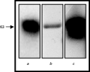
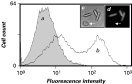
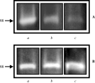
In previous studies (Santos et al. 2002a, 2003), we showed that Herpetomonas samuelpessoai produced a large amount of a surface-located metallopeptidase (60–70 kDa) that presented similar biochemical properties with the gp63 of Leishmania spp., which is a well-known virulence factor expressed by these digenetic human parasites (Yao et al. 2003). In order to confirm that the cell-surface and the released peptidase produced by H. samuelpessoai is a member of the gp63 family, cellular and secretory extracts were probed with anti-gp63 polyclonal antibody by Western blotting (Fig. 1). This antibody strongly recognized a single 63 kDa polypeptide band in L. amazonensis lysate (Fig. 1, lane c), as well as a similar molecular mass component in the cellular extract (Fig. 1, lane a) and in the released polypeptides (Fig. 1, lane b) from H. samuelpessoai. In addition, flow cytometry analysis provided measurements for the relative levels of H. samuelpessoai surface gp63-like molecules (Fig. 2). Fluorescence microscopy corroborated with the fact that the anti-gp63 antibody recognized a similar molecule on the cell-surface of H. samuelpessoai (Fig. 2, inset). Nevertheless, some cells were not fluorescently labelled with the anti-gp63 antibody, corroborating the flow cytometry analysis in which 2 distinct populations were clearly observed (Fig. 2).

Fig. 1. Western blotting showing the leishmanolysin-like polypeptide detected in the whole cellular extract (a) and in the released polypeptides (b) from Herpetomonas samuelpessoai cells. The total cellular extract of promastigotes from Leishmania amazonensis was used as a positive control (c). The number on the left indicates the apparent molecular mass of the reactive polypeptide, expressed in kilodaltons.

Fig. 2. Flow cytometry analysis showing the anti-leishmanolysin antibody binding to the cell-surface of Herpetomonas samuelpessoai. The cells were incubated in the absence (a) and in the presence (b) of anti-gp63 antibody as described in the Material and Methods section, and then analysed by flow cytometry. Phase contrast (c) and fluorescence microscopy images of parasites sequentially incubated with anti-gp63 and FITC-secondary antibodies (d) are shown in the inset. Note that in the FACS analysis 2 distinct populations were clearly observed, 1 of them was inaccessible to the anti-gp63 antibody. Corroborating this result, fluorescence microscopy showed that some cells were not labelled with this antibody (arrow in c and d).
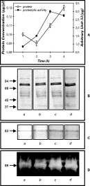
Figure 3 shows the time-course of protein and peptidase released by H. samuelpessoai cells during the 4 h incubation in PBS. The results demonstrated that H. samuelpessoai secreted several polypeptides and an extracellular peptidase of 66 kDa. The total protein content increased linearly from 2 to 4 h and the maximal peptidase liberation was detected in the third hour of incubation with PBS (Fig. 3A). The cellular viability was assessed throughout the incubation period in PBS by monitoring the mobility of cells and trypan blue cell dye exclusion. Non-motile or dead cells were not detected during the 4 h incubation in PBS (data not shown). In addition, lactate dehydrogenase, an intracellular enzyme, was not found in the supernatant fluids, indicating that the extracellular peptidase detected in this study was not released by autolysis (data not shown). SDS–PAGE analysis showed that at least 12 polypeptides, ranging from 40 to 100 kDa, were released by the parasite cells (Fig. 3B). The slight augmentation in the 66 kDa peptidase was also apparent when the supernatant fluids (from 1 to 3 h) were examined by gelatin-SDS–PAGE (Fig. 3D) or by immunoblotting using the anti-leishmanolysin antibody (Fig. 3C).

Fig. 3. Quantitative measurements of proteolytic activity, using gelatin as soluble substrate, and protein released to the extracellular medium (A). SDS–PAGE showing the secretory protein profile revealed by silver staining (B), Western blotting showing the leishmanolysin-like polypeptide (C) and gelatin-SDS–PAGE evidencing the 66 kDa extracellular peptidase (D) released from Herpetomonas samuelpessoai during 1 (a), 2 (b), 3 (c) and 4 h (d) of incubation in isotonic PBS. The numbers on the left indicate the apparent molecular masses, expressed in kilodaltons.
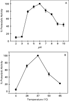
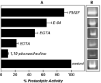
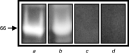
The pH dependence of the 66 kDa extracellular peptidase was determined by densitometric measurements of the digestion halo after gelatin-SDS–PAGE (Fig. 4A). On the gelatin substrate, the enzyme exhibited the maximal activity at pH 6·0. However, the enzyme activity decreased markedly at pH values below 4·0 and above 8·0 (Fig. 4A). The optimum temperature of 66 kDa peptidase was 37 °C, and a decrease by approximately 80% in its maximum activity was detected at 65 °C (Fig. 4B). This extracellular peptidase had its activity inhibited (≈90%) by 10 mM 1,10-phenanthroline, a zinc-metallopeptidase inhibitor (Fig. 5). However, a total inhibition on the 66 kDa peptidase activity was observed when the gel strip was incubated in the presence of 20 mM 1,10-phenanthroline (data not shown). The metallopeptidase inhibitors EDTA and EGTA also promoted a significant decrease on the 66 kDa activity. E-64, a cysteine peptidase inhibitor, weakly inhibited the enzyme (Fig. 5), this could indicate the presence of critical thiol group(s) near the active site as previously suggested (Melo et al. 2002; Santos et al. 2002a). PMSF did not interfere significantly with the enzyme behaviour (Fig. 5). The 66 kDa metallopeptidase exhibited selective substrate utilization on SDS–PAGE, being active with gelatin and casein, while haemoglobin and BSA were not hydrolysed (Fig. 6).

Fig. 4. Effect of pH (A) and temperature (B) on the 66 kDa peptidase secreted by Herpetomonas samuelpessoai after 3 h of PBS incubation. The gel strips were incubated in different buffers (pH 2·0–10·0), as described in the Materials and Methods section, for 20 h at 37 °C. Alternatively, the gel strips were incubated in phosphate buffer (pH 6·0) for 20 h at different temperatures (ranging from 4 to 65 °C). Proteolytic activity of each control system is shown as 100%. The percentage values represent means±standard error of 3 independent densitometric measurements of the digestion halos after gelatin-SDS–PAGE analysis.

Fig. 5. Gelatin-SDS–PAGE showing the modulation of the 66 kDa extracellular proteolytic activity from Herpetomonas samuelpessoai, when the gel strips were incubated in the absence (control) and in the presence of different proteolytic inhibitors: 10 mM 1,10-phenanthroline, 10 mM EDTA, 10 mM EGTA, 10 mM PMSF and 10 μM E-64 (B). The graphic represents the densitometric measurements of the digestion halos (A). Proteolytic activity of control is shown as 100%. The percentage values represent means±standard error of 3 independent measurements.

Fig. 6. Effect of different protein substrates incorporated into 10% SDS–PAGE on the 66 kDa extracellular peptidase activity: gelatin (a), casein (b), BSA (c) and haemoglobin (d). The gel strips were incubated in phosphate buffer (pH 6·0) for 20 h at 37 °C. The number on the left indicates the apparent molecular mass, expressed in kilodaltons, of the peptidase activity.
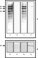
Previously, we showed that H. samuelpessoai cells express at least 4 major surface polypeptides and that living parasites normally released these 4 polypeptides to the extracellular milieu, including the 66 kDa surface metallopeptidase activity (Santos et al. 2001b, 2002a). Conversely, we showed that paraformaldehyde-fixed parasites were incapable of releasing proteins (data not shown) and surface polypeptides to the extracellular environment (Fig. 7A, lane b). However, when the fixed parasites were treated with BtPLC, we observed in the supernatant reaction mixture at least 4 bands (Fig. 7A, lane d), corresponding to the major cell-surface polypeptides, which were recognized by anti-CRD antibody, including a 63 kDa polypeptide component (Fig. 7B, lane d1) that also reacted with the anti-gp63 antibody (Fig. 7B, lane d2), showing that these polypeptides were anchored to the parasite surface via GPI anchor. Control fixed cells, which were not treated with BtPLC, did not produce any positive reaction when probed with the 2 tested antibodies (Fig. 7B, lanes b1 and b2).

Fig. 7. Detection of polypeptides GPI-anchored to the cell-surface of Herpetomonas samuelpessoai. Formaldehyde-fixed Herpetomonas samuelpessoai promastigotes were biotin-labelled with Sulfo-NHS-LC-biotin for 30 min. Then parasite cells were non-treated (a) or treated (c) for 1 h with BtPLC. After this period, the supernatants (reaction mixtures) were obtained (b, d) after centrifugation. Cellular extracts and supernatants were electrophoretically transferred to nitrocellulose membranes and then revealed with AACP to show the cell-surface composition (a, c) as well as the released polypeptides (b, d) (A). The reaction mixture supernatants of both non-treated (b) and BtPLC-treated cells (d) were probed with anti-CRD (1) and anti-gp63 (2) antibodies (B). The number on the left indicates the apparent molecular mass, expressed in kilodaltons.
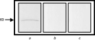
Here, we also looked for a probable reminiscent GPI-anchor in the released polypeptides, especially in the gp63-like component of H. samuelpessoai. The anti-CRD antibody, which recognizes the inositol cyclic phosphate on the reminiscent glycosyl moiety of the protein, failed in recognizing any secreted polypeptide from living H. samuelpessoai (Fig. 8, lane b). A comparable result was obtained when the PBS-derived supernatant was treated with BtPLC (Fig. 8, lane c). Moreover, as similarly demonstrated in Leishmania species (McGwire et al. 2002), 1,10-phenanthroline-treated H. samuelpessoai cells showed a considerably reduction in release of 66 kDa peptidase to the extracellular medium (Fig. 9A). Inhibition of the proteolytic secretion was dose dependent, increasing from 56 to 87% as 1,10-phenanthroline concentration rose from 1 to 10 mM (Fig. 9A). On the other hand, p-CMPS, a PLC inhibitor, at 0·1 and 1 μM did not interfere with the 66 kDa peptidase secretion (Fig. 9B). Additionally, these 2 inhibitors did not affect the parasite viability when tested under the conditions employed in this work. Collectively, these results corroborated with the fact that the metallopeptidase of 66 kDa detected in the extracellular environment could be released on the cell-surface from H. samuelpessoai through proteolysis instead of phospholipolysis.

Fig. 8. Detection of reminiscent GPI-anchor in the extracellular leishmanolysin-like protein of Herpetomonas samuelpessoai. PBS-conditioned supernatant was incubated in the absence (a, b) and in the presence of BtPLC (c) for 1 h at 37 °C. These mixtures were submitted to SDS–PAGE and then the polypeptides were transferred to nitrocellulose membranes. The membranes were incubated separately with anti-gp63 (a) and anti-CRD (b, c) antibodies. The number on the left indicates the apparent molecular mass, expressed in kilodaltons.

Fig. 9. Effect of metallopeptidase and PLC inhibitors on the 66 kDa secretion by living Herpetomonas samuelpessoai cell. Parasites were treated for 30 min in the absence (a) and in the presence of 1 (b) and 10 mM 1,10-phenanthroline (c), then cells were exhaustively washed in PBS, centrifuged and then allowed to secrete for an additional hour (A). Otherwise, cells were incubated for 1 h in the absence (a) and in the presence of 0·1 (b) and 1 μM p-CMPS (c), and the supernatants were obtained after centrifugation and filtration steps (B). All these supernatants were analysed on 10% gelatin-SDS–PAGE, to evidence the proteolytic activity. The number on the left indicates the apparent molecular mass, expressed in kilodaltons, of the peptidase activity.
DISCUSSION
A variety of microorganisms possess membrane-bound and/or secreted peptidases that aid in their interaction with host tissues. In parasites of the genus Leishmania, leishmanolysin molecules on the surface of promastigotes probably form a significant part of the interface between the invading parasite cell and the mammalian host at the time of infection (Yao et al. 2003). Approximately 75% of gp63 from Leishmania species are located on the cell-surface according to surface biotinylation, fluorescence microscopy and immunoelectron microscopy (Weise et al. 2000). Previous studies (Etges, 1992; Santos et al. 2003) showed a cell-surface located gp63-like molecule in H. samuelpessoai by biochemical enzymatic characterization. Here, we corroborated those early studies demonstrating an immunological cross-reactivity between H. samuelpessoai and anti-leishmanolysin antibody using Western blotting, flow cytometry and fluorescence microscopy analyses. Intriguingly, 2 distinct populations with different affinities for the anti-gp63 antibody were clearly identified in H. samuelpessoai, indicating that gp63-like molecules are not equally expressed on the surface of parasite cells. A first explanation for this observation would be that the lack of equal expression is correlated to the H. samuelpessoai growth phase, since flagellate cultures were not synchronized. Furthermore, the occurrence of distinct subpopulations could alternatively denote a different expression of surface gp63-like molecules in the promastigote, paramastigote and opisthomastigote developmental stages (Santos et al. 2003) or even a diminished accessibility to external ligands in cell subsets, as previously reported for other cell-surface molecules expressed in H. samuelpessoai (Santos et al. 2002b).
In addition, several groups recently reported that a significant proportion of leishmanolysin is released by Leishmania promastigotes (Weise et al. 2000; McGwire et al. 2002; Yao et al. 2002; Jaffe and Dwyer, 2003). In this context, we also showed the expression of a leishmanolysin-like activity secreted by living H. samuelpessoai cells. As previously proposed, the existence of a leishmanolysin homologue in monoxenous trypanosomatids, which are insect parasites with no mammalian host, suggests a primary role in the midgut of the insect vector. Interestingly, the prevailing pH range of the insect gut (pH 6–7), the normal habitat for Herpetomonas (McGhee and Cosgrove, 1980), coincides with the pH range in which higher values for proteolytic activity were observed in this paper. However, there is disagreement about the pH optimum of Leishmania gp63. Using native proteins as substrates, Chaudhuri and Chang (1988) observed a pH optimum of 3·0–4·0 for L. mexicana amazonensis promastigote gp63, Tzinia and Soteriadou (1991) reported 2 peaks of activity at pH 5·8 and pH 7·2, for the cleavage of insulin in 7 different Leishmania strains, while Etges et al. (1986) found that L. major promastigote gp63 was most active against azocasein above pH 7·0. Some of the differences may be explained by the use of native, globular substrates versus denatured, linear substrates (Yao et al. 2003). Additionally, in a previous study, we reported that H. samuelpessoai produced a cell-surface metallopeptidase of 60–70 kDa that presented a broad spectrum of activity ranging from pH 5·0 to 10·0 (Santos et al. 2003).
In H. samuelpessoai, a differential expression of cell-surface metallopeptidase at 26 and 37 °C was recently determined (Santos et al. 2003). Here, we also compared the activity of the 66 kDa extracellular metallopeptidase at different temperatures. Similarly, at 26 °C (insect vector temperature), this proteolytic activity was lower than in the vertebrate temperature (37 °C). The different optimal temperatures for these enzymes might reflect adaptations of the parasite to the distinct environments it might confront during its life-cycle. Accordingly, Jansen, Carreira and Deane (1988) demonstrated that the scent glands of experimentally infected opossum Didelphis marsupialis support growth of H. samuelpessoai for many months. In addition, H. samuelpessoai grows well at 37 °C in complex and chemically defined media (Roitman, Roitman and Azevedo, 1972). Consequently, the evolutionary proximity of H. samuelpessoai to important human pathogens, including Leishmania species (Hughes and Piontkivska, 2003), is possibly reflected in the similarity of some aspects of the basic cellular machinery. Therefore, biochemical and immunological similarities between the Leishmania gp63 enzyme and that of H. samuelpessoai suggests that the metallopeptidase was represented in a common ancestor that predated invasion of the vertebrate host. This is evidence that the peptidase activity is important for the survival of the trypanosomatid inside its invertebrate vector, but it does not necessarily preclude it from fulfilling its role in the vertebrate host (Yao et al. 2003).
The effect of inhibitors on the proteolytic activity provides the reliable information concerning the catalytic type of a peptidase. In this sense, extracellular leishmanolysin-like enzyme from H. samuelpessoai demonstrated close similarities to the cell-surface gp63 from Leishmania, including sensitivity towards the metal-chelating 1,10-phenanthroline and modulation by divalent cations (data not shown). Other lower trypanosomatids also release peptidases that can be inhibited by 1,10-phenanthroline (Santos et al. 1999, 2001a, 2005; D'Avila-Levy et al. 2001; Melo et al. 2002; Almeida et al. 2003; Jaffe and Dwyer, 2003; Vermelho et al. 2003). E-64, a cysteine peptidase inhibitor diminished the 66 kDa metallopeptidase activity secreted by H. samuelpessoai. Likewise, the cysteine residue detected in the L. major gp63 protein demonstrated involvement in a cysteine switch mechanism of peptidase activity, binding the zinc atom at the active site and inhibiting enzyme activity (MacDonald et al. 1995). Moreover, leishmanolysin is an enzyme capable of degrading many protein substrates including gelatin, casein, azocasein, albumin, haemoglobin and fibrinogen (Bouvier et al. 1990). Indeed, its ability to hydrolyse such a range of substrates has made it a convenient protein to interact with molecules encountered in diverse host or vector environments. In contrast, the extracellular leishmanolysin-like molecule released from H. samuelpessoai hydrolysed gelatin and casein, but not BSA and haemoglobin, showing a more evident strict substrate specificity under the conditions employed in our experiments.
The extracellular gp63-like enzyme identified herein showed similar biochemical properties with the major metallopeptidase associated to the surface of H. samuelpessoai (Santos et al. 2003). The surface and released molecules synthesized by H. samuelpessoai could form the interface between the parasites and the microenvironments provided by its vector. In addition, this fact suggests that the metallopeptidase may be released into the culture medium by a mechanism that could be similar to that observed for the Leishmania gp63, since H. samuelpessoai surface metallopeptidase is also GPI-anchored to the plasma membrane (Etges, 1992; Schneider and Glaser, 1993; Santos et al. 2002a). Although the GPI anchor can be cleaved by BtPLC revealing the CRD epitope, evidence using anti-CRD antibody indicates that the GPI anchor is absent or not enzymatically cleaved during the release of surface gp63-like molecule from H. samuelpessoai cells. McGwire and co-workers (2002) demonstrated that release of GPI-anchored gp63 into the extracellular medium was dramatically reduced by 1,10-phenanthroline or in the case of a leishmanolysin mutation at the zinc-binding motif. This result suggests that gp63 release is dependent on autoproteolysis, consistent with early reports that gp63 is able to cleave a synthetic peptide substrate corresponding to the pro-peptide cleavage site (Bouvier et al. 1990) and with a later study of L. major leishmanolysin active site mutants (MacDonald et al. 1995). In this sense, we also showed that 1,10-phenanthroline significantly inhibited the release of the 66 kDa metallopeptidase, which suggests a resembling mechanism of secretion in these 2 distinct trypanosomatids. Corroborating these findings, the PLC inhibitor p-CMPS did not alter the 66 kDa metallo-enzyme secretion pattern.
Recently, gp63-like activity has been described in other human pathogenic trypanosomatids, including T. brucei and T. cruzi. In the former, it was suggested that a metallopeptidase surface activity is responsible for the shedding of variant surface glycoprotein (VSG) during cellular differentiation (Bangs et al. 2001). In fact, one of the T. brucei gp63 families is involved in the release of transgenic VSG from procyclic cells (Lacount et al. 2003). T. cruzi possesses a family of gp63 genes composed of multiple groups (Cuevas, Cazzulo and Sanchez, 2003). Two of these groups, Tcgp63-I and –II, are present as high-copy-number genes and antibodies against Tcgp63-I partially blocked the infection of Vero cells by trypomastigotes, which suggests a possible role for this metallopeptidase during the infection process in vitro (Cuevas et al. 2003).
A myriad of functions can be hypothesized for extracellularly released metallopeptidases. For instance, secreted gp63 could allow the parasites to evade a variety of anti-microbial factors in the extracellular environment, including degradation of serine peptidases that are mediators in many aspects of invertebrate immunity such as haemolymph coagulation, activation of anti-microbial peptide synthesis and melanotic encapsulation (Gorman and Paskewitz, 2001). Furthermore, within the sand fly vector, released gp63 could favour the nutrient utilization present in the bloodmeal during the early stages of development, as well as to protect the parasite against the sand fly digestive enzymes (Schlein et al. 1990). We are currently testing the potential role(s) of the extracellular gp63-like molecule from H. samuelpessoai and L. amazonensis in these processes.
This work was supported by grants from Fundação Universitária José Bonifácio (FUJB), Financiadora de Estudos e Projetos (FINEP), Conselho Nacional de Desenvolvimento Científico e Tecnológico (CNPq), Conselho de Ensino e Pesquisa para Graduados da Universidade Federal do Rio de Janeiro (CEPG–UFRJ): Fundação de Amparo à Pesquisa do Estado do Rio de Janeiro (FAPERJ) and Programa de Apoio a Núcleos de Excelência (PRONEX). The authors thank Dr Marta Helena Branquinha (Departamento de Microbiologia Geral, IMPPG – UFRJ) for the useful critical English review as well as for the valuable suggestions on the manuscript, Cláudia Masini d'Avila-Levy for helping with the photographs and Elise Ayumi Hayashi for the assistance with FACS. The authors are indebted to Dr Kwang-Poo Chang (University of Health Sciences, Chicago Medical School, USA) for donating the valuable anti-gp63 antibody and Dr Maria Lucia S. Guther and Dr Michael A. J. Ferguson (Department of Biochemistry, University of Dundee, U K) for donating the anti-CRD antibody.