Implications
Appetite control by the brain is a critical component of the normal growth and development process as well as of major importance for reproduction. From an understanding of how these mechanisms function, we can develop specific protocols to support appetite in circumstances where feed intake is compromised. For example, development of a melanocortin-4 receptor antagonist that can cross the blood–brain barrier provides the opportunity to increase feed intake in normal animals, as well as to prevent the reduction in feed intake and perhaps body mass wasting in disease conditions. Other areas to consider might be early lactation or in metabolic disease circumstances. Thus, the study of brain control of appetite could have a major impact on improved growth and minimizing losses due to disease or other external stresses.
Introduction
Appetite control is a complex process whereby multiple stimulatory and inhibitory inputs are integrated in the hypothalamus to yield an increase or decrease in feed intake. Initial lesioning studies in rodents determined a critical role for the arcuate nucleus (ARC) and ventromedial nucleus (VMN) in the control of appetite. Similar studies of lesions to the paraventricular nucleus (PVN) caused hyperphagia, while lesions of the lateral hypothalamic area (LHA) produced anorexia. These studies have been expanded upon to provide a current concept whereby the ARC contains first-order neurons that in turn activate second-order neurons in the LHA to activate feeding of the PVN to inhibit feed intake (Schwartz et al., Reference Schwartz, Woods, Porte, Seely and Baskin2000). These neural pathways have been extensively described for the rodent species (Schwartz et al., Reference Schwartz, Woods, Porte, Seely and Baskin2000; Valassi et al., Reference Valassi, Scacchi and Cavagnini2008), and though less studied, are no less important in ruminants. In the ARC, there resides a grouping of neurons that release either orexigenic or anorexigenic neurotransmitters. The orexigenic neurotransmitters found in the ARC are neuropeptide Y (NPY) and agouti-related protein (AgRP), while the primary anorexigenic neurotransmitter found in the ARC is α-melanocyte-stimulating hormone (α-MSH), a product of the proopiomelanocortin (POMC) gene. The second-order neurons are located in the LHA and consist of neurons containing melanin-concentrating hormone (MCH) and orexin (ORX). These appetite-regulating neurons are in turn impacted by a diverse array of modifiers (both stimulatory and inhibitory) ranging from hormones such as leptin, ghrelin and insulin to inflammatory cytokines to nutritional molecules such as glucose, fatty acids, amino acids and volatile fatty acids to nerve impulses from the liver and intestine (for overviews of the rodent literature, see Schwartz et al., Reference Schwartz, Woods, Porte, Seely and Baskin2000; Valassi et al., Reference Valassi, Scacchi and Cavagnini2008).
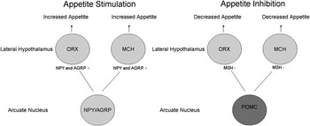
Following an orexigenic stimulus, NPY and AgRP are released from their axon terminals adjacent to second-order neurons in the LHA to activate neurons containing either MCH or cells containing ORX. These neurotransmitters are in turn released from their axon terminals to activate higher order centers to alter feed intake. In the event that the net activation within the ARC is inhibitory to feed intake, α-MSH is released in the LHA to decrease activation of MCH and ORX and subsequently reduce feed intake (Figure 1). Though a simplistic description of the neural architecture and the process regulating appetite, this provides a framework to test this model in ruminant species.
Abbreviated outline of the neural pathways regulating appetite in the hypothalamus. Neuropeptide Y (NPY), Agouti-related protein (AGRP), melanin-concentrating hormone (MCH), Orexin (ORX), α-melanocyte-stimulating hormone MSH, proopiomelanocortin (POMC). NPY, AgRP, ORX, MCH circles represent stimulatory cells and the POMC circles represent inhibitory cells. Reprinted from Sartin et al. (Reference Sartin, Wagner, Marks, Daniels, McMahon, Obese and Partridge2005).

The remainder of this review will focus on the hypothalamus and the regulation of these pathways in sheep with attention to attempts to activate orexigenic and anorexigenic pathways to influence feed intake.
Studies in sheep indicated that unlike the rodent models, feeding activated c-Fos expression in the supraoptic nucleus (Chaillou et al., Reference Chaillou, Baumont, Tramu and Tillet2000). In addition, underfeeding produced c-Fos expression in the PVN, ARC, VMN and dorsomedial nuclei, indicating the importance of these areas in the sheep for appetite control, similar to data in rats.
Neuropeptide Y
Beginning with the first-order neurons in this pathway, there is a potent orexigenic neurotransmitter, NPY. There is both immunohistochemical evidence for NPY protein expression and in situ hybridization evidence for NPY gene expression localized to the ovine hypothalamus. In colchicine-treated sheep, NPY neurons were found distributed between two populations of cells within the ARC, median eminence, pituitary stalk, the dorsomedian and dorsocaudal nuclei, and in the periventricular nucleus (PeVN; Polkowska et al., Reference Polkowska, Wańkowska and Wójcik-Gładysz2006a), but were absent in the magnocellular neurons of the paraventricular and supraoptic nuclei (Chaillou et al., Reference Chaillou, Baumont, Chilliard and Tillet2002; Chaillou and Tillet, Reference Chaillou and Tillet2005). There is also a rich interconnection between the ARC and VMN in sheep (Qi et al., Reference Qi, Iqbal, Oldfield and Clarke2008). In addition, numerous studies have demonstrated expression of NPY mRNA in the ARC (Clarke et al., Reference Clarke, Scott, Rao, Pompolo and Baker-Gibb2000; Adam et al., Reference Adam, Archer, Findlay, Thomas and Marie2002; Pillon et al., Reference Pillon, Caraty, Fabian-Nys and Bruneau2003). Interestingly, in vitro autoradiography (Williams et al., Reference Williams, Adam, Mercer, Moar, Slater, Hunter, Findlay and Hoggard1999) and dual label immunohistochemistry (Iqbal et al., Reference Iqbal, Pompolo, Murakami, Grouzmann, Sakurai, Meister and Clarke2001b) indicate that leptin receptors are expressed by NPY neurons, suggesting a mechanism for leptin to reduce feed intake in ruminants.
In order to demonstrate a role for NPY as an appetite regulator in ruminants, the effect of alteration in nutritional stimuli should be reflected by changes in NPY neuron activation. For example, reducing caloric intake by fasting should provide a stimulus to NPY. Indeed, feeding a protein-restricted energy balanced diet (8% v. 18% protein) resulted in an increase in immunohistochemical staining for NPY in the PeVN and ARC (Polkowska and Gladysz, Reference Polkowska and Gladysz2001). In other studies, negative energy balance created by a 4-day fast resulted in an increase in NPY gene expression in the ARC (Adam et al., Reference Adam, Archer, Findlay, Thomas and Marie2002) with similar data obtained in castrate male sheep in low body condition (Archer et al., Reference Archer, Rhind, Findlay, Kyle, Thomas, Marie and Adam2002a). In another study comparing lean ewes to fat ewes, the number of neurons positive for NPY was increased by 225% as well as an increase in NPY-positive cells co-expressing leptin receptors (Kurose et al., Reference Kurose, Iqbal, Rao, Murata, Hasegawa, Terashima, Kojima, Kangawa and Clarke2005). A related study employed a daily 4-h leptin infusion for four consecutive days that was found to reduce the immunoreactive staining density for NPY (Polkowska et al., Reference Polkowska, Wójcik-Gładysz and Wańkowska2006b). Finally, short-day photoperiod exposure in ovariectomized ewes or in rams reduced the number of cells positive for NPY mRNA (by in situ hybridization) as well as the number of silver grains per cell, suggesting reduced NPY expression during a period that corresponds to reduced voluntary feed intake (Clarke et al., Reference Clarke, Scott, Rao, Pompolo and Baker-Gibb2000, Reference Clarke, Rao, Chilliard, Delavaud and Lincoln2003). Similar effects were observed with wethers (Dobbins et al., Reference Dobbins, Lubbers, Jackson, Kuehl and Hileman2004). By contrast, the effect of fasting to increase NPY expression appeared to be restricted to long-day photoperiod exposed sheep (Archer et al., Reference Archer, Findlay, McMillen, Rhind and Adam2004). Moreover, McShane et al. (Reference McShane, May, Miner and Keisler1992) found that undernutrition elevated cerebrospinal concentrations of NPY, consistent with the gene and protein expression data. In a study of negative energy balance produced as a consequence of lactation, Sorenson et al. (Reference Sorenson, Adam, Findlay, Marie, Thomas, Travers and Vernon2002) found that the negative energy balance of lactation was associated with an increase in NPY expression in the ARC and dorsomedial hypothalamus. Thus, the evidence suggests that NPY is present in the area of the hypothalamus that initiates feed intake and is regulated in a manner consistent with expected changes due to an increase or a reduction in calorie intake. In addition to feed intake regulation, the NPY fasting response may correlate to increased growth hormone (GH) and decreased luteinizing hormone (LH). Fasting is a well-known stimulus to GH that can increase the expression of NPY. In an interesting study linking appetite to the growth axis, growth hormone-releasing hormone was found to colocalize with NPY in the ARC during periods of undernutrition (Iqbal et al., Reference Iqbal, Manley, Ciofi and Clarke2005). Moreover, NPY is a known stimulus to GH release in sheep (McMahon et al., Reference McMahon, Buxton, Elsasser, Gunter, Sanders, Steele and Sartin1999; Morrison et al., Reference Morrison, Daniel, Hampton, Buff, McShane, Thomas and Keisler2003) and cattle (Thomas et al., Reference Thomas, Gazal, Williams, Stanko and Keisler1999) and an inhibitor of LH (Morrison et al., Reference Morrison, Daniel, Hampton, Buff, McShane, Thomas and Keisler2003), suggesting that the effects of fasting may be linked to GH and LH by way of the NPY neuron.
Fetal sheep also express NPY within the hypothalamus at least by day 110 of gestation, which led to an interesting hypothesis that fetal nutrition may imprint on developing neural circuitry in the hypothalamus and have serious consequences in later life (Mülhaüsler et al., Reference Mülhaüsler, McMillen, Rouzaud, Findlay, Marrocco, Rhind and Adam2004). The role of fetal NPY was further illustrated by lateral ventricle injections of NPY into fetal sheep, which resulted in increased swallowing behavior (El-Haddad et al., Reference El-Haddad, Ismail, Guerra, Day and Ross2003).
For NPY to be considered an appetite regulator, it must also be capable of stimulating an increase in feed intake in ad libitum fed sheep. Indeed, intracerebroventricular (ICV) injection of NPY in sheep resulted in a pronounced increase in feed intake (Miner et al., Reference Miner, Della-Fera, Paterson and Baile1989; Sartin et al., Reference Sartin, Dyer, Matteri, Buxton, Buonomo, Shores, Baker, Osbourne, Braden and Steele2001; Wagner et al., Reference Wagner, McMahon, Marks, Daniel, Steele and Sartin2004; Whitlock et al., Reference Whitlock, Daniel, McMahon, Buonomo, Wagner, Steele and Sartin2005). An interesting study of the NPY receptor types mediating feed intake (Clarke et al., Reference Clarke, Backholer and Tilbrook2005) indicated that the major receptor mediating the increased intake due to NPY was the NPY-Y1 receptor, and to a lesser degree, the Y2 receptor. This effect of NPY to increase feed intake was also significant in the presence of rumen distention or propionate infusion (Miner et al., Reference Miner, Della-Fera and Paterson1990). In a related study, endotoxin was used to inhibit feed intake in sheep (as a model for disease) and was coupled with NPY infusion ICV (McMahon et al., Reference McMahon, Buxton, Elsasser, Gunter, Sanders, Steele and Sartin1999). The infusion of NPY in continually endotoxin-treated sheep normalized feed intake during the period of infusion. More importantly, feed intake remained at normal levels even after the removal of NPY. Therefore, NPY has been characterized as a neurotransmitter of the appetite system that regulated increased feed intake and is in turn regulated by nutritional as well as photoperiod manipulations in sheep.
Agouti-related protein
AgRP mRNA (Henry et al., Reference Henry, Rao, Ikenasio, Mountjoy, Tilbrook and Clarke2001a; Adam et al., Reference Adam, Archer, Findlay, Thomas and Marie2002; Archer et al., Reference Archer, Rhind, Findlay, Kyle, Thomas, Marie and Adam2002a) and AgRP protein (Wagner et al., Reference Wagner, McMahon, Marks, Daniel, Steele and Sartin2004) were localized to the ARC nucleus in sheep. Cloning of a partial sequence of AgRP mRNA indicated a high homology with bovine AgRP but the comparison of the human message indicated only about 56% homology for the portion cloned (Wagner et al., Reference Wagner, McMahon, Marks, Daniel, Steele and Sartin2004). Upregulation of AgRP mRNA was observed in chronically underfed ewes (Henry et al., Reference Henry, Rao, Ikenasio, Mountjoy, Tilbrook and Clarke2001a; Archer et al., Reference Archer, Findlay, McMillen, Rhind and Adam2004 and Reference Archer, Rhind, Findlay, Kyle, Barber and Adam2005; Relling et al., Reference Relling, Pate, Reynolds and Loerch2010) or castrate male sheep fasted for 4 days (Adam et al., Reference Adam, Archer, Findlay, Thomas and Marie2002). In addition, dual label immunohistochemistry (for AgRP and c-Fos) indicated that fasting for 3 days increased the number of AgRP-positive neurons and the activation of AgRP neurons in the ARC (percentage of AgRP neurons also expressing c-Fos; Wagner et al., Reference Wagner, McMahon, Marks, Daniel, Steele and Sartin2004). Gene expression for AgRP was also increased in sheep with a low body condition score compared to sheep with a high body condition score (Archer et al., Reference Archer, Findlay, McMillen, Rhind and Adam2004). Interestingly, feed restriction did not alter mRNA expression for the receptor targets of AgRP (Iqbal et al., Reference Iqbal, Pompolo, Dumont, Wu, Mountjoy, Henry and Clarke2001a), the melanocortin-3 receptor (MC3R) and the MC4R. Similarly, lambs born from ewes fed 160% of metabolizable energy requirements had more body fat and a lower expression of AgRP message (Mülhaüsler et al., Reference Mülhaüsler, Adam, Findlay, Duffield and McMillen2006). The infusion of glucose into fetal sheep in late gestation (day 130 to 140) did not affect the expression of AgRP in the fetal ARC, though there were increases in body fat in the glucose-infused sheep (Mülhaüsler et al., Reference Mülhaüsler, Adam, Marrocco, Findlay, Roberts, McFarlane, Kauter and McMillen2005). These studies reflect a functioning appetite regulatory system in the fetus and lend support to the hypothesis that fetal nutrient exposure may have an outcome in postnatal life.
The effects of AgRP on feed intake are thought to be mediated largely via the MC4R, where AgRP inhibits the activity of the receptor to prevent α-MSH from activating the receptor and inhibiting appetite (Ellacott and Cone, Reference Ellacott and Cone2004). Thus, AgRP increases appetite by inhibition of a receptor that inhibits appetite (or an inverse agonist). Injection of AgRP into the lateral ventricle of ad libitum fed sheep resulted in an increase in cumulative feed intake. The effect was present between 4 and 12 h, but unlike other animal models, the effect on feed intake did not persist to 24 h (Wagner et al., Reference Wagner, McMahon, Marks, Daniel, Steele and Sartin2004). Examination of whether altered AgRP expression contributed to reduced feed intake after endotoxin injection determined that there were no changes in AgRP expression during the early phase of appetite suppression (Sartin et al., Reference Sartin, Marks, McMahon, Daniel, Levasseur, Wagner and Whitlock2008). In another experiment, AgRP injected before endotoxin prevented the inhibition of appetite (Sartin et al., Reference Sartin, Marks, McMahon, Daniel, Levasseur, Wagner and Whitlock2008) characteristic of endotoxin treatment. This suggests that MC4R is a major component of the mechanism for inhibition of feed intake in diseased ruminants and suggests a possible means of reversing the negative impact of disease on feeding and perhaps on subsequent body wasting associated with disease (Sartin et al., Reference Sartin, Wagner, Marks, Daniels, McMahon, Obese and Partridge2005).
AgRP is a potent neurotransmitter regulating feed intake in sheep. The synthesis of AgRP is regulated by nutritionally relevant stimuli and AgRP can activate feeding in sheep that are satiated. Thus, like NPY, this neurotransmitter is clearly an appetite regulator in sheep, and perhaps may provide a unique opportunity to manipulate appetite in disease, stress and/or other metabolic problems related to a reduction in feed intake.
Melanin-concentrating hormone
The expression of MCH is pronounced in the lateral hypothalamic area, based on mRNA expression (Henry et al., Reference Henry, Tilbrook, Dunshea, Rao, Blache, Martin and Clarke2000) and immunohistochemical data evidence of the MCH protein (Tillet et al., Reference Tillet, Batailler and Fellman1996; Chaillou et al., Reference Chaillou, Baumont, Fellman, Tramu and Tillet2003; Whitlock et al., Reference Whitlock, Daniel, McMahon, Buonomo, Wagner, Steele and Sartin2005). Cloning of a partial ovine MCH sequence indicates a high homology to other species (Whitlock et al., Reference Whitlock, Daniel, McMahon, Buonomo, Wagner, Steele and Sartin2005). While localization is similar to other species, there are some minor differences in localization (Tillet et al., Reference Tillet, Batailler and Fellman1996). In sheep, MCH has been found to be localized to the supramammillary nucleus, nucleus medialis thalami and nucleus reunions (Tillet et al., Reference Tillet, Batailler and Fellman1996). Of further interest are studies which indicate that MCH neurons from the LHA provide input to the ARC and VMN (Qi et al., Reference Qi, Iqbal, Oldfield and Clarke2008). This adds a possible layer of reciprocal control of appetite, metabolic and endocrine control within the hypothalamus.
Comparison of gene expression for MCH in thin v. fat ewes (Henry et al., Reference Henry, Tilbrook, Dunshea, Rao, Blache, Martin and Clarke2000; Anukulkitch et al., Reference Anukulkitch, Rao, Dunchea and Clarke2009) indicated more mRNA expression for MCH in thin ewes (as well as lower insulin, lactate and free fatty acids in thin ewes), while neurotransmitter gene expression in first-order neurons (NPY and POMC) was unaffected. Moreover, leptin receptors were expressed in all MCH neurons in the LHA (Iqbal et al., Reference Iqbal, Pompolo, Murakami, Grouzmann, Sakurai, Meister and Clarke2001b), though in the absence of the ARC there was no effect of leptin injection on MCH neuron activation (as measured by c-Fos staining; Qi et al., Reference Qi, Henry, Oldfield and Clarke2010). However, even though the presence of leptin receptors suggests that effects of fasting might regulate MCH neurons, undernutrition did not alter the number of MCH-positive neurons as examined by immunohistochemistry (Chaillou et al., Reference Chaillou, Baumont, Fellman, Tramu and Tillet2003). Similarly, short-term fasting of 3 days also had no effect on MCH mRNA in the whole hypothalamus or on the number of MCH-positive neurons in the LHA or on the percentage of neurons co-expressing c-Fos with MCH, suggesting that these MCH neurons were not affected by the nutritional stimulus (Whitlock et al., Reference Whitlock, Daniel, McMahon, Buonomo, Wagner, Steele and Sartin2005). These studies contrast with studies in laboratory animals that suggest that fasting increases MCH (Schwartz et al., Reference Schwartz, Woods, Porte, Seely and Baskin2000).
The physiological effects of MCH have also been examined. Parkes (Reference Parkes1996) infused MCH into the lateral ventricle of the sheep brain for 24 h. The infusion of MCH increased serum osmolality, urine volume, urine sodium and potassium concentrations. Examination of appropriate endocrine responses showed that MCH increased serum glucose, decreased plasma aldosterone concentrations and had no effect on serum protein, adrenocorticotropin, cortisol, vasopressin, renin, endothelin or atrial natriuretic peptide. These changes suggest that MCH may regulate both diuretic and natriuretic functions in sheep (Parkes, Reference Parkes1996). Finally, MCH injection into the lateral ventricle of the brain increased feed intake, but there was no dose–response effect. A similar increase in feed intake was found between a single injection of MCH into the ventricle of the brain and a molar-equivalent dose of NPY (Whitlock et al., Reference Whitlock, Daniel, McMahon, Buonomo, Wagner, Steele and Sartin2005). However, infusion of MCH did not increase feed intake, though an infusion of NPY produced a significant increase in feed intake, thus indicating that MCH regulates feed intake as predicted by the model and suggests a possible downregulation of the MCH receptor when confronted with prolonged high levels of MCH. Therefore, MCH appears to be a very potent regulator of appetite in sheep, though evidence for interactions with endocrine or metabolic systems have not been extensively examined in sheep.
Orexin
ORX expression has been demonstrated in neurons of the LHA (Iqbal et al., Reference Iqbal, Pompolo, Sakurai and Clarke2001c; Qi et al., Reference Qi, Iqbal, Oldfield and Clarke2008) as well as the dorsomedial nucleus, zona incerta, perifornical area and anterior hypothalamic area (Iqbal et al., Reference Iqbal, Pompolo, Sakurai and Clarke2001c). The receptor for orexin (OX1R) was localized to the ARC, median eminence, lateral hypothalamic nuclei and ventral portion of the preoptic area (Zhang et al., Reference Zhang, Blache, Veroe, Adam, Blackberry, Findlay, Eidne and Martin2005). Interestingly, ORX neurons originating in the LHA provide input to both the ARC and VMN (Qi et al., Reference Qi, Iqbal, Oldfield and Clarke2008). Combined, these studies link ORX to regions of the hypothalamus related to feed intake, endocrine, metabolic and reproductive regulation.
Use of nutritional manipulation by chronic undernutrition v. controls indicated no effect on ORX gene expression (Iqbal et al., Reference Iqbal, Henry, Pompolo, Rao and Clarke2003). However, ORX gene expression was negatively associated with adiposity in sheep selected for fatness or lean body condition (Anukulkitch et al., Reference Anukulkitch, Rao, Periera, McEwan and Clarke2010). ORX gene expression was enhanced in short-day photoperiod when appetite is typically lower (Archer et al., Reference Archer, Findlay, Rhind, Mercer and Adam2002b). Similarly, following sheep throughout periods of seasonal appetite changes again demonstrated a negative correlation of ORX to adiposity (Anukulkitch et al., Reference Anukulkitch, Rao, Periera, McEwan and Clarke2010). Related is the demonstration that 100% of ORX neurons express the leptin receptor, suggesting a means of communicating nutritional status to the appetite centers of the hypothalamus to integrate appetite signals (Iqbal et al., Reference Iqbal, Pompolo, Sakurai and Clarke2001c). Moreover, leptin activation of c-Fos in ORX neurons was demonstrated in the absence of the ARC, suggesting a direct action of leptin on second-order neurons in sheep (Qi et al., Reference Qi, Henry, Oldfield and Clarke2010), which differs slightly with the model proposed for laboratory animals.
ORX is related to the regulation of feed intake and, indeed, injection of ORX B into the lateral ventricles of the sheep brain (but not IV injection) enhanced short-term feed intake (Sartin et al., Reference Sartin, Dyer, Matteri, Buxton, Buonomo, Shores, Baker, Osbourne, Braden and Steele2001). There have also been hypotheses of a relationship to hypothalamic control of reproduction, where ORX neurons terminate close to gonadotropin releasing hormone neurons (Iqbal et al., Reference Iqbal, Pompolo, Sakurai and Clarke2001c; Qi et al., Reference Qi, Henry, Oldfield and Clarke2010). In addition to reproduction, ORX neurons project to the PeVN adjacent to somatostatin neurons (Iqbal et al., Reference Iqbal, Manley, Ciofi and Clarke2005), suggesting a possible role in regulating GH. Indeed, ORX A and B receptors are expressed in somatotropes and both molecules activate calcium channels in the pituitary via protein kinase C mechanisms (Xu et al., Reference Xu, Wang, Yan, Hernandez, Gong, Boon, Murata, Ueta and Chen2002 and Reference Xu, Roh, Gong, Hernandez, Ueta and Chen2003). ORX B will also release GH from isolated pituitary cells. Interestingly, there was an increase in plasma cortisol, along with an anecdotal report of increased water intake. However, ICV injection of ORX B in sheep had no effect on plasma LH, GH or insulin concentrations (Sartin et al., Reference Sartin, Dyer, Matteri, Buxton, Buonomo, Shores, Baker, Osbourne, Braden and Steele2001), though perhaps in steroid-treated or photoperiod-manipulated sheep, there may have been a different outcome. Thus, ORX also appears to be a molecule that cannot only enhance feed intake, but may also integrate appetite control with reproduction and metabolic endocrinology in sheep.
Proopiomelanocortin/α-melanocyte-stimulating hormone
Based on the appetite model in Figure 1, neurons containing POMC should be localized to the ARC. However, it must be emphasized that POMC expression does not always correlate to POMC neurons regulating appetite. In sheep, in situ hybridization studies clearly localize POMC to the ARC (Henry et al., Reference Henry, Tilbrook, Dunshea, Rao, Blache, Martin and Clarke2000; Clarke et al., Reference Clarke, Scott, Rao, Pompolo and Baker-Gibb2000; Lincoln et al., Reference Lincoln, Rhind, Pompolo and Clarke2001; Adam et al., Reference Adam, Archer, Findlay, Thomas and Marie2002). In addition, immunohistochemical evidence for α-MSH (product of the POMC gene) was likewise found in the ARC (Sartin et al., Reference Sartin, Marks, McMahon, Daniel, Levasseur, Wagner and Whitlock2008). In one study, POMC gene expression in the ARC was found to be unaffected by a short-term (4-day) fast (Adam et al., Reference Adam, Archer, Findlay, Thomas and Marie2002). In a study of feed intake and adiposity, castrate male sheep with estradiol implants were fed (4 weeks) to maintain body condition or to produce low or high body condition (Archer et al., Reference Archer, Rhind, Findlay, Kyle, Thomas, Marie and Adam2002a). The expression of POMC was similar across all groups. In another study of long-term changes in nutrition, there were no differences in thin v. fat ewes in terms of POMC gene expression (Henry et al., Reference Henry, Tilbrook, Dunshea, Rao, Blache, Martin and Clarke2000). And finally, another 4-week trial found no effects of feed restriction on POMC gene expression (Relling et al., Reference Relling, Pate, Reynolds and Loerch2010). By contrast, McShane et al. (Reference McShane, Petersen, McCrone and Keisler1993) observed a 52% decrease in POMC expression with a long-term feed restriction in ewes (restricted ewes were fed 30% of requirements for 7 weeks), so that with sufficient energy reductions it was possible to impact POMC. Based on these data, it is interesting to note that an effect of fasting to reduce POMC gene expression is a more common finding in laboratory animals as compared to the minimal effects on POMC expression in sheep. This may indicate that the grazing animal has developed a different strategy to regulate the inhibition of appetite compared to intermittent eaters such as the rodent.
Additional studies have examined photoperiod and the interaction between photoperiod and nutritional effects on expression of the POMC gene. Clarke et al. (Reference Clarke, Scott, Rao, Pompolo and Baker-Gibb2000) compared variations in POMC gene expression with voluntary feed intake and natural photoperiod. In the ARC, NPY, and feed intake changed in the opposite direction to POMC. In another study contrasting long-day photoperiod and short-day photoperiod, Clarke et al. (Reference Clarke, Rao, Chilliard, Delavaud and Lincoln2003) found that there was a decrease in gene expression for POMC in the medial ARC in long-day photoperiod exposed rams. Anukulkitch et al. (Reference Anukulkitch, Rao, Dunchea and Clarke2009) also found that POMC gene expression in ewes was reduced during long-day photoperiod exposure, the opposite of that seen with NPY gene expression. Castrate rams supplemented with testosterone were also found to have a reduced POMC gene expression during long-day photoperiods (Hileman et al., Reference Hileman, Kuehl and Jackson1998). A photoperiod study conducted for 11 weeks (Archer et al., Reference Archer, Findlay, McMillen, Rhind and Adam2004) found no effect of photoperiod alone on POMC gene expression in castrate male sheep supplemented with estradiol and either ad libitum fed or fasted. However, when intact rams were switched from a long- to a short-day photoperiod, POMC gene expression was increased. When this photoperiod strategy was combined with feed restriction, POMC gene expression was reduced in the short-day photoperiod group. Thus, POMC neuron regulation in the sheep requires both the appropriate photoperiod and reproductive steroid background as well as nutritional changes before regulation of POMC neurons was consistently observed. Moreover, POMC is a precursor to multiple products, which makes interpretation of general observations difficult in the absence of knowledge of the protein products coupled to the POMC expression. This may, in part, explain the variable POMC response to fasting observed in this species.
Fetal sheep also have POMC gene expression in the ARC (Mülhaüsler et al., Reference Mülhaüsler, McMillen, Rouzaud, Findlay, Marrocco, Rhind and Adam2004) in mid- to late gestation. Glucose infusion to late gestation fetal sheep increased fat mass in the sheep, accompanied by increased POMC gene expression (Mülhaüsler et al., Reference Mülhaüsler, Adam, Marrocco, Findlay, Roberts, McFarlane, Kauter and McMillen2005). Similar increases in fetal POMC expression were obtained by increased nutrition delivered to the mother, which was related to elevated fetal glucose levels (Adam et al., Reference Adam, Findlay and Miller2006). These studies imply that maternal nutrition may program later postnatal development within the appetite regulatory pathway. Indeed, increased maternal nutrition (160% of controls) was found to result in increased plasma glucose and body fat after birth and this was accompanied by increased POMC expression (Mülhaüsler et al., Reference Mülhaüsler, Adam, Findlay, Duffield and McMillen2006), thus providing direct evidence for fetal environment having a role in programming of appetite regulatory genes in postnatal life.
Effects of treatments on α-MSH (protein product of the POMC gene) synthesis or release or effects of treatment with α-MSH have been less studied in sheep. In one study, injection of a melanocortin agonist, MTII (which activates the same receptor as α-MSH), upregulated kisspeptin and LH release (Backholer et al., Reference Backholer, Smith and Clarke2009). In another study, α-MSH immunohistochemistry indicated that with time after endotoxin injection and associated with prolonged reductions (6 h) in feed intake, there is a reduction in α-MSH staining and a reduction in dual labeling for c-Fos, suggesting that α-MSH neurons were not being activated under these circumstances (Sartin et al., Reference Sartin, Marks, McMahon, Daniel, Levasseur, Wagner and Whitlock2008). This reduced expression of POMC and α-MSH neuron activation was accompanied by an increase in AgRP gene expression and c-Fos staining. These changes at 6 h are consistent with an increase in appetite stimulation and correlate to the time when sheep first eat following endotoxin injection. While speculative at this time, perhaps these changes may account for the subsequent compensatory feeding that occurs between 24 and 48 h when cumulative feed intake between the controls and endotoxin-treated sheep no longer differs.
The POMC neuron is typically activated with feeding and inhibited by fasting. This is further complemented by the opposing changes in the endogenous MC4R antagonist, AgRP. An elevation in AgRP inhibits MC4R activity and prevents feed intake inhibition. Thus, the POMC neuron represents the initiation of a potent inhibitory system regulating appetite, though in sheep the POMC response is influenced by photoperiod and steroid exposure.
A summary of neurotransmitters and appetite control in sheep is shown in Table 1.
Summary of neurotransmitters and appetite control in sheep

? = information not available.
Leptin
Leptin is a product of the fat cell and is transported from the blood to the cerebrospinal fluid (CSF) to inhibit NPY and AgRP neurons, which would result in reduced feed intake. Leptin will also activate POMC neurons to inhibit feed intake. In ruminants, leptin gene expression and immunohistochemistry indicate that leptin is synthesized by the fat cell (Dyer et al., Reference Dyer, Simmons, Matteri and Keisler1997; Kumar et al., Reference Kumar, Francis, Suttie and Thompson1998; Daniel et al., Reference Daniel, Elsasser, Morrison, Keisler, Whitlock, Steele, Pugh and Sartin2003). Circulating leptin concentrations are episodic in nature and are increased by obesity and decreased by fasting, though there were no apparent photoperiod effects on plasma leptin concentrations (Kumar et al., Reference Kumar, Francis, Suttie and Thompson1998; Marie et al., Reference Marie, Findlay, Thomas and Adam2001; Daniel et al., Reference Daniel, Whitlock, Baker, Steele, Morrison, Keisler and Sartin2002; Ehrhardt et al., Reference Ehrhardt, Greenwood, Bell and Boisclair2003; Altmann et al., Reference Altmann, Sauerwein and Borell2005; Chilliard et al., Reference Chilliard, Delavaud and Bonnet2005; Delavaud et al., Reference Delavaud, Bocquier, Baumont, Chaillou, Ban-Tokuda and Chilliard2007). Feeding high fat or carbohydrate meals resulted in increased plasma leptin concentrations (Yildiz et al., Reference Yildiz, Blache, Celebi, Kaya, Saatci, Cenesiz and Guven2003) suggesting that higher energy content rather than the source of energy was important. After release from the adipocyte, leptin crosses the blood–brain barrier to enter the hypothalamus as indicated by its entry into the CSF compartment (Thomas et al., Reference Thomas, Preston, Wilson, Farrell and Segal2001; Adam et al., Reference Adam, Findlay and Miller2006), though amounts of leptin entering the CSF were greater in long-day as opposed to short-day photoperiods (Adam et al., Reference Adam, Findlay and Miller2006). Interestingly, plasma and CSF levels only correlated in long-day photoperiod exposed sheep, suggesting a photoperiod influence on leptin transport that corresponds to reported sensitivity in POMC gene expression to photoperiod (Clarke et al., Reference Clarke, Rao, Chilliard, Delavaud and Lincoln2003; Archer et al., Reference Archer, Findlay, McMillen, Rhind and Adam2004; Anukulkitch et al., Reference Anukulkitch, Rao, Dunchea and Clarke2009).
Treatment of sheep with ICV but not IV leptin resulted in an inhibition of feed intake (Henry et al., Reference Henry, Goding, Alexander, Tilbrook, Canny, Dunshea, Rao, Mansell and Clarke1999 and Reference Henry, Goding, Tilbrook, Dunshea and Clarke2001b; Clarke et al., Reference Clarke, Tilbrook, Turner, Doughton and Goding2001; Morrison et al., Reference Morrison, Daniel, Holmberg, Djiane, Raver, Gertler and Keisler2001 and Reference Morrison, Wood, McFadin, Whiley and Keisler2002; Miller et al., Reference Miller, Findlay, Morrison, Raver and Adam2002 and Reference Miller, Harrison, Bennett, Findlay and Adam2007), though the effect could be related to the time of year and whether the sheep were well fed or fasted (thin body condition; Clarke et al., Reference Clarke, Tilbrook, Turner, Doughton and Goding2001). Interestingly, some of the leptin receptors in the ARC were found to be localized to NPY neurons (Williams et al., Reference Williams, Adam, Mercer, Moar, Slater, Hunter, Findlay and Hoggard1999; Kurose et al., Reference Kurose, Iqbal, Rao, Murata, Hasegawa, Terashima, Kojima, Kangawa and Clarke2005) and leptin decreased NPY gene expression (Henry et al., Reference Henry, Goding, Alexander, Tilbrook, Canny, Dunshea, Rao, Mansell and Clarke1999) and immunostaining (Polkowska et al., Reference Polkowska, Wójcik-Gładysz and Wańkowska2006b) in the ARC. There was an increase in the number of NPY neurons expressing leptin receptors in lean sheep (Kurose et al., Reference Kurose, Iqbal, Rao, Murata, Hasegawa, Terashima, Kojima, Kangawa and Clarke2005) and leptin receptor gene expression was increased in long-day exposed sheep (Clarke et al., Reference Clarke, Rao, Chilliard, Delavaud and Lincoln2003). Thus, the evidence suggests that leptin inhibits feed intake, in part by reducing NPY expression as suggested by the model (Figure 1). Interestingly, unlike studies in rodent models, lesions of the ARC do not fully remove the inhibitory effects of leptin in sheep (Qi et al., Reference Qi, Henry, Oldfield and Clarke2010). In this model, leptin increased c-Fos expression in the dorsomedial nucleus, VMN and PVN. While lesions of the ARC prevented c-Fos labeling in MCH neurons following leptin treatment, ORX neurons in the lateral hypothalamus and dorsomedial nucleus were still evident. These data provide evidence that all actions of leptin are not mediated exclusively through first-order to second-order neurons in sheep (Qi et al., Reference Qi, Henry, Oldfield and Clarke2010).
Fetal sheep also have circulating leptin, which is regulated in the fetus (McMillen et al., Reference McMillen, Edwards, Duffiels and Mülhaüsler2006). With high nutrient intake in the pregnant ewe, there is an expected increase in plasma leptin. However, the increased leptin is not reflected in fetal circulating leptin (Mülhaüsler et al., Reference Mülhaüsler, Adam, Findlay, Duffield and McMillen2006).
Inhibition of feed intake is a hallmark of disease, leading to the hypothesis that an increase in leptin during disease might provide a mechanism for the disease-associated reduction in feed intake. However, in a well-characterized endotoxin injection model of disease in sheep, there was no evidence of an increase in circulating leptin, in spite of significant reductions in feed intake (Soliman et al., Reference Soliman, Abdelhady, Fattou, Ishioka, Kitamura, Kimura and Saito2001; Daniel et al., Reference Daniel, Elsasser, Morrison, Keisler, Whitlock, Steele, Pugh and Sartin2003), though tumor necrosis factor and the endotoxin receptor (CD14) were both found to be expressed in the ovine adipocyte (Daniel et al., Reference Daniel, Elsasser, Morrison, Keisler, Whitlock, Steele, Pugh and Sartin2003) as expected. In parasitized lambs, one study suggested that leptin might be a partial factor in reduced feed intake (Zaralis et al., Reference Zaralis, Tolkamp, Houdijk, Wylie and Kyriazakis2008), though in other studies, plasma leptin concentrations were unchanged by the infection (Greer et al., Reference Greer, Boisclair, Stankiewicz, McAnulty, Jay and Sykes2009; Zaralis et al., Reference Zaralis, Tolkamp, Houdijk, Wylie and Kyriazakis2009). However, no studies have examined the effects of disease on CSF leptin levels, which could change by altered uptake of leptin independently of changes in plasma levels. While leptin may have a role in disease suppression of appetite in other species, the evidence is lacking in sheep, suggesting that some other mechanism may be in place to mediate peripheral disease effects on hypothalamic neurons regulating appetite.
Conclusion
This review did not make an attempt to contrast the sheep and other models of appetite control (for these contrasts, see Clarke, Reference Clarke2008). It should be noted that as a grazing animal with continuous nutrient absorption, the nutritional demands differ with regard to intermittent eaters. Indeed, there are key differences in these animal models, especially with regard to factors such as photoperiod, on the one hand, and GH regulation by NPY on the other. The expression of NPY, AgRP, MCH and ORX was found in the proper locations in the hypothalamus for appetite control. Moreover, first-order neurons (NPY and AgRP) are responsive to altered nutrition and all four of these key neurotransmitters increase feed intake. Thus, in general terms, the simple model for neurotransmitter regulation of appetite in Figure 1 is a reasonable fit to use for framing hypotheses in ruminant species. However, studies such as those providing the indication that leptin inhibition of appetite was not exclusively transmitted through first-order neurons as well as the lowered sensitivity of POMC neurons (unless modified by photoperiod) for activation indicates definitive species differences with laboratory animals and indicates a clear need for further study of appetite in farm animal species.
Acknowledgments
This work is based on an invited presentation at the 60th Annual Meeting of the European Association for Animal Production held in Barcelona, Spain during August 2009. This project was supported by National Research Initiative Competitive Grant no. 2004-35206-14136 from the USDA Cooperative State Research, Education and Extension Service.