Introduction
Extracorporeal membrane oxygenation is a short-term mechanical circulatory support system used in patients with severe cardiopulmonary failure unresponsive to conventional therapies. Despite its life-saving potential, extracorporeal membrane oxygenation is invasive and associated with complications such as bleeding and neurological injury, which occur more frequently in paediatric patients than in adults. Reference Tonna, Boonstra and MacLaren1 Timely weaning from extracorporeal membrane oxygenation is critical; however, evidence to guide the optimal timing for safe discontinuation remains limited. Although studies have attempted to identify echocardiographic predictors of successful extracorporeal membrane oxygenation weaning in adults with cardiogenic shock, Reference Aissaoui, Luyt and Leprince2,Reference Kim, Jang and Park3 similar investigations in paediatric patients are lacking.
The high arterial blood flow during venoarterial extracorporeal membrane oxygenation increases left ventricular afterload, thereby limiting the accuracy of echocardiographic assessment of ventricular function. Therefore, accurate evaluation of weaning feasibility requires echocardiography under conditions of reduced extracorporeal membrane oxygenation flow. Recent studies have demonstrated a correlation between increased tissue Doppler velocities and successful extracorporeal membrane oxygenation weaning in adults with cardiogenic shock when the extracorporeal membrane oxygenation flow is reduced to 30–50% of full flow. Reference Kim, Jang and Park3 However, to our knowledge, no comparable study has been conducted in paediatric patients.
This study aimed to identify echocardiographic parameters predictive of successful extracorporeal membrane oxygenation weaning in paediatric patients supported with venoarterial extracorporeal membrane oxygenation for cardiogenic shock, as well as in patients with CHD requiring post-cardiotomy extracorporeal membrane oxygenation.
Material and methods
Study design
We conducted a retrospective analysis of medical records to examine the clinical information and echocardiographic findings of patients who underwent venoarterial extracorporeal membrane oxygenation at Seoul National University Children’s Hospital from March 2018 to September 2023. This study was approved by the Institutional Review Board (IRB) of the Seoul National University Hospital on October 26, 2023 (IRB number: H-2310-080-1475). The study procedures were conducted in accordance with the Declaration of Helsinki and the ethical standards of the IRB. The requirement for informed consent was waived owing to the retrospective nature of the study and the absence of any clinical intervention.
Participants
This study included paediatric patients who underwent venoarterial extracorporeal membrane oxygenation for cardiogenic shock and patients with CHD requiring post-cardiotomy extracorporeal membrane oxygenation support. Exclusion criteria included patients who received venovenous extracorporeal membrane oxygenation, those on venoarterial extracorporeal membrane oxygenation for non-cardiac indications (e.g., respiratory failure or septic shock), and those with functional single ventricles.
Main outcome measures
The primary outcome was successful weaning from extracorporeal membrane oxygenation. Unsuccessful weaning was defined as failure to discontinue extracorporeal membrane oxygenation support, including cases of death on extracorporeal membrane oxygenation, conversion to a durable ventricular assist device, or heart transplantation.
Data collection
Data related to extracorporeal membrane oxygenation were collected, including extracorporeal membrane oxygenation indication, duration, cannulation method, presence of left heart decompression, and initial and reduced extracorporeal membrane oxygenation flow. Additionally, patient-related data (sex, age at extracorporeal membrane oxygenation initiation, weight, underlying disease, occurrence of cardiac arrest before extracorporeal membrane oxygenation, need for renal replacement therapy, vital signs, inotropic support, and laboratory findings at the time of weaning from extracorporeal membrane oxygenation) were collected.
Echocardiographic measurements were performed by a single experienced paediatric cardiologist using an advanced echocardiograph with a matrix probe (Vivid E95; General Electric Medical Systems, Milwaukee, WI, USA). Measurements were attempted at the following four time points: before extracorporeal membrane oxygenation initiation, immediately after extracorporeal membrane oxygenation initiation, before extracorporeal membrane oxygenation weaning, and under reduced extracorporeal membrane oxygenation flow prior to the weaning trial. However, due to unstable vital signs or time constraints, it was not possible to perform assessments at all time points for all patients.
The evaluated parameters included the following:
(a) Left ventricular functional parameters: (i) systolic function: ejection fraction (EF) by the biplane Simpson method using apical 4-chamber and 2-chamber views, systolic myocardial velocity at the lateral mitral annulus (LV lateral S′), and left ventricular peak global longitudinal strain; (ii) diastolic function: early diastolic transmitral flow velocity (E velocity), late diastolic transmitral flow velocity during atrial contraction (A velocity), the ratio of early to late diastolic transmitral flow velocity (E/A ratio), and early and late diastolic myocardial velocities at the lateral mitral annulus (LV lateral E′ and A′); and (iii) global function: left ventricular Tei index.
(b) Right ventricular functional parameters: (i) systolic function: tricuspid annular plane systolic excursion, systolic myocardial velocity at the lateral tricuspid annulus (RV lateral S′), and right ventricular peak global longitudinal strain. These parameters were measured according to the guidelines of the American Society of Echocardiography. Reference Lopez, Colan and Frommelt4,Reference Rudski, Lai and Afilalo5 Myocardial velocities were measured using pulsed-wave tissue Doppler imaging in the apical four-chamber view. Reference Harada, Tamura and Toyono6 Two-dimensional left ventricular and right ventricular peak systolic longitudinal strains were measured using speckle-tracking echocardiography. Reference Amundsen, Helle-Valle and Edvardsen7
(c) Cardiac output (CO) was calculated by the conventional method from the velocity time integral at the left ventricular outflow tract and left ventricular outflow tract diameter, according to the following equation: CO = LVOT area × LVOT VTI × heart rate, where LVOT area = 3.14 × (LVOT diameter2/4). Reference Lang, Badano and Afilalo8,Reference Cecconi, Lillo and Chiara9 Cardiac index was calculated by dividing cardiac output by the patients’ body surface area. Reference Evangelista, Garcia del Castillo and Gonzalez-Alujas10
Patients’ vital signs, laboratory findings, and inotropic usage were reviewed when performing the last echocardiography on extracorporeal membrane oxygenation support. Inotropic usage was quantified as follows: dopamine + dobutamine + 100 × epinephrine + 100 × norepinephrine + 100 × isoproterenol + 15 × milrinone. Reference Hsu, Chi and Yu11
Statistical analysis
Data were analysed using SAS statistical software (SAS System for Windows, version 9.4; SAS Institute, Cary, NC, USA) and R (version 4.3.3; R Foundation for Statistical Computing, Vienna, Austria) with assistance from the Medical Research Collaborating Center of Seoul National University Hospital. Categorical variables are presented as frequencies and percentages, and continuous variables are expressed as means ± standard deviations.
The proportions between the successful and unsuccessful extracorporeal membrane oxygenation weaning groups were compared using either the chi-square test or the Fisher exact test, depending on the expected frequencies. The mean values of clinical information and echocardiographic findings between the two groups were compared using an independent t-test or a Wilcoxon rank-sum test based on normality assumptions. Associations between repeated echocardiographic measurements were analysed using a linear mixed model. Depending on the normality assumptions, between-group differences at two time points were assessed using paired t-tests or Wilcoxon signed-rank tests.
Optimal cutoff values were determined using receiver operating characteristic curve analysis, with area under the receiver operating characteristic curve (AUROC), sensitivity, specificity, positive predictive value, and negative predictive value. Univariate logistic regression analysis was also conducted on the receiver operating characteristic-derived cutoff values for extracorporeal membrane oxygenation weaning.
Results
Between March 2018 and September 2023, 65 venoarterial extracorporeal membrane oxygenation procedures were performed at our hospital. After excluding 13 patients with functional single ventricles and 6 patients lacking echocardiographic data, the data from 46 patients were analysed. The mean age at extracorporeal membrane oxygenation initiation was 9.6 ± 13.9 years, and the mean body weight was 25.98 ± 24.95 kg. The mean duration of extracorporeal membrane oxygenation support was 12.3 ± 12.1 days. Venoarterial extracorporeal membrane oxygenation was indicated in 25 patients with cardiogenic shock from various aetiologies (cardiogenic shock group) and in 21 patients because of unsuccessful cardiopulmonary bypass weaning after cardiac surgery for CHD (post-cardiotomy extracorporeal membrane oxygenation group).
Extracorporeal membrane oxygenation weaning was successful in 31 patients (67.4%) and unsuccessful in 15 patients (32.6%). Of the patients with unsuccessful extracorporeal membrane oxygenation weaning, five underwent ventricular assist device implantation. Overall, ten patients in the unsuccessful weaning group died, including two who subsequently underwent heart transplantation.
No significant differences in sex distribution, age, height, or weight were observed between the successful and unsuccessful weaning groups (Table 1). However, there were differences in the underlying disease and extracorporeal membrane oxygenation indication, with higher rates of CHDs and post-cardiotomy extracorporeal membrane oxygenation in the successful weaning group than in the unsuccessful weaning group. The successful weaning group had significantly higher systolic blood pressure and pulse pressure, lower central venous pressure, and lower inotropic equivalent at the time of the last echocardiography than the unsuccessful weaning group (Supplementary Table S1).
Clinical characteristics

† = Chi-square test; †† = Fisher exact test; * = t test; ** = Wilcoxon rank-sum test.
The values represent the number of cases. The percentage of the study population is presented in parenthesis.
CHD = congenital heart disease; CPR = cardiopulmonary resuscitation; ECMO = extracorporeal membrane oxygenation.
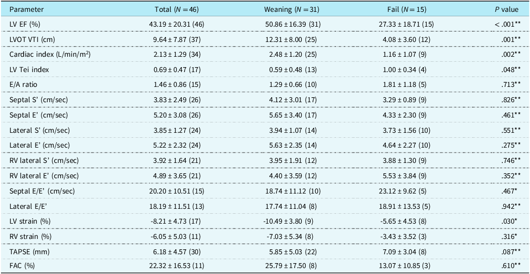
The last echocardiography before the extracorporeal membrane oxygenation weaning trial showed a significantly higher left ventricular ejection fraction and left ventricular outflow tract velocity time integral in the successful weaning group than in the unsuccessful weaning group (Table 2). The cutoff values for predicting successful extracorporeal membrane oxygenation weaning were 43.03% for left ventricular ejection fraction (AUROC: 0.833, sensitivity: 74.2%, specificity: 86.7%) and 4.45 cm for left ventricular outflow tract velocity time integral (AUROC: 0.855, sensitivity: 92.0%, specificity: 66.7%) (Fig 1). Univariate logistic regression analysis showed that the odds ratio of successful weaning with left ventricular ejection fraction > 43.03% compared with left ventricular ejection fraction < 43.03% was 18.687 (95% confidence interval [CI]: 3.44, 101.49; p = 0.001). For left ventricular outflow tract velocity time integral > 4.45 cm compared with left ventricular outflow tract velocity time integral < 4.45 cm, the odds ratio of successful weaning was 7.906 (95% CI: 1.95, 32.02; p = 0.005). Subgroup analyses for the cardiogenic shock and post-cardiotomy extracorporeal membrane oxygenation groups showed different cutoff values: left ventricular ejection fraction of 38.95% and 43.18%, left ventricular outflow tract velocity time integral of 11.65 cm and 6.54 cm, respectively (Supplementary Table S2).
Cutoff values for predicting successful extracorporeal membrane oxygenation (ECMO) weaning. Receiver operating characteristic curves depicting the cutoff values for predicting successful ECMO weaning. ( a ) The cutoff value for left ventricular ejection fraction was 43.03%. ( b ) The cutoff value for the velocity time integral of the left ventricular outflow tract was 4.45 cm. Sens = sensitivity, Spec: specificity; PV+ = positive predictive value; PV− = negative predictive value.

Echocardiographic data before considering weaning of extracorporeal membrane oxygenation

* = t test; ** = Wilcoxon rank-sum test.
The values are presented as means ± standard deviations. The numbers in parentheses denote the sample size for each echocardiographic parameter. E = early diastolic transmitral flow velocity; E/A = ratio of early to late diastolic transmitral flow velocity; ECMO = extracorporeal membrane oxygenation; EF = ejection fraction; FAC = fractional area change; LV = left ventricular; LVOT = left ventricular outflow tract; RV = right ventricular; S’ = systolic myocardial velocity; TAPSE = tricuspid annular plane systolic excursion; VTI = velocity time integral.
In the cardiogenic shock group, cardiac function tended to worsen immediately after extracorporeal membrane oxygenation initiation and then gradually improved (Supplementary Figure S1). In the post-cardiotomy extracorporeal membrane oxygenation group, echocardiographic data before extracorporeal membrane oxygenation initiation was unavailable. As a result, comparisons were made between the data obtained immediately after extracorporeal membrane oxygenation initiation and those from the last echocardiography before the extracorporeal membrane oxygenation weaning trial in both groups. The successful weaning group showed a 14.9% increase in left ventricular ejection fraction, while the unsuccessful weaning group did not demonstrate a change in left ventricular ejection fraction (Supplementary Table S3). Both the cardiogenic shock group and the post-cardiotomy extracorporeal membrane oxygenation group showed significantly greater increases in left ventricular ejection fraction in the successful weaning group than in the unsuccessful weaning group.
Cardiac function was reevaluated with reduced extracorporeal membrane oxygenation flow in 14 patients (Supplementary Table S4). When extracorporeal membrane oxygenation flow was reduced to an average of 54.42% of the initial flow, left ventricular ejection fraction increased by an average of 5.34%, and left ventricular outflow tract velocity time integral increased by an average of 1.14 cm in the successful weaning group. Statistical analysis of the differences in changes of left ventricular ejection fraction and left ventricular outflow tract velocity time integral between the successful and unsuccessful weaning groups was not possible because of the small number of patients in the unsuccessful group.
Discussion
This study demonstrated that higher left ventricular ejection fraction and left ventricular outflow tract velocity time integral values were strongly associated with successful extracorporeal membrane oxygenation weaning. The optimal cutoff values for left ventricular ejection fraction and left ventricular outflow tract velocity time integral were 43.03% and 4.45 cm, respectively, with significant differences in weaning success rates above these thresholds. These findings are consistent with established adult data, underscoring the importance of stable systolic function and adequate left ventricular outflow tract velocity time integral for successful weaning. Reference Charbonneau, Chahinian and Bebawi12 Specifically, our study indicates that successful weaning can occur at an left ventricular outflow tract velocity time integral threshold of 4.45 cm, which is lower than the 10 cm threshold referenced in an adult study. Reference Aissaoui, Luyt and Leprince2 This difference may partly reflect the smaller body surface area and stroke volume in paediatric patients compared with adults. Nevertheless, the Extracorporeal Life Support Organization guidelines for paediatric cardiac failure cite an adult study in which an left ventricular outflow tract velocity time integral exceeding 10 cm was associated with successful extracorporeal membrane oxygenation weaning, Reference Brown, Moynihan and Deatrick13 highlighting the lack of established paediatric thresholds and the importance of developing age- and size-adjusted criteria.
As previously noted, venoarterial extracorporeal membrane oxygenation increases left ventricular afterload, limiting the reliability of conventional echocardiographic parameters under full-flow conditions. This observation underscores the value of flow-reduction trials for assessing native cardiac function. Accordingly, the cutoff values for left ventricular ejection fraction and left ventricular outflow tract velocity time integral identified in this study to predict successful weaning should be regarded as exploratory indicators rather than definitive thresholds, given that flow reduction was not performed during the acquisition of these measurements. A systematic approach to weaning, involving gradual reduction of extracorporeal membrane oxygenation support while closely monitoring haemodynamic responses, has been shown to be effective. Reference Fried, Masoumi and Takeda14 Such a strategy may facilitate timely weaning decisions and potentially reduce extracorporeal membrane oxygenation duration and related complications.
Using echocardiography to guide extracorporeal membrane oxygenation weaning decisions is particularly beneficial in paediatric settings, where clinical judgment is challenging due to variability in patients’ size and underlying conditions. Although patients in the successful weaning group exhibited more favourable haemodynamic parameters, there are no established haemodynamic thresholds that reliably predict successful extracorporeal membrane oxygenation discontinuation. Therefore, echocardiographic assessment provides an objective complement to clinical evaluation, offering quantitative evidence of myocardial recovery, thereby helping clinicians proceed with greater confidence when determining readiness for extracorporeal membrane oxygenation discontinuation. In this study, due to limitations in the echocardiographic window and the unstable condition of patients undergoing extracorporeal membrane oxygenation, it was not possible to measure all echocardiographic parameters at all four time points for every patient. Consequently, we included only left ventricular ejection fraction and left ventricular outflow tract velocity time integral in the ROC curve analysis, as these parameters were measured in at least 80% of patients. Additionally, the left ventricular Tei index and global strain, measured in 17 patients (37%), were significantly lower in the group successfully weaned from extracorporeal membrane oxygenation than in the group that was not (Table 2). This finding suggests that, where feasible, serial follow-up of these parameters during extracorporeal membrane oxygenation could be informative.
Although our study provides valuable insights, it shares common limitations with other retrospective analyses, including potential selection and recording biases. The single-centre design may also limit the generalisability of our findings. In addition, the heterogeneity between cardiogenic shock and post-cardiotomy patients and the relatively small sample size reduced the statistical power of subgroup analyses. Finally, the cutoff values were derived retrospectively and have not been externally validated.
Future multicentre, prospective studies are needed to validate these echocardiographic thresholds in a broader paediatric population, including patients with CHD. In addition, recent evidence from adult venoarterial extracorporeal membrane oxygenation populations suggests that integrating echocardiographic findings with other clinical, biomarker, and microcirculatory parameters may improve prediction of weaning outcomes. Reference Charbonneau, Chahinian and Bebawi12 Such an approach could help establish a more comprehensive and reliable framework for assessing readiness for extracorporeal membrane oxygenation discontinuation.
In conclusion, echocardiographic monitoring plays a crucial role in the management of venoarterial extracorporeal membrane oxygenation and provides essential data to guide weaning decisions. Our study validates that left ventricular ejection fraction and left ventricular outflow tract velocity time integral are useful predictors of weaning success. These parameters provide a practical and reliable basis for guiding the weaning process and improving outcomes in this high-risk population.
Supplementary material
The supplementary material for this article can be found at https://doi.org/10.1017/S1047951125111062
Data, Material, and/or Code Availability
The datasets generated and/or analysed during the current study are available from Dr Hye Won Kwon (email: [drhwkwon@snu.ac.kr]) on reasonable request.
Acknowledgements
We gratefully acknowledge the support of the Medical Research Collaborating Center of Seoul National University Hospital for their assistance with statistical analysis.
The authors also used ChatGPT (OpenAI, San Francisco, CA, USA) to assist with language editing and figure design (Supplementary Figure S1). The authors reviewed and verified all AI-assisted content to ensure accuracy and integrity in accordance with the Cambridge University Press AI Contributions to Research Content Policy.
Author contribution
Conceptualisation: Hye Won Kwon, Jae Gun Kwak, Woong-Han Kim
Data curation: Hye Won Kwon, Jae Gun Kwak
Formal analysis: Hye Won Kwon, Jae Gun Kwak
Investigation: Hye Won Kwon, Jae Gun Kwak, Sungkyu Cho
Methodology: Hye Won Kwon, Mi Kyoung Song, Sang Yun Lee
Project administration: Hye Won Kwon, Gi Beom Kim
Resources: Jae Gun Kwak, Sungkyu Cho, Woong-Han Kim
Supervision: Gi Beom Kim, Eun Jung Bae, Woong-Han Kim
Validation: Mi Kyoung Song, Sang Yun Lee
Writing-original draft: Hye Won Kwon
Writing-review and editing: Hye Won Kwon, Jae Gun Kwak, Sungkyu Cho, Woong-Han Kim, Mi Kyoung Song, Sang Yun Lee, Gi Beom Kim, Eun Jung Bae
Competing interests
The authors declare no conflicts of interest.
Ethical standard
The procedures followed during the study were in accordance with the Declaration of Helsinki and the ethical standards of the Institutional Committee on Human Experimentation. The study was approved by the Institutional Review Board of the Seoul National University Hospital on October 26, 2023 (IRB number: H-2310-080-1475).
Consent to Participate
The requirement for informed consent was waived owing to the retrospective nature of the study and the absence of any clinical intervention.