Implications
Economic analyses demonstrate that a 10% increase in tenderness of beef would add $150 million to the income of the US beef industry. An improvement is associated with the degradation of cytoskeleton proteins and the activation of caspases. This experiment demonstrated that caspase-9 was activated at 5 h in beef and some physiological factors, for example, the ratio of bcl-2 and bax proteins and ATP content affected the activation of caspase-9. The work helps us to further understand the tenderization mechanism and contributes to marketing strategies to improve meat quality.
Introduction
The quality of meat most appreciated by consumers is tenderness, particularly in the case of beef (Hocquette et al., Reference Hocquette, Botreau, Picard, Jacquet, Pethick and Scollan2012). An improvement in meat tenderness during conditioning is associated with continuous degradation of cytoskeleton proteins (Hopkins and Thompson, Reference Hopkins and Thompson2002). These proteins are considered commonly as nebulin, titin, integrin, desmin, troponin T, etc. (Fritz and Greaser, Reference Fritz and Greaser1991; Kolczak et al., Reference Kolczak, Pospiech, Palka and Lacki2003).
Ouali et al. (Reference Ouali, Herrera-Mendez, Coulis, Becila, Boudjellal, Aubry and Sentandreu2006) suggested that the cysteine protease family of caspases is in a better position than other proteolytic enzymes to cleave cytoskeleton, as this is their primary function in vivo. Kemp et al. (Reference Kemp, Bardsley and Parr2006a) indicated that caspase-mediated cleavage takes place in the porcine muscles during conditioning. Kemp et al. (Reference Kemp, Parr, Bardsley and Buttery2006b) detected caspase-3 (20 kDa) and caspase-8 (18 kDa) active fragments in the porcine muscles. Our previously published results indicated that there was a significant increase in caspase-3 activities and that apoptosis did exist in the bovine skeletal muscles during conditioning (Cao et al., Reference Cao, Sun, Zhou, Xu, Peng and Hu2010 and Reference Cao, Ou, Zou, Ye, Zhang, Khan, Pan and Zhou2013). Recently, in vitro proteolytic and chemical promoters treating experiments have shown that caspases were involved in postmortem proteolysis (Chen et al., Reference Chen, Feng, Lu, Xu, Zhou, Li and Guo2011; Huang et al., Reference Huang, Huang, Zhou, Xu and Xue2011 and Reference Huang, Huang, Xue, Xu and Zhou2011).
Caspases are a family of cysteine proteases, which play essential roles in apoptosis (programmed cell death; Adams, Reference Adams2003). Caspase-9 has been reported to be one of the most important caspases among the family members, as activated caspase-9 can further activate other initiator caspases, such as caspase-8 and -7, and effector caspases, such as caspase-3 (Thornberry and Lazebnik, Reference Thornberry and Lazebnik1998). Our previously published results indicated that calcium chloride injection had no significant effect on the caspase activities in the bovine skeletal muscles during conditioning (Cao et al., Reference Cao, Yu, Khan, Shao, Xiang and Zhou2012). However, the activation of caspase-9 and the affected physiological factors during conditioning are far from being understood.
Medical reports demonstrated that the activating steps of caspase-9 are ATP-dependent and that the availability of intracellular ATP and the release of cytochrome c from the mitochondria into the cytosol are necessary for the activation of caspase-9 (Phaneuf and Leeuwenburgh, Reference Phaneuf and Leeuwenburgh2002; Delivoria-Papadopoulos et al., Reference Delivoria-Papadopoulos, Gorn, Ashraf and Mishra2007). In addition, the balance between pro-apoptotic bax and anti-apoptotic bcl-2 proteins in normal tissue determines the activation of caspase-9 and the fate of cells during ischemia condition (Kluck et al., Reference Kluck, Bossy-Wetzel, Green and Newmeyer1997). In an effort to further understand the biochemical mechanism and the activating process of caspase-9 in the postmortem muscles, this study was conducted to detect the expression of anti-apoptotic bcl-2 and pro-apoptotic bax proteins in the fresh tissues and determine the postmortem changes in the ATP content, caspase-9 activity and cytosol cytochrome c during conditioning in the longissimus thoracis (LT), Psoas minor (PMi) and Semitendinosus (STN) muscles of bulls.
Material and methods
Animals and samples
Animals were humanely handled as described previously (Cao et al., Reference Cao, Sun, Zhou, Xu, Peng and Hu2010). Five crossbred bulls (Simmental×local Chinese yellow cattle in the Anhwei province) with ages from 22 to 24 months (body weight 443±48 kg) were slaughtered in a local abattoir. A core sample (∼20 g) was taken from the LT muscle (from the 7th to 8th thoracic vertebrae), PMi muscle and STN muscle (near the middle of length and between 5 to 10 cm below sarcolemma) from the right side of carcasses at 30 min after slaughter and exsanguinations, frozen rapidly and stored in liquid nitrogen until bcl-2 family proteins, caspase-9 activity and ATP content analyses. In addition, partly fresh muscle samples were used for cytosolic protein isolation and cytochrome c determination. The samples at 30 min were considered as samples at 0 h postmortem. After samples at 0 h were taken, the whole LT muscles were dissected from the 5th to 11th thoracic vertebrae on the right side of carcasses; the whole PMi and STN muscles were dissected immediately, exposed to air and stored at the temperature of 4°C from the right side of carcasses. In terms of different sampling points (0, 3, 5, 8, 14, 18, 24, 39, 96 and 168 h), about 20 g of the muscle samples were taken randomly, over-wrapped with aluminum foil, frozen and stored in liquid nitrogen for later ATP content and caspase-9 activity analysis. Extra fresh muscle samples were taken at 5, 24 and 96 h and used for cytosolic protein isolation and cytochrome c determination.
Reagents and standards
Protease inhibitor cocktail was obtained from Roche Applied Science (Indianapolis, IN, USA); BCA protein assay kit was obtained from Pierce (Rockford, IL, USA); reconstituted N-acetyl-Leu-Glu-His-Asp-7-amino-4- methylcoumarin (Ac-LEHD-AMC) was obtained from Biomol (Plymoth Meeting, PA, USA). The standard of ATP, Phenylmethylsulfonyl fluoride, methanol, sodium deoxycholate, Fast DAB (diaminobenzidintetrahydrochloride) and Metal enhancer (CoCl2) were obtained from Sigma (St. Louis, MO, USA); Tetrabutylammonium hydrogen sulfate, Potassium dihydrogen orthophosphate and Dipotassium hydrogen orthophosphate were obtained from Nanjing Pharmaceutical Co., Ltd (Nanjing, China). All chemicals used were of ACS grade, except the standard of ATP and the methanol were of HPLC grade. Primary antibodies were obtained from the following sources: rabbit anti-bax was from Stressgen (Victoria, Canada); mouse anti-cytochrome c was from BioLegend (San Diego, CA, USA); rabbit anti-bcl-2 was from Cell Signaling (Beverly, MA, USA); goat anti-glyceraldehyde 3-phosphate dehydrogenase (GAPDH) was from Genscript (Nanjing, China). Horseradish peroxidase (HRP)-conjugated mouse anti-goat, HRP-conjugated mouse anti-rabbit and HRP-conjugated rabbit anti-mouse antibodies were from Genscript.
Measurement of caspase-9 activity
Caspase-9 activity was determined as described by Viemann et al. (Reference Viemann, Barczyk, Vogl, Fischer, Sunderkötter, Schulze-Osthoff and Roth2007). Two hundred milligrams of sample was taken from the whole frozen sample (20 g), pulverized and homogenized on ice in 0.5 ml lyses buffer comprising 100 mM HEPES (pH 7.5), 10% sucrose, 0.1% NP-40, 10 mM DTT and a protease inhibitor cocktail. Homogenates were centrifuged at 18 000×g at 4°C for 30 min by a Beckman Coulter Allegra 25 R Centrifuge (Fullerton, CA, USA). Twenty microliters of supernatant was combined with 0.2 ml of protease assay buffer (10% sucrose, 0.1% CHAPS, 100 mM HEPES, pH 7.5); and then added 5 μl each reconstituted Ac-LEHD-AMC substrates. The mixture was incubated at 37°C for 1 h and fluorescence values were determined by using excitation and emission wavelengths of 360 and 460 nm by a 96-Well Plate Tecan M200 Reader (Männedorf, Switzerland), respectively. Results were calculated as relative intensity of fluorescence per min per milligram of meat.
ATP content measurement
The ATP content of samples was determined by HPLC as described previously with modifications (Veciana-Nogués et al., Reference Veciana-Nogués, Izquierdo-Pulido and Vidal-Carou1997). Eight hundred milligrams of sample was taken from the whole frozen sample (20 g) and homogenized in 3 ml 7% perchloric acid. After centrifugation at 15 000×g at 4°C for 10 min, the supernatant was neutralized with 1.44 ml 0.85 M K2CO3, centrifuged again as described above and filtrated through a 0.2 μm syringe filter (Millipore, Billerica, MA, USA). The ATP content was measured at a wavelength of 254 nm on an Agilent 1100 series, using a reversed-phase C18 column (Zorbax SB 4 mm×250 mm i.d., 5 μm particle size; Agilent, Santa Clara, CA, USA), at a flow rate of 1 ml/min at 25°C. Mobile phase A was phosphate buffer (2.5 mM tetrabutylammonium hydrogen sulfate, 0.04 M potassium dihydrogen orthophosphate and 0.06 M dipotassium hydrogen orthophosphate, pH 7.0). Mobile phase B was methanol. The elution program was as follows: 0 min, 98% A, 2% B; 8 min 98% A, 2% B; 8.5 min 90% A, 10% B; 19 min 90% A, 10% B; 20 min 98% A, 2% B; 24 min 98% A, 2% B. Peaks were identified and quantified by the comparison of retention time and peak area with known external standard of ATP.
The isolation of cytosolic protein fractions
Cytosolic protein fractions were isolated as described previously with modifications (Marzetti et al., Reference Marzetti, Lees, Manini, Buford, Aranda, Calvani, Capuani, Marsiske, Lott, Vandenborne, Bernabei, Pahor, Leeuwenburgh and Wohlgemuth2012). Briefly, fresh muscle samples (800 mg) at 0, 5, 24 and 96 h postmortem were homogenized in 5 ml of pre-chilled isolation buffer (0.225 M mannitol, 0.075 M sucrose, 0.2% bovine serum albumin, 1 mM EDTA, pH 7.4) using a Potter–Elvehjem glass homogenizer (Wheaton, Fisher Scientific, Pittsburgh, PA, USA). Samples were subsequently centrifuged at 1000×g at 4°C for 15 min. The supernatants were centrifuged at 14 000×g at 4°C for 20 min. The supernatants were prepared by further centrifugation at 100 000×g for 1 h at 4°C. The protein contents of supernatants were determined using a BCA protein assay kit; protein samples were mixed 1 : 1 with 2× electrophoresis sample buffer (1.0 M Tris-HCl, pH 6.7, 15% glycerol, 5% SDS, 10% β-mercaptoethanol, 0.2% bromophenol blue), heated at 90°C for 4 min and stored at −80°C for cytosolic cytochrome c determination.
Proteins extraction from whole tissues
The muscle samples at 0 h postmortem as described were used for protein extraction and bcl-2 family’s protein level determination. Proteins were extracted from whole tissues with modifications as described previously (Havaki et al., Reference Havaki, Kouloukoussa, Amawi, Drosos, Arvanitis, Goutas, Vlachodimitropoulos, Vassilaros, Katsantoni, Voloudakis-Baltatzis, Aleporou-Marinou, Kittas and Marinos2007). The frozen muscles (400 mg) in liquid nitrogen were homogenized in 3 ml of RIPA buffer (50 mM Tris-HCl pH 7.4, 150 mM NaCl, 2 mM EDTA, 1% NP-40, 0.1% SDS, 1% sodium deoxycholate, 1 mM Phenylmethylsulfonyl fluoride) with a mortar (Nanjing University Instrument Plant, Nanjing, China). The samples were centrifuged at 14 000×g for 10 min at 4°C. The protein contents of supernatants were determined using a BCA protein assay kit; protein samples were mixed 1 : 1 with 2× electrophoresis sample buffer, heated at 90°C for 4 min, and stored at −80°C for bax and bcl-2 protein level determination.
SDS-PAGE and Western blotting
The bax and bcl-2, as well as cytosolic cytochrome c expression levels, were determined by SDS-PAGE and Western blotting with modifications as described previously (Vendelin et al., Reference Vendelin, Pulkkinen, Rehn, Pirskanen, Räisänen-Sokolowski, Laitinen, Haahtela, Saarialho-Kere, Laitinen and Kere2006). Protein samples (100 μg) were run on a 12.5% SDS-PAGE and transferred to Polyvinylidene fluoride membranes (Immobilon-P, Millipore, MA, USA). Membranes were blocked in Tris-buffered saline containing 0.05% Tween-20 (TBST) solution (50 mM Tris, 150 mM NaCl, 0.05% Tween-20, pH 7.4) containing 5% dry milk for 1 h at 25°C and incubated with primary antibody for 24 h at 4°C. The primary antibodies were diluted 1 : 200 for bax and bcl-2 and 1 : 500 for cytochrome c and GAPDH with TBST solution containing 5% dry milk. The appropriate HRP-conjugated secondary antibodies were diluted 1 : 1000 with TBST solution containing 5% dry milk. Membranes were incubated with the appropriate HRP-conjugated secondary antibodies for 1 h at 25°C. The blot was developed with Sigma Fast DAB and Metal enhancer as described previously (Nagasaka et al., Reference Nagasaka, Okamoto and Ushio2006). Specialized software and a microcomputer (Quantity One Quantitation Software, Bio-Rad Laboratories, Philadelphia, PA, USA) were used to quantify Western blot data.
Statistical analyses
Statistical analyses were performed by using the one-way ANOVA procedure (Duncan’s Multiple Range Test) of SAS 8.0 (SAS, USA) to determine whether the expression of bcl-2 family (bcl-2 and bax) proteins and the ratio of bax to bcl-2 are significantly different among fresh LT, PMi and STN muscles. Two-way ANOVA procedure (muscle and time were chosen as main effects) was performed to determine whether the caspase-9 activity, ATP content and cytosol cytochrome c levels changed significantly during the postmortem period among muscles.
Results and discussion
The caspase-9 activity
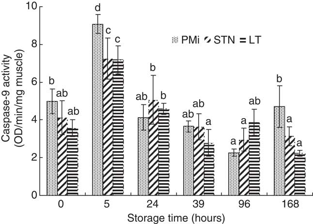
The change in caspase-9 activity in the PMi, STN and LT muscles during conditioning was shown in Figure 1. The highest activities of caspase-9 appeared at 5 h. From 0 to 5 h storage, caspase-9 activity increased significantly in the muscles, and a significant decrease was observed from 5 to 24 h in caspase-9 activity (P<0.001).
Caspase-9 activities in the PMi, STN and LT muscles during 168 h storage. Caspase activities are reported as arbitrary absorbance units/min per mg muscles. Error bars are standard errors of the mean (s.e.m.). The statistical comparisons for every muscle were made within five samples. aDifferent letters above columns indicate the significant differences (P<0.05). LT=longissimus thoracis; STN=semitendinosus; PMi=psoas minor.

The increase in caspase-9 activity is consistent with the results in the hypoxic cerebral cortical tissue reported by Khurana et al. (Reference Khurana, Ashraf, Mishra and Delivoria-Papadopoulos2002). These results demonstrate that postmortem ischemia/hypoxia environment induces the activation of caspase-9 in the muscles.
The ATP contents
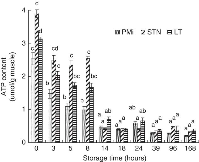
The change in the ATP contents in the PMi, STN and LT muscles during conditioning was shown in Figure 2. There was a significant decrease in the ATP content from 0 to 3 h and from 8 to 14 h postmortem in the PMi and LT muscles (P<0.0001), whereas from 0 to 5 h and from 8 to 14 h postmortem in the STN muscle (P<0.0001). There is still 60.2%, 55.3% and 43.1% ATP content in the STN, LT and PMi muscles at 5 h compared with the one at 0 h. After 18 h postmortem, there was no significant change in the ATP level in the three muscles (P>0.05). During postmortem ischemia, intracellular ATP is rapidly degraded as a result of insufficient oxygen supply and rapid consumption of glycogen (Du et al., Reference Du, Shen and Zhu2005). Several reports have shown that apoptosis is composed of several ATP-dependent steps and that the availability of intracellular ATP determines whether cells undergo apoptosis or necrosis (Fujimura et al., Reference Fujimura, Morita-Fujimura, Murakami, Kawase and Chan1998). Delivoria-Papadopoulos et al. (Reference Delivoria-Papadopoulos, Gorn, Ashraf and Mishra2007) demonstrated that active caspase-9 density increased by 30%, 45% and 60% in the presence of ATP, cytochrome c and ATP+cytochrome c, respectively, in the hypoxic condition. Liu et al. (Reference Liu, Kim, Yang, Jemmerson and Wang1996) indicated that the depletion of ATP prevented the activation of caspase-9.
HPLC analysis in the changes in the ATP content in the PMi, STN and LT muscles of bulls during 168 h storage. Error bars are standard errors of the mean (s.e.m.). The statistical comparisons for every muscle were made within five samples. aDifferent letters above columns indicate the significant differences (P<0.05). LT=longissimus thoracis; STN=semitendinosus; PMi=psoas minor.

Our results suggested that the available intracellular ATP before 5 h postmortem could be favorable for the activation of caspase-9, as the activation was composed of several ATP-dependent steps (Delivoria-Papadopoulos et al., Reference Delivoria-Papadopoulos, Gorn, Ashraf and Mishra2007). However, we supposed that caspase-9 activity could be prevented subsequently because of the decreased ATP level.
The quantitative analysis of cytosolic cytochrome c level
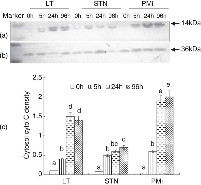
As shown in Figure 3, the 14 kDa cytochrome c protein was determined in cytosolic protein fractions by SDS-PAGE and immunoblotting with specific antibody (Figure 3a). GAPDH protein levels were determined as a standardized internal control (Figure 3b). The changes in the cytosolic cytochrome c level were visualized by Western blot analysis as shown in Figure 3c. There was a 4.1-fold and 14.9-fold increase in the cytosolic cytochrome c level at 5 and 24 h, respectively, than the one at 0 h postmortem in the LT muscle (P<0.0001); there is a 6.25-fold and 7.5-fold increase in the cytosolic cytochrome c level at 5 and 96 h, respectively, than the one at 0 h in the STN muscle (P<0.0001); and there is a 9.8-fold and 31.2-fold increase in the cytosolic cytochrome c level at 5 and 24 h, respectively, than the one at 0 h in the PMi muscle (P<0.0001). At 24 h postmortem, there was the highest value of cytosol cytochrome c found in the PMi, followed by STN and a very low expression was displayed in the LT muscle (P<0.001).
Western blot analysis of relative changes in the cytosolic cytochrome c protein levels in the PMi, STN and LT muscles during 96 h storage. The 36 kDa GAPDH (a) and 14 kDa cytochrome c protein (b) in the PMi, STN and LT muscles. The results are reported as ratios to the GAPDH protein density (c). Error bars are standard errors of the mean (s.e.m.). The statistical comparisons for every muscle were made within five samples. aDifferent letters above columns indicate the significant differences (P<0.05). LT=longissimus thoracis; STN=semitendinosus; PMi=psoas minor; GAPDH=glyceraldehyde 3-phosphate dehydrogenase.

Numerous death-inducing signals, such as reactive oxidant intermediates, decreases in mitochondrial redox status and increases in intracellular calcium, could trigger the mitochondria to release caspase-activating protein cytochrome c (Phaneuf and Leeuwenburgh, Reference Phaneuf and Leeuwenburgh2002). Soeda et al. (Reference Soeda, Miyagawa, Sano, Masumoto, Taniguchi and Kawasaki2001) reported that the cytochrome c in cytosolic fraction was barely detected in the normal tissue, but was detected after 15 min of ischemia, increasing in a time-dependent manner in the hepatocytes and sinusoidal endothelial cells. The increase in the cytosolic cytochrome c level at 5 and 24 h in the LT and PMi muscles and at 5 and 96 h in the STN muscle could be associated with the promoted mitochondrial permeability transition (MPT) after the bulls were bled.
Mitochondrial-mediated apoptosis can be executed via the release of cytochrome c from the mitochondria into cytosol, subsequent the formation of apoptosome and activation of caspase-9 (Kluck et al., Reference Kluck, Bossy-Wetzel, Green and Newmeyer1997). It has been demonstrated that the formation of cytochrome c–Apaf-1 complex is necessary for the activation of caspase-9 and is dependent on hydrolysis of ATP (Zou et al., Reference Zou, Henzel, Liu, Lutschg and Wang1997). The increase in the cytosolic cytochrome c level should be responsible for the increase in caspase-9 activity. We suppose that the relationship between the increase in cytosolic cytochrome c and caspase-9 activity could be limited by the rapid postmortem depletion of ATP.
Bcl-2 family protein expression
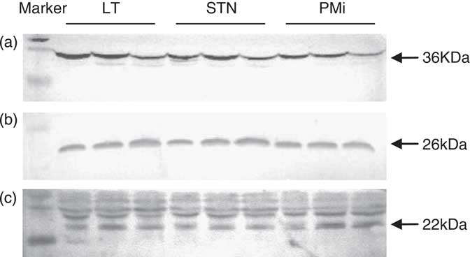
As shown in Figure 4, the GAPDH protein levels were determined as a standardized internal control (Figure 4a); 26 kDa bcl-2 protein (Figure 4b) and 22 kDa bax protein (Figure 4c) were detected in whole-muscle lysates by SDS-PAGE and immunoblotting with specific antibodies. Levels in bcl-2 and bax expression and the ratio of bax to bcl-2 were visualized by Western blot analysis, as shown in Table 1. There were higher expression levels in bax (P<0.01) and bcl-2 (P<0.05) proteins and higher ratio of bax to bcl-2 (P<0.01) in the PMi muscle than the ones in the LT and STN muscles.
Western blot analysis of GAPDH (a), bcl-2 (b) and bax (c) protein in the PMi, STN and LT muscles of bulls at 0 h. GAPDH=glyceraldehyde 3-phosphate dehydrogenase; LT=longissimus thoracis; STN=semitendinosus; PMi=psoas minor.

The effect of muscles on the density ratio for bax to bcl-2, bcl-2 to GAPDH and bax to GAPDH

GAPDH=glyceraldehyde 3-phosphate dehydrogenase.
The statistical comparisons for every muscle were done within three samples from the five samples.
a Different letters within the same line indicate the significant differences (P<0.05).
b The blots were stripped and probed with GAPDH to document equal protein loading. LT (longissimus thoracis), STN (semitendinosus), PMi (psoas minor).
The different bax and bcl-2 expression levels in different normal skeletal muscles of rats has been associated with the activation of caspases under subsequent chronic hypoxia and aging conditions (Rice and Blough, Reference Rice and Blough2006). In the cell, if there is no higher ratio of bax to bcl-2, more mitochondria still cannot result in the higher cytosolic cytochrome c level, as cytochrome c just can be released through the specific mitochondrial pore formed by bax homodimerization, whereas bcl-2 can prevent the release and block the pore by forming a heterodimer with bax protein (Gross et al., Reference Gross, Jockel, Wei and Korsmeyer1998 and Reference Gross, McDonnell and Korsmeyer1999). Pastorino et al. (Reference Pastorino, Chen, Tafani, Snyder and Farber1998) demonstrated that the overexpression of bax induced MPT and accompanied by the increase of cytosolic cytochrome c level and caspase-9 activation. It has been suggested that the bax to bcl-2 ratio may be more important than either the promoter alone in determining apoptosis (Stoetzer et al., Reference Stoetzer, Nussler, Darsow, Gullis, Pelka-Fleischer, Scheel and Wilmanns1996). On the basis of others’ reports and our observations, we concluded that the more expression of cytosolic cytochrome c at 24 h in the PMi muscle than that in the other muscles should be explained by the high ratio of bax to bcl-2.
Conclusion
Postmortem ischemia/hypoxia environment induces the activation of caspase-9; available intracellular ATP before 5 h could be favorable for the activation of caspase-9; the ascend of caspase-9 activity could be attributed to the increase of cytosolic cytochrome c and could be limited by the postmortem depletion of ATP; the higher ratio of bax to bcl-2 in the fresh PMi muscle than the other muscles contributed to the more expression of cytosolic cytochrome c in samples at 24 h.
Acknowledgments
This work was financially supported by National Natural Science Foundation (No. 31101309), The Public Welfare Project of Zhejiang Province (2012C22058), Agricultural Research Project of Ningbo City (2011C10021), Scientific Research Fund of Zhejiang Provincial Education Department (No. Y201121121), Subject Program in Ningbo University (No. xkl11099) and K.C. Wong Magna Fund of Ningbo University and Sponsored by Start Research and Fund projects Fund at Ningbo University.