

Introduction
Stroke, also known as a brain attack, is when poor blood flow to the brain leads to cell death (Donnan, Fisher, Macleod, & Davis, Reference Donnan, Fisher, Macleod and Davis2008). Most cerebral strokes are ischemic resulting from the occlusion of a major cerebral artery by a thrombus or an embolism, leading to decreased blood flow and necrosis in that area (Gibson, Reference Gibson2013). As a major cause of death and long-term debilitation (Mozaffarian et al., Reference Mozaffarian, Benjamin, Go, Arnett, Blaha, Cushman and Howard2015), acute lacunar stroke (Als) has exacted a heavy toll in disability and even death worldwide. Characteristic signs and symptoms of Als may include an inability to move or feel one side of the body (Schulz et al., Reference Schulz, Koch, Zimerman, Wessel, Bönstrup, Thomalla and Hummel2015), vision problems (Sand et al., Reference Sand, Wilhelmsen, Næss, Midelfart, Thomassen and Hoff2016) and/or problems understanding or speaking (Donnan et al., Reference Donnan, Fisher, Macleod and Davis2008). Aphasia is the chronic difficulty in communicating in more than 20% of patients post stroke (Pedersen, Stig Jørgensen, Nakayama, Raaschou, & Olsen, Reference Pedersen, Stig Jørgensen, Nakayama, Raaschou and Olsen1995). Therefore, understanding the mechanism of aphasia includes clinical and theoretical implications for diagnosis and prognosis.
Diffusion tensor imaging (DTI) has been employed to investigate the anatomical correlates of specific items in speech impairments such as repetition, comprehension or naming disturbances underlining the role of white matter (WM) tracts (Kümmerer et al., Reference Kümmerer, Hartwigsen, Kellmeyer, Glauche, Mader, Klöppel and Saur2013; Marchina et al., Reference Marchina, Zhu, Norton, Zipse, Wan and Schlaug2011). Breier and his colleagues employed DTI to determine the relationship between damage to specific WM tracts and linguistic deficits after left hemisphere stroke (Breier, Hasan, Zhang, Men, & Papanicolaou, Reference Breier, Hasan, Zhang, Men and Papanicolaou2008). They found lower fractional anisotropy (FA) values in the left superior longitudinal fasciculus (SLF) and arcuate fasciculi were correlated with a decreased ability to repeat spoken language. In addition, lower FA values in the left arcuate fasciculus were associated with comprehension deficits after stroke (Breier et al., Reference Breier, Hasan, Zhang, Men and Papanicolaou2008). Consistently, a combined disconnection of the dorsal and ventral pathways (i.e., the arcuate and inferior fronto-occipital fasciculi) was found to be associated with aphasia severity in chronic stroke patients (Rosso et al., Reference Rosso, Vargas, Valabregue, Arbizu, Henry-Amar, Leger and Samson2015). However, the results may be biased by the WM fibers analysis method, which was carried out by manually drawing them or with the region of interest analysis. Recently, tract-based spatial statistics (TBSS) focus on the centres of all fiber bundles that are common to the participants (the most compact WM skeleton), thus improving the probability that the given space voxels contain data from the same part of the same WM tract among each participant (Yu et al., Reference Yu, Yuan, Zhang, Liu, Dong, Jin and Zhao2016). Therefore, TBSS is more sensitive and robust for detecting WM abnormalities compared with previous ROI analyses (Yuan et al., Reference Yuan, Qin, Wang, Zeng, Zhao, Yang and von Deneen2011; Yu et al., Reference Yu, Yuan, Zhang, Liu, Dong, Jin and Zhao2016). The lack of healthy controls (HC) also limits the conclusions that can be drawn. More importantly, FA has high sensitivity to microstructural changes that reflect axonal cell membrane structures and myelin sheaths, but is not very specific to the types of changes (Thomason & Thompson, Reference Thomason and Thompson2011). As a supplement, mean diffusivity (MD), axial diffusivity (AD) and radial diffusivity (RD) may provide more specific information about the structure of axonal cell membranes and myelin sheaths (Thomason & Thompson, Reference Thomason and Thompson2011).
Several lines of evidence from neuropsychological and brain imaging studies suggested that disrupted WM fiber connectivity included the main biological correlates of aphasia involved in the mechanisms of the symptoms (Catani & Mesulam, Reference Catani and Mesulam2008; Galantucci et al., Reference Galantucci, Tartaglia, Wilson, Henry, Filippi, Agosta and Gorno-Tempini2011; Glasser & Rilling, Reference Glasser and Rilling2008; Schlaug, Marchina, & Norton, Reference Schlaug, Marchina and Norton2009). To define the pathophysiological features of WM abnormalities in acute ischemic stroke patients, which include myelination, demyelination, axonal loss, or inflammation, the TBSS method and multiple DTI-derived indices analysis were used in the present study (Thomason & Thompson, Reference Thomason and Thompson2011), which have been proven to be more sensitive and robust (Lee, Han, Kim, & Renshaw, Reference Lee, Han, Kim and Renshaw2013; Yu et al., Reference Yu, Yuan, Zhao, Dong, Liu, Yang and Xue2013; Reference Yu, Yuan, Zhang, Liu, Dong, Jin and Zhao2016; Yuan et al., Reference Yuan, Qin, Wang, Zeng, Zhao, Yang and von Deneen2011). Besides, correlation analyses were carried out to investigate the relationship between WM changes and aphasia severity of Als patients. We hypothesized that the multiple DTI-derived indices analysis could provide more specific pathophysiological features of WM abnormalities in Als patients.
Materials and Methods
All research procedures were approved by the Ethical Committee of the Second Hospital of Hebei Medical University and were conducted in accordance with the ethical standards as laid down in the 1964 Declaration of Helsinki and its later amendments or comparable ethical standards. Informed consent was obtained from all individual participants included in the study. The data sets generated and/or analysed during the current study are not publicly available because this is a pilot study and we are still conducting further research on them, but are available from the corresponding author upon reasonable request.
Subjects
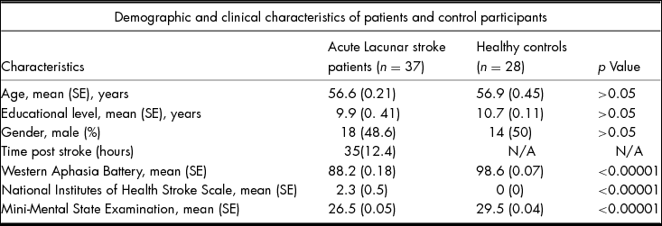
A group of 37 patients, containing 18 males and 19 females, were diagnosed with Als for the first time at the intensive care unit of the Department of Neurology in the Second Hospital of Hebei Medical University between May 2014 and November 2015. Patient ages ranged between 32 and 77 years, with a mean of 56.6 ± 0.21 years. All patients were diagnosed with Als for the first time based on full stroke investigation, including brain MRI, imaging of the extracerebral arteries, arteries and electrocardiogram. Lacunar stroke was defined as a clinical lacunar syndrome, with an anatomically compatible lesion on MRI seen as either acute on diffusion-weighted imaging or low signal on T1 or fluid-attenuated inversion recovery imaging and with a diameter <15 mm. All patients were given the Western Aphasia Battery (WAB) and the composite indices were extracted as the aphasia quotient (AQ) to assess the severity of aphasia (Berthier, Reference Berthier2005). We focused on aphasia induced by Als, therefore, AQ < 93.8 was another inclusion criteria for the patients. Exclusion criteria were as follows: stenosis >50% in the extracranial or intracranial cerebral vessels or previous carotid endarterectomy; cardioembolic source of stroke; cortical infarct on MRI; subcortical infarct >15 mm in diameter, as these can be caused by embolic mechanisms (striatocapsular infarcts); and any other specific cause of stroke (e.g., lupus anticoagulant, cerebral vasculitis, dissection and monogenic cause of stroke). In addition, 28 HC without aphasia (AQ > 93.8) were also recruited and had undergone thorough physical examinations during the same period (age range: 25–75 years, 56.9 ± 0.45 years). National Institutes of Health Stroke Scale (NIHSS) and Mini-Mental State Examination (MMSE) were administered as well. The participants with dementia were excluded according to MMSE < 24. More detailed demographic information is reported in Table 1.
Demographic and Clinical Characteristics of Acute Lacunar Stroke Patients and Healthy Controls

DTI Imaging Acquisition
A high-resolution T1-weighted image (TR = 1900 ms, TE = 2.53 ms, FA = 9°, matrix = 256 × 256, FOV = 256 × 256 mm2, slice number = 176, voxel size = 1 × 1 × 1 mm3) was acquired via a 3-Tesla MRI system (EXCITE, General Electric, Milwaukee, Wisc.) for each subject. Imaging was performed in the department of Radiology; the Second Hospital of Hebei Medical University in China, with a volumetric three-dimensional spoiled gradient recall sequence. The diffusion sensitizing gradients were applied along 60 non-collinear directions (b = 1000 s/mm2) with an acquisition without diffusion weighing (b = 0 s/mm2). The imaging parameters were 75 continuous axial slices with a slice thickness of 2 mm and no gap, field of view (FOV) = 256 × 256; TR = 9400 ms; TE = 84 ms; matrix size = 128 × 128, resulting in 2 mm isotropic voxels.
Data Analysis
DTI data were processed by using the FMRIB Software Library (FSL) software 4.1.9 (Oxford Centre for Functional Magnetic Resonance Imaging of the Brain Software Library, http://www.fmrib.ox.ac.uk/fsl) according to previous studies (Lee, Han, Kim, & Renshaw, Reference Lee, Han, Kim and Renshaw2013; Yu et al., Reference Yu, Yuan, Zhang, Liu, Dong, Jin and Zhao2016). After the eddy current distortion correction, simple head motion and brain extraction of raw diffusion data were carried out. For each subject, the FA and MD maps were calculated by fitting a tensor model at each voxel. The AD image was the 1st eigenvalue (L1) map for each participant. The RD image was calculated by the mean of the 2nd and 3rd eigenvalue maps. Then, TBSS analyses were performed between Als patients and HC. In more detail, all of the participants’ FA images were nonlinearly registered to the FMRIB58-FA standard-space template. The average image of all aligned FA images was created and thinned to create a skeletonized mean FA image with the threshold at 0.2. Each aligned FA image of all participants was then projected onto this skeleton. Next, using the protocol of non-FA Images in TBSS, the MD, AD and RD images were aligned into MNI space and projected onto the mean FA skeleton. Finally, the permutation-based nonparametric inferences within the framework of the general linear model were performed to investigate the differences between the two groups. The threshold-free cluster enhancement with the family-wise error (FWE) correction for multiple comparison corrections was used to detect significant results (p < 0.05, FWE corrected, 5000 permutations). A series of Pearson's correlation analyses was performed to examine the correlations between the FA values of the abnormal fibers and aphasia severity, NIHSS and MMSE of Als patients.
Results
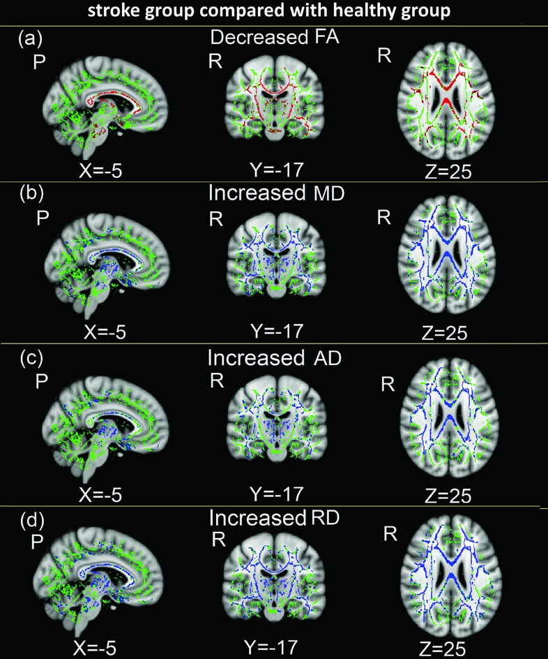
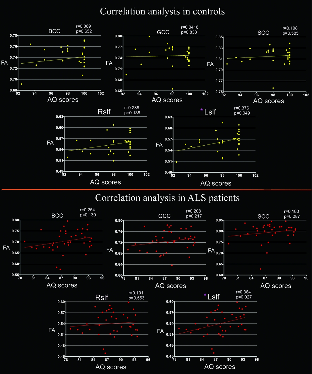
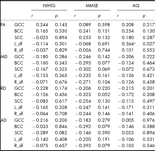
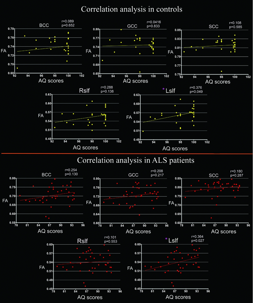
The age, education and gender distribution was not different between Als patients and HC (Table 1). Relative to HC, Als patients showed a significantly lower FA and higher MD, RD and AD in multiple brain regions, i.e., decreased FA and increased MD, RD and AD in the genu, body and splenium of the corpus callosum (CC), posterior and anterior limb of the internal capsule, external capsule, SLF and corona radiata in the bilateral hemisphere (Figure 1). In no region did patients show a higher FA or lower RD and MD than control subjects. The AQ scores were significantly correlated with FA values of the left SLF in both HC and Als patients (Figure 2). No other significant correlations were found. Detailed information is given in Table 2.
Compared with healthy controls, acute lacunar stroke (Als) patients showed significantly reduced fractional anisotropy (FA), increased axial diffusivity (AD), mean diffusivity (MD) and radial diffusivity (RD) in multiple white matter fibers (p < 0.05, family-wise error (FWE) corrected). No increased FA, reduced AD, MD and RD of white matter (WM) fibers were observed.

The aphasia quotient (AQ) scores were significantly correlated with fractional anisotropy (FA) values of the left superior longitudinal fasciculus (SLF) in both healthy controls and acute lacunar stroke (Als) patients (Figure 2). No other significant correlations were found.
Correlation Results of the DTI Findings and Behavioral Variates in Patients

*p < 0.05.
GCC: genu of the corpus callosum.
BCC: body of the corpus callosum.
SCC: splenium of the corpus callosum.
SLF: superior longitudinal fasciculus.
NIHSS: National Institutes of Health Stroke Scale.
MMSE: Mini-Mental State Examination.
L: left.
R: right.
AQ: aphasia quotient.
FA: fractional anisotropy.
MD: mean diffusivity.
AD: axial diffusivity.
RD: radial diffusivity.
Discussion
The high incidence of aphasia had been detected in Als patients (Engelter et al., Reference Engelter, Gostynski, Papa, Frei, Born, Ajdacic-Gross and Lyrer2006; Pedersen et al., Reference Pedersen, Stig Jørgensen, Nakayama, Raaschou and Olsen1995). WM fibers involved in aphasia have theoretical and clinical implications for diagnosis and prognosis. Although DTI methods have been employed to investigate the anatomical correlates of aphasia in Als patients (Breier et al., Reference Breier, Hasan, Zhang, Men and Papanicolaou2008; Rosso et al., Reference Rosso, Vargas, Valabregue, Arbizu, Henry-Amar, Leger and Samson2015), the ROI-based method and lack of HC do not fully explain the microstructure of WM tracts in Als patients. In the present study, the TBSS method and multiple DTI-derived indices analysis were utilized to specify the neurophysiological feature of WM integrity in Als patients. We not only observed reduced FA, but also detected increased MD, RD and AD in multiple brain WM regions of a sample of Als patients (Figure 1). The FA findings were partially consistent with previous studies (Breier et al., Reference Breier, Hasan, Zhang, Men and Papanicolaou2008; Rosso et al., Reference Rosso, Vargas, Valabregue, Arbizu, Henry-Amar, Leger and Samson2015) and improved our understanding of the disruption of WM integrity in Als patients by adding multi-DTI parameter results (Figure 1). FA has high sensitivity to microstructural changes that reflect axonal cell membrane structures and myelin sheaths, but is not very specific to the types of changes, such as myelination, maturation, demyelination, axon loss, gliosis or inflammation (Yu et al., Reference Yu, Yuan, Zhang, Liu, Dong, Jin and Zhao2016). MD, AD and RD may provide more information about the structure of axonal cell membranes and myelin sheaths (Yu et al., Reference Yu, Yuan, Zhang, Liu, Dong, Jin and Zhao2016). A decrease in AD suggests axonal loss or loss of bundle coherence, and an increase in RD suggests disrupted myelination. MD is a measure of the average molecular motion, independent of tissue directionality. WM abnormalities with myelin sheath disruption result in reduced FA and increased MD. Higher MD and AD values probably mean axonal loss of WM in Als patients. The extent of the abnormalities hints at a more severe disruption of WM integrity in the CC, corona radiata and SLF, where lower FA values were accompanied by higher MD, AD and RD. Increased RD is thought to signify increased space between fibers, suggesting demyelination or dysmyelination, and a decrease in parallel diffusivity suggests axonal injury (Benedetti et al., Reference Benedetti, Yeh, Bellani, Radaelli, Nicoletti, Poletti and Scotti2011; Thomason & Thompson, Reference Thomason and Thompson2011); this finding proposes disrupted integrity of myelin sheaths among these fiber tracts.
Furthermore, the correlation analysis revealed a special connection between the FA values of the SLF in the left hemisphere and AQ scores in Als patients and HC (Figure 2). The SLF joins the areas in the lateral frontal cortex that promote language, including Broca's area, with areas of the parietal and temporal cortices, including Wernicke's area, that maintain receptive language function (Catani, Howard, Pajevic, & Jones, Reference Catani, Howard, Pajevic and Jones2002; Catani & Jones, Reference Catani and Jones2005). Broca's area including the pars opercularis and triangularis, stores memories of movements needed for speaking (Embick, Marantz, Miyashita, O'Neil, & Sakai, Reference Embick, Marantz, Miyashita, O'Neil and Sakai2000; Hagoort, Reference Hagoort2005; Roth, Nadeau, Hollingsworth, Marie Cimino-Knight, & Heilman, Reference Roth, Nadeau, Hollingsworth, Marie Cimino-Knight and Heilman2006). Wernicke's area is believed to be involved in the “storage of phonologic representations or memories of word sounds” (Just, Carpenter, Keller, Eddy, & Thulborn, Reference Just, Carpenter, Keller, Eddy and Thulborn1996; Knecht et al., Reference Knecht, Dräger, Deppe, Bobe, Lohmann, Flöel and Henningsen2000). In the left hemisphere, SLF fibers connect the temporal and frontal cortices involved in receptive and expressive language that form a portion of the distributed language network (Catani & Jones, Reference Catani and Jones2005). Moreover, a dual pathway network for language had revealed that sublexical repetition of speech is subserved by a dorsal pathway, which is connected to the superior temporal lobe and premotor cortices in the frontal lobe via the arcuate and SLF. In contrast, higher-level language comprehension is processed by a ventral pathway connecting the middle temporal lobe and the ventrolateral prefrontal cortex via the extreme capsule (Saur et al., Reference Saur, Kreher, Schnell, Kümmerer, Kellmeyer, Vry and Abel2008). Consistently, SLF damage may cause obstacles in repeating words among Als patients despite the function of their comprehension was fairly intact (Benson et al., Reference Benson, Sheremata, Bouchard, Segarra, Price and Geschwind1973). Brier et al. found lower FA values of the SLF in stroke patients with language dysfunction and detected a specific relationship between aphasia severity and damage of WM tracts (Breier et al., Reference Breier, Hasan, Zhang, Men and Papanicolaou2008). Consistent with previous results, the findings of the current study validated the reduced FA values of the SLF in stroke patients (Figure 1). We enhanced our understanding of the disrupted integrity of myelin sheaths of the SLF by revealing reduced FA with increased MD and RD. Moreover, we observed a significant correlation between the FA values of the SLF in both Als patients and HC (Figure 2). For this specific correlation, after including age as a covariate, the correlation remains significant. Therefore, the relationship is not due to a normal aging effect.
Besides, the findings of the current study included other key WM tracts contributing to the functional integrity of the brain (Figure 2). As the largest WM brain structure, the CC consists of neural fibers under the brain cortex at the longitudinal fissure connecting the left and right cerebral hemispheres (Huang et al., Reference Huang, Zhang, Jiang, Wakana, Poetscher, Miller and Mori2005). It mainly communicates between the two hemispheres of the brain and enhances inter-hemispheric communication (Hofer & Frahm, Reference Hofer and Frahm2006), which has been implicated in brain diseases (Bi et al., Reference Bi, Yuan, Feng, Xing, Li, Wang and Qin2015; Yuan et al., Reference Yuan, Qin, Liu, Zhao, Yu, Zhao and von Deneen2012). A previous study found that degeneration of the CC constituted as an important factor having influence on cortical reorganization and motor outcome following subcortical stroke (Wang et al., Reference Wang, Tittgemeyer, Imperati, Diekhoff, Ameli, Fink and Grefkes2012). The internal capsule communicates with the basal ganglia, separating the caudate nucleus and thalamus from the putamen and globus pallidus (Wakana, Jiang, Nagae-Poetscher, Van Zijl, & Mori, Reference Wakana, Jiang, Nagae-Poetscher, Van Zijl and Mori2004). The internal capsule contains both ascending and descending axons and fibers going to and coming from the cerebral cortex (Wakana et al., Reference Wakana, Jiang, Nagae-Poetscher, Van Zijl and Mori2004). Atypical aphasia syndromes commonly result from circumscribed non-haemorrhagic infarctions of the anterior limb of the internal capsule in the dominant hemisphere (Damasio, Damasio, Rizzo, Varney, & Gersh, Reference Damasio, Damasio, Rizzo, Varney and Gersh1982). The WM of the external capsule contains fibers known as corticocortical association fibers (Wakana et al., Reference Wakana, Jiang, Nagae-Poetscher, Van Zijl and Mori2004), which are responsible for connecting the cerebral cortex to another cortical area. The corticospinal tract carries motor information from the primary motor cortex to the lower motor neurons in the spinal cord (Wakana et al., Reference Wakana, Jiang, Nagae-Poetscher, Van Zijl and Mori2004). The corona radiata travels ventrally as the internal capsule and dorsally as the semi-oval center (Wakanaet al., Reference Wakana, Jiang, Nagae-Poetscher, Van Zijl and Mori2004). Motor fibers from small infarcts are somatotopically arranged in the human corona radiata (Cho et al., Reference Cho, Kim, Kim, Kim, Lee and Jang2007). Compromised inter-hemispheric connections involved in communication and limbic, large frontal, parietal and fronto-occipital connections could define the dysfunctions in Als patients. A more comprehensive experimental design is necessary to depict the accurate roles of abnormal DTI properties of such fibers in Als patients.
Conclusion
In the present study, the TBSS method with multiple DTI-derived indices analysis was employed collectively to specify the neurophysiological feature of WM integrity in Als patients. We not only observed reduced FA, but also increased MD, RD and AD in multiple brain WM fibers in Als patients, such as the CC, SLF, internal capsule, external capsule and corona radiata, which suggested disrupted integrity of myelin sheaths among these fiber tracts. The AQ scores were significantly correlated with FA values of the left SLF in both HC and Als patients. It is hoped that the multi-parameter TBSS can shed new insights into the pathology of patients with Als patients.
Limitations
The study is limited in its cross-sectional design that may not infer the causality of the relationships between WM integrity abnormalities and aphasia in Als patients. More longitudinal studies with a larger sample size would be required to improve the understanding of the neuroimaging findings in the current study. The time interval between the onset of stroke and DTI abnormalities can provide more clinical implications for the mechanisms of aphasia induced by stroke. In the future, we will collect this important information to investigate the relationship between these variables.
Acknowledgements
All authors contributed equally to the design, data collection and analysis and article writing/editing.
Financial Support
This work was supported by the National Natural Science Foundation of China under Grant No. 81501543.
Conflict of Interest
All authors declare that they have no conflict of interest to disclose.
Ethical Standards
The authors assert that all procedures contributing to this work comply with the ethical standards of the relevant national and institutional committees on human experimentation and with the Helsinki Declaration of 1975, as revised in 2008.