Congenital scoliosis is the most common congenital deformity of the spine; however, its effects on cardiac function are not well understood. Previous studiesReference Beals, Robbins and Rolfe 1 , Reference Basu, Elsebaie and Noordeen 2 have mainly focused on the prevalence of CHD in patients with scoliosis. Congenital scoliosis is usually accompanied by rib abnormalities, which may contribute to a crowded middle mediastinum. In addition, certain pliable structures such as the main bronchus, descending aorta, and pulmonary artery can be displaced and compressed.Reference Doi, Harimaya, Matsumoto and Iwamoto 3 - Reference Donnelly, Bisset and McDermott 5 Currently, the lung function of patients with scoliosis has been well documented,Reference Bowen, Scaduto and Banuelos 6 , Reference McPhail, Howells and Boesch 7 and their pulmonary function is routinely tested, although little attention has been paid to their cardiac function.
Patients with advanced congenital scoliosis typically have clinical signs of cardiac dysfunction.Reference Arlet, Odent and Aebi 8 This dysfunction is commonly assumed to be a result of chronic hypoxaemia and pulmonary artery hypertension. Our hypothesis is that newer echocardiographic techniques, namely, speckle tracking echocardiography, can identify abnormal relationships between ventricular and atrial mechanics in these patients, thereby suggesting additional causative factors for cardiac dysfunction.
Materials and methods
Patients
Patients who had been diagnosed with congenital scoliosis in the spine clinic or hospitalised for surgery and who fulfilled the following criteria were recruited: isolated congenital scoliosis, normal cardiac structure, sinus rhythm, and no clinical heart failure. The exclusion criteria included muscle disease and symptoms of cardiac dysfunction. The patient’s diagnosis and Cobb’s angle9 were retrieved from the case notes.
A total of 26 patients fulfilled the criteria and were recruited. Of the patients, one with significant sinus arrhythmia and two with a suboptimal acoustic window were excluded, and thus 23 patients were included in the final analysis. A total of 22 healthy patients were recruited as controls. These controls were healthy volunteers; patients of the cardiac clinic with a functional murmur, non-specific chest pain, and/or palpitations without an organic cause; or healthy siblings of the patients.
The body weight and height of the patients were measured, and their body surface area was calculated accordingly. All patients underwent an echocardiographic assessment as described below. Our study was approved by the ethics committee, and all adult patients and parents of minors provided written informed consent.
Conventional and Doppler echocardiographic assessments
Echocardiographic assessments were made using the Vivid 7 ultrasound system (GE Vingmed Ultrasound AS, Horten, Norway). Average echocardiographic indices measured from three cardiac cycles were used in the statistical analyses.
M-mode assessment from the parasternal short-axis view at the level of the papillary muscle was performed to measure left ventricular end-systolic and end-diastolic dimensions and shortening fraction. The apical four-chamber view was used to measure right ventricular end-systolic and end-diastolic dimensions. A pulsed-wave Doppler examination was conducted from the apical four-chamber view to obtain trans-mitral and trans-tricuspid early and late diastolic inflow velocities; the ratio of early to late diastolic inflow velocity was calculated. Tissue Doppler echocardiography was performed with the sample volume positioned at the basal left and right ventricular free wall/atrioventricular annular junction to obtain the following indices: peak systolic myocardial velocity, early and late diastolic myocardial velocities, the ratio of early to late diastolic myocardial velocities, and the ratio of early diastolic inflow to myocardial velocities.
Speckle tracking echocardiography
Left and right ventricular mechanics were assessed by two-dimensional speckle tracking echocardiography as previously reported.Reference Chow, Liang, Cheung, Lam and Cheung 10 , Reference Cheung, Liang, Lam and Cheung 11 Two-dimensional echocardiographic recordings were analysed off-line using two-dimensional strain software (EchoPAC; GE Vingmed Ultrasound, Horten, Norway).
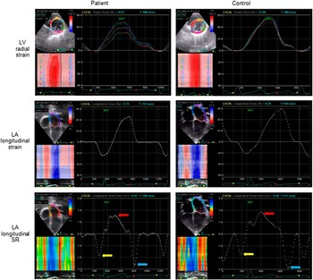
Because some of the patients had abnormal heart locations and morphology, the optimal left ventricular apical two-chamber and three-chamber views could not be acquired from every patient. Therefore, global left and right ventricular longitudinal systolic strain and systolic and diastolic strain rates were determined from the apical four-chamber view, whereas left ventricular circumferential and radial strain and strain rates were assessed in the parasternal short-axis view at the level of the papillary muscle (Fig 1, upper panel). The left and right ventricular free walls and ventricular septum were divided, respectively, into three segments, basal, mid, and apical, to quantify the regional longitudinal strain. Because the ventricular septum contributes to both left and right ventricular global function,Reference Buckberg, Hoffman, Mahajan, Saleh and Coghlan 12 this structure was included in the assessment of left and right ventricular global strain and strain rate. The left ventricular short axis was divided into six segments – anteroseptal, anterior, lateral, posterior, inferior, and septal – to assess regional circumferential and radial strain. The coefficients of variation for intra- and inter-observer measurements were 4.72 and 5.17%, respectively, for left ventricular global longitudinal strain, 5.68 and 6.95%, respectively, for global radial strain, and 5.34 and 7.96%, respectively, for global circumferential strain.
Representative LV radial and LA longitudinal strain and SR curves in a patient and control patient. The LA active contractile strain rate (yellow arrow), conduit strain rate (blue arrow), and reservoir strain rate (red arrow) are highlighted. LA=left atrial; LV=left ventricular; SR=strain rate.

The mechanics of left atrial deformation were also assessed using speckle tracking echocardiography with tracing of the left atrial contours. The onset of the p wave was used as the reference point for determining peak positive strain, peak negative strain, and global strain (Fig 1, middle panel). The left atrial active contractile strain rate, conduit strain rate, and reservoir strain rate were then derived accordingly (Fig 1, lower panel).Reference Debonnaire, Leong and Witkowski 13 The coefficients of variation for intra- and inter-observer measurements were 6.2 and 7.3%, respectively, for atrial global strain, 8.5 and 8.9%, respectively, for active contractile strain rate, 7.3 and 9.5%, respectively, for conduit strain rate, and 7.6 and 10.5%, respectively, for reservoir strain rate.
Statistical analysis
Data are expressed as the means±standard deviations and medians with ranges where appropriate. The absolute values for strain and strain rate were used to facilitate interpretation and analysis. Left and right ventricular dimensions were indexed by body surface area. Differences in demographic and echocardiographic parameters between the groups were compared using an unpaired Student’s t test and Fisher’s exact test where appropriate. Pearson’s correlation analysis was used to study relationships between Cobb’s angle and echocardiographic parameters and between right and left ventricular parameters and left ventricular and atrial parameters. A p-value<0.05 was considered to be statistically significant. All statistical analyses were performed using SPSS version 16.0 (SPSS Inc., Chicago, Illinois, United States of America).
Results
Patients
A total of 23 patients, of which 11 were male, were studied. The median age of the patients was 14 years – with a range from 2 to 22 years – and the median Cobb’s angle was 61° – with a range from 29 to 150°. The age (12.8±5.0 versus 12.8±4.9 years, p=0.97), sex distribution (11M/12F versus 11M/11F, p=0.88), body weight (35.2±12.3 versus 39.2±16.4 kg, p=0.36), and height (140.7±22.3 versus 148±22.4 cm, p=0.28) of the patients were similar between the patients and the controls.
Left ventricular size and mechanics
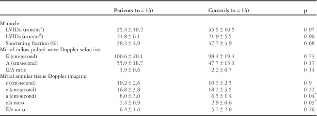
Table 1 summarises the left ventricular echocardiographic parameters of the patients and controls. The M-mode-derived left ventricular dimensions and shortening fraction were similar between the patients and controls (all p>0.05). Doppler studies showed that the patients had a significantly greater annular late diastolic myocardial velocity (p=0.04) and a lower ratio of early to late diastolic myocardial velocities (p=0.03) than the controls; the controls and patients had similar mitral early and late inflow velocities, a similar ratio of mitral early inflow velocity to mitral late inflow velocity, similar annular early diastolic myocardial velocities, and a similar ratio of early diastolic inflow to myocardial velocities (all p>0.05).
Left ventricular M-mode and Doppler parameters.

A=late diastolic inflow velocity; a=late diastolic myocardial velocity; E=early diastolic inflow velocity; e=early diastolic myocardial velocity; LVIDd=left ventricular end-diastolic dimension; LVIDs=left ventricular end-systolic dimension; s=systolic myocardial velocity
* Statistically significant
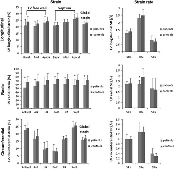
The strain parameters of the left ventricle are shown in Figure 2. Significant differences were observed in the radial dimension but not in the longitudinal and circumferential dimensions. The patients had a significantly lower radial global (p=0.04), segmental systolic strain (anteroseptal p=0.049 and septal p=0.04), and early diastolic strain rate (p=0.03) than the controls.
Comparisons of LV systolic longitudinal, radial and circumferential strain and systolic and diastolic strain rates in patients and controls. Ant=anterior; Antsept=anteroseptal; Inf=inferior; Lat=lateral; LV=left ventricular; Post=posterior; Sept=septal; SRa=late diastolic strain rate; SRe=early diastolic strain rate; SRs=systolic strain rate. *p<0.05 versus controls.

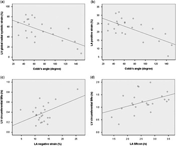
The left ventricular global radial systolic strain of the patients was negatively correlated with their Cobb’s angle (r=−0.65, p=0.001) (Fig 4a).
Right ventricular size and mechanics
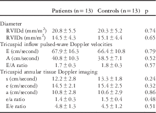
Table 2 summarises the right ventricular echocardiographic parameters in the patients and controls. No significant differences in right ventricular dimensions, pulsed-wave Doppler, and tissue Doppler parameters were found between the patients and the controls.
Right ventricular diameter and Doppler parameters.

A=late diastolic inflow velocity; a=late diastolic myocardial velocity; E=early diastolic inflow velocity; e=early diastolic myocardial velocity; RVIDd=right ventricular end-diastolic dimension; RVIDs=right ventricular end-systolic dimension; s=systolic myocardial velocity
p>0.05
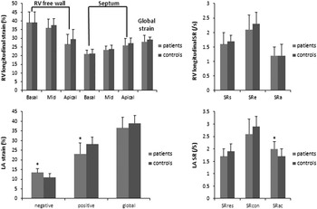
Figure 3 (upper panel) compares the right ventricular longitudinal strain and strain rates of the patients and controls. The patients tended to have lower strain and systolic and early diastolic strain rate, but these differences were not significant (all p>0.05).
Comparisons of RV and LA longitudinal strain and strain rates in patients and controls. LA=left atrial; RV=right ventricular; SR=strain rate; SRa=late diastolic strain rate; SRac=active contractile strain rate; SRcon=conduit strain rate; SRe=early diastolic strain rate; SRres=reservoir strain rate; SRs=systolic strain rate. *p<0.05 versus controls.

The correlation analyses showed no significant correlations between right ventricular parameters and Cobb’s angle.
Left and right ventricular interaction
No significant correlations between left and right ventricular echocardiographic parameters were identified.
Left atrial mechanics
The comparisons of the global atrial strain and strain rates of the patients and controls are shown in Figure 3 (lower panel). The patients had significantly greater atrial negative strain and active contractile strain rate and a lower positive strain than the controls. To identify the role of left atrial active contraction during deformation, the negative/global strain ratio was calculated.Reference Khoo, Smallhorn, Kaneko, Kutty, Altamirano and Tham 14 This strain ratio was significantly greater in the patients than in the controls (0.4±0.1 versus 0.3±0.05, p=0.003).
The Cobb’s angle of the patients was negatively correlated with their left atrial positive strain (r=−0.68, p<0.001) (Fig 4b), global strain (r=−0.5, p=0.02), and conduit strain rate (r=−0.55, p=0.01) and positively correlated with left atrial negative/global strain ratio (r=0.64, p=0.001).
Scatter plots showing correlations between ( a ) Cobb’s angle and LV global radial systolic strain, ( b ) Cobb’s angle and LA positive strain, ( c ) LA negative strain and LV circumferential late diastolic SRa, and ( d ) LA SRcon and LV circumferential SRe. LA=left atrial; LV=left ventricular; SRa=late diastolic strain rate; SRcon=conduit strain rate; SRe=early diastolic strain rate.

Left atrial and ventricular interaction
The left atrial negative strain of the patients was positively correlated with their left ventricular circumferential late diastolic strain rate (r=0.46, p=0.01) (Fig 4c) and left ventricular longitudinal early diastolic strain rate (r=0.42, p=0.01). The left atrial conduit strain rate was positively correlated with the left ventricular circumferential early diastolic strain rate (r=0.42, p=0.03) (Fig 4d) and the left atrial active contractile strain rate was positively correlated with the left ventricular longitudinal late diastolic strain rate (r=−0.4, p=0.03).
Discussion
The present study evaluated left ventricular radial deformation and left atrial mechanics in patients with isolated congenital scoliosis. We found that both of these parameters were correlated with Cobb’s angle, thus providing evidence of abnormal left atrial and ventricular interactions in these patients. To the best of our knowledge, this study is the first to comprehensively evaluate the mechanical performance of and interaction between the both ventricles and the left atrium and to highlight subclinical cardiac abnormalities in patients with congenital scoliosis.
Congenital scoliosis, a curvature of the spine occurring in utero, is often associated with defects in other organs. Cardiac abnormalities commonly occur in patients with this disorder, and a higher prevalence of CHD has been documented in these patients.Reference Beals, Robbins and Rolfe 1 , Reference Basu, Elsebaie and Noordeen 2 Congenital scoliosis is thought to alter the morphology and decrease the volume of the thoracic cavity and compress certain pliable structures, which could influence cardiopulmonary function. Many studiesReference Bowen, Scaduto and Banuelos 6 , Reference McPhail, Howells and Boesch 7 , Reference Day, Upadhyay, Ho, Leong and lp 15 have assessed lung function in patients with congenital scoliosis, and the lung function of every patient is tested before surgical intervention. However, the cardiac function of these patients is often not evaluated. We believe that there are two reasons for this: first, cardiac dysfunction in patients with advanced scoliosis has been hypothesised to be induced by lung disease; second, it is difficult to assess cardiac function accurately in these patients, as their hearts are displaced and tend to have an abnormal morphology.
The cardiac dysfunction mentioned above has been previously defined as right ventricular hypertrophy with or without congestive heart failure secondary to increased pulmonary vascular resistance.Reference Swank, Winter and Moe 16 Our previous study also confirmed that right ventricular dysfunction can occur in patients with severe idiopathic scoliosis.Reference Li, Yang and Li 17 In our present study, none of the patients presented with symptoms of cardiac dysfunction, and the dimensions of their right ventricle and the longitudinal deformation of their myocardium were within normal ranges. However, changes in the left ventricle and left atrium of these patients indicate that mild cardiac functional abnormalities may occur very early in the course of their disease, even when the size and function of their right ventricle are normal.
The correlation between Cobb’s angle and the left ventricular global radial systolic strain suggests that the curvature of the spine may cause left ventricular dysfunction. The myocardial deformation abnormality observed only in the radial dimension and not in the longitudinal dimension further suggests that the shape of the thoracic cavity limits cardiac motion. The decrease in the left ventricular radial early diastolic strain rate and in the ratio of the annular early diastolic myocardial velocity to the late diastolic myocardial velocity indicate impaired left ventricular diastolic function; this impairment could be explained by reduced pulmonary arterial blood flow and a resultant decreased pulmonary venous return causing inadequate left ventricular diastolic filling.Reference Primiano, Nussbaum and Hirschfeld 18 A study by Liang et alReference Liang, Qiu and Shen 19 showed that patients with congenital scoliosis have normal left ventricular function. However, this study only evaluated left ventricular ejection fractions derived from M-mode echocardiography. Our study also showed a normal left ventricular ejection fraction; however, speckle tracking echocardiography and tissue Doppler identified subclinical changes in myocardial performance.
No studies have been conducted on the left atrial function in patients with congenital scoliosis. Our findings have shown that left atrial mechanics are altered significantly in these patients; more specifically, these patients have a significantly decreased positive strain and increased negative strain, negative/global strain ratio and active contractile strain rate. We believe that these results might be explained by the posterior position of the left atrium and its connection to the pulmonary veins. The correlation between left atrial positive strain and Cobb’s angle suggests that patients with congenital scoliosis may experience reduced pulmonary venous return and therefore less left atrial reservoir enlargement or direct compression of the left atrium. Similarly, the significant correlations between left atrial negative strain, conduit strain rate, active contractile strain rate, and left ventricular parameters provide evidence of a significant interaction between the left atria and ventricle. In our study, the patients presented with significantly greater left atrial negative strain, negative/global strain ratio, and active contractile strain rate than the controls, which indicates an increased reliance on active left atrial contraction for left ventricular filling. These changes may also represent a compensatory mechanism associated with left ventricular diastolic dysfunction.
The difficulties associated with the evaluation of cardiac function in patients with congenital scoliosis can be overcome with the use of speckle tracking echocardiography, as this modality can track myocardial motion regardless of cardiac location or morphology.Reference Leitman, Lysyansky and Sidenko 20 In our study, speckle tracking echocardiography and complete analyses with low variability could be performed in all but two of the patients.
Congenital scoliosis is increasingly being detected in utero;Reference Prada, Sellars, Spaeth, Kline-Fath, Crombleholme and Hopkin 21 therefore, strategies to comprehensively assess patients with this disorder are needed. We found that patients with congenital scoliosis may have abnormal left ventricular and left atrial mechanics; this finding may provide evidence that there are early markers of subclinical heart dysfunction that can be evaluated when considering early surgical or medical interventions.
Limitations
Several limitations to this study warrant discussion. First, the cross-sectional nature of the present study does not provide information on the time course during which abnormal cardiac function develops. Second, we assessed left ventricular longitudinal myocardial deformation in the apical four-chamber view only and radial and circumferential myocardial deformation at the level of the papillary muscle; thus, some segments of the heart may not have been detected. Third, patients with clinical heart failure were not enrolled. Finally, the need for high-quality echocardiographic images made speckle tracking techniques22 ,23 more challenging in this patient cohort; however, most of the cases in our study could be satisfactorily tracked.
Conclusions
Impaired left ventricular and altered left atrial mechanics occur relatively early in patients with congenital scoliosis and the extent of these changes are correlated with the severity of their scoliosis. Our findings provide evidence of preclinical heart dysfunction in patients with congenital scoliosis.
Acknowledgement
We thank Prof. Huishen Wang for initiating the study and critically reviewing the manuscript.
Financial Support
Our study was supported by grants from the Guangdong Natural Science Foundation (S2012010008338) and Educational Foundation (20130171110048).
Conflicts of Interest
None.
Ethical Standards
The authors assert that all procedures performed in this work comply with the ethical standards of the Chinese National Guidelines on Human Experimentation and with the Helsinki Declaration of 1975, as revised in 2008; this study was also approved by our hospital institutional committees.