Introduction
Spiculopteragia houdemeri (Schwartz, Reference Schwartz1926) (syn. S. yamashitai Ohbayashi, Reference Ohbayashi1966) is an abomasal trichostrongylid recorded from members of the Cervidae family in East Asia, such as barking deer (Muntiacus muntjak) and sambar (Rusa unicolor) in Vietnam (Dróżdż, Reference Dróżdż1965, Reference Dróżdż1973), and sika deer (Cervus nippon) in Japan (Ohbayashi, Reference Ohbayashi1966; Yamaguchi et al., Reference Yamaguchi, Kamiya and Kudo1977; Kitamura et al., Reference Kitamura, Yokohata, Suzuki and Kamiya1997). This species has also been recorded from sika deer introduced into Europe (Rehbein et al., Reference Rehbein, Visser and Lutz2003; Rehbein & Visser, Reference Rehbein and Visser2007). Most of these recordings reported the dominance of this species in the abomasum with a minor or scarce population of another Ostertagiinae species, S. andreevae (Dróżdż, Reference Dróżdż1965) (syn. Rinadia andreevae Dróżdż, Reference Dróżdż1965 and R. japonica Ohbayashi, Reference Ohbayashi1966). Dróżdż (Reference Dróżdż1995) described such a consistent co-occurrence, or specific combination, of two species of the subfamily Ostertagiinae (Trichostrongylidae). Based on the common co-occurrence of a dominant (major) and a scarce (minor) species, significant correlation between the intensity of infection of these two species, and regular recovery of monomorphic females, Dróżdż compiled a list of 19 polymorphic species (special combination of major and minor morphs) of the genera Ostertagia, Orloffia, Teladorsagia, Marshallagia and Spiculopteragia. Failure of the genetic isolation of, or experimental hybridization between Teladorsagia circumcincta, T. trifurcata and T. davtiani (Suarez & Cabaret, Reference Suarez and Cabaret1992), and pure infection experiments of lambs with T. circumcincta (Copland, Reference Copland1965; Morales & Cabaret, Reference Morales and Cabaret1985) or of calves with Cooperia oncophora (Isenstein, Reference Isenstein1971), support the idea that major and minor morphs actually represent a single species. Indeed, male dimorphism or polymorphism of a single species can often be seen in the Ostertagiinae subfamily (Lancaster & Hong, Reference Lancaster and Hong1981; Lichtenfels & Hoberg, Reference Lichtenfels and Hoberg1993; Lichtenfels et al., Reference Lichtenfels, Hoberg and Zarlenga1997).
Recent molecular techniques such as allozyme analyses, or genetic analyses of the internal transcribed spacer (ITS) region of the ribosomal RNA gene (rDNA) or of mitochondrial DNA (mtDNA) genes also support the aforementioned hypothesis (Andrews & Beveridge, Reference Andrews and Beveridge1990; Gasnier et al., Reference Gasnier, Cabaret and Suarez1993; Humbert & Cabaret, Reference Humbert and Cabaret1995; Stevenson et al., Reference Stevenson, Gasser and Chilton1996; Zarlenga et al., Reference Zarlenga, Hoberg, Stringfellow and Lichtenfels1998; Dallas et al., Reference Dallas, Irvine and Halvorsen2000; Santín-Durán et al., Reference Santín-Durán, de la Fuente, Alunda, Rosenthal and Hoberg2002; Liénard et al., Reference Liénard, Depaquit and Ferté2006). Furthermore, cryptic species have been noted in such polymorphic species in the Ostertagiinae subfamily irrespective of morphotype, e.g. T. circumcincta, by use of certain molecular markers, allozyme profiles, morphology and host preference (Gasnier & Cabaret, Reference Gasnier and Cabaret1996; Leignel et al., Reference Leignel, Cabaret and Humbert2002).
In the present study, all possible morphs of male and female Spiculopteragia were collected from the abomasa of sika deer and Japanese serows (Capricornis crispus) on the mainland of Japan, i.e. Honshu and Shikoku Islands. Representatives of all the morphological variants were subjected to genetic analyses based on the ITS region of rDNA and the cytochrome c oxidase subunit I (COI) region of mtDNA.
Materials and methods
Collection and examination of ruminants for parasites
Sika deer (total number = 181) were collected in the western part of the mainland of Japan, Honshu Island: 132 deer from Hyogo Prefecture during the period 15 July 2009 to 19 May 2010; 24 deer from Wakayama Prefecture during the period 25 March 2009 to 14 April 2010; and 25 deer from Yamaguchi Prefecture during the period 6 September 2009 to 18 April 2010. The body weight of the deer ranged from 15 to 65 kg. The deer population in Japan has been increasing in recent years, causing ecological and social problems. Most of the collected deer were hunted under a control programme for harmful birds and mammals established by each prefecture, and the rest were killed by road traffic accidents. Six Japanese serows killed by road traffic accidents were collected in Shikoku Island (Tokushima and Kochi Prefectures) and Honshu Island (Wakayama Prefecture) during the period 18 March 2009 to 11 May 2011.
Frozen viscera were transported to the parasitology laboratory, and helminth parasites were collected according to standard procedures. Briefly, organs were separated, placed in different vessels, opened and washed in tap water. Following repeated washings, the sediments were checked under a dissection microscope. Collected parasites were washed in physiological saline, and preserved in either neutral-buffered 10% formalin or 70% alcohol. In addition to the nematode samples from Japanese wild ruminants, the following three trichostrongylid species were examined: (1) Haemonchus longistipes collected from the abomasum of an Arabian camel (Camelus dromedarius) in Cairo, Egypt (Albastin abattoir, August 2009); (2) H. contortus collected from the abomasum of a sheep (Ovis aries) near Cairo, Egypt (El-mahalla El-Kurba abattoir, September 2009); and (3) Ostertagia ostertagi collected from the abomasum of a Holstein-Friesian breed cow (Bos taurus) at an abattoir in Yamaguchi, Japan (3 April 2010).
The collected parasites were categorized by sex and certain morphological characters under a dissection microscope. Intensive morphological analysis of formalin-fixed specimens of S. houdemeri was performed under a differential interference contrast microscope. Figures were drawn with the aid of a camera lucida. Measurements were performed on these drawn figures using a digital curvimeter type S (Uchida Yoko, Tokyo, Japan) when necessary. Observations of the synlophe at midbody were made on cross-sections of worms obtained by free-hand cuts made with a disposable blade and mounted in glycerin jelly. The collected specimens were deposited in the National Science Museum, Tokyo, Japan under specimen numbers NSMT-As3698–As3901.
DNA extraction, polymerase chain reaction and sequencing
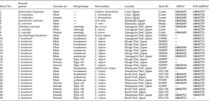
Male and female morphological variants of different origins (table 1) were cut into three parts, and the anterior and posterior parts were kept in 70% alcohol. The middle part of each worm was used for DNA extraction using an Illustra™ tissue and cells genomicPrep Mini Spin Kit (GE Healthcare UK, Buckinghamshire, UK), according to the manufacturer's instructions. Polymerase chain reaction (PCR) amplification of the entire ITS1, 5.8S and ITS2 region of rDNA was performed using a combination of a forward primer (NC5; 5′-GTAGGTGAACCTGCGGAAGGATCATT-3′) and a reverse primer (NC2; 5′-TTAGTTTCTTTTCCTCCGCT-3′) flanking the 3′-terminus of 18S and the 5′-terminus of 28S, respectively, according to Newton et al. (Reference Newton, Chilton, Beveridge, Hoste, Nansen and Gasser1998). PCRs were conducted in a thermal cycler using the following cycling protocol: 4 min at 94°C, then 40 cycles at 94°C for 30 s, 52°C for 45 s, and 72°C for 1 min, followed by a final extension at 72°C for 10 min. PCR products for sequencing were purified using a High Pure PCR Cleanup Micro Kit (Roche Diagnostics GmbH, Mannheim, Germany). After direct sequencing of PCR amplicons using the two amplification primers and another primer (NC13; 5′-ATCGATGAAGAACGCAGC-3′), sequences were assembled manually with the aid of the CLUSTAL W multiple alignment programme (Thompson et al., Reference Thompson, Higgins and Gibson1994). For a part of the rDNA segments, the amplicon was cloned into a plasmid vector, pTA2 (Target Clone™; Toyobo, Osaka, Japan), and transformed into Escherichia coli JM109 (Toyobo), according to the manufacturer's instructions. Following propagation, plasmid DNA was extracted using a NucleoSpin® Plasmid kit (Macherey-Nagel GmbH, Düren, Germany), and inserts from multiple independent clones were sequenced using universal M13 forward and reverse primers. Four or more clones were sequenced for each worm.
Nematodes used for genetic analyses.

* Accession no. at the DDBJ/EMBL/GenBank databases.
The COI region of parasite mtDNA was amplified by a primer pair of BpCoxI-F1 (5′-TTTGGTCATCCTGAGGTTTATATT-3′) and BpCoxI-R1 (5′-ATGAAAATGTCTAACTACATAATAAGTATC-3′) designed in-house. PCRs were conducted in a thermal cycler using the following cycling protocol: 3 min at 94°C, then 40 cycles at 94°C for 45 s, 52°C for 1 min and 72°C for 1 min, followed by a final extension at 72°C for 7 min. In addition to direct sequencing of amplicons, a part of them was sequenced after genomic cloning as detailed above.
The nucleotide sequences reported in the present study are available in the DDBJ/EMBL/GenBank databases under the accession numbers AB682683–AB682732 (table 1).
Phylogenetic analysis
For phylogenetic analysis, the newly obtained ITS2 rDNA sequences for different morphotypes of S. houdemeri and those of related trichostrongylid species obtained from the DDBJ/EMBL/GenBank databases were aligned using the CLUSTAL W multiple alignment program (Thompson et al., Reference Thompson, Higgins and Gibson1994), with subsequent manual adjustment. Regions judged to be poorly aligned, and characters with a gap in any sequences, were excluded from subsequent analyses (Smythe et al., Reference Smythe, Sanderson and Nadler2006); 213 characters, of which 94 were variable, remained for subsequent analysis. Maximum likelihood (ML) analysis was performed with the program PhyML (Guindon & Gascuel, Reference Guindon and Gascuel2003; Dereeper et al., Reference Dereeper, Guignon, Blanc, Audic, Buffet, Chevenet, Dufayard, Guindon, Fefort, Lescot, Claverie and Gascuel2008) provided on the ‘phylogeny.fr’ website (http://www.phylogeny.fr/). The probability of inferred branches was assessed by the approximate likelihood ratio test (aLRT), an alternative to the non-parametric bootstrap estimation of branch support (Anisimova & Gascuel, Reference Anisimova and Gascuel2006). Similarly, the newly obtained COI sequences of morphotypes of S. houdemeri and those of related trichostrongylid species were analysed; 316 characters, of which 111 were variable, were used in this analysis.
Results
Morphology of S. houdemeri with different origins
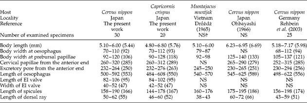
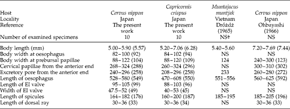
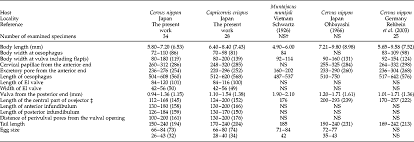
Male specimens of Spiculopteragia were divided into two morphotypes, i.e. morphs houdemeri and andreevae, corresponding to previous records of S. houdemeri (Schwartz, Reference Schwartz1926) (syn. S. yamashitai) and S. andreevae (Dróżdż, Reference Dróżdż1965) (syn. R. andreevae, R. japonica) from Asian cervids or wild bovids (Dróżdż, Reference Dróżdż1965; Ohbayashi, Reference Ohbayashi1966). Morph andreevae was characterized by a short, conical, thick dorsal lobe supported by a short dorsal ray (which was not bifurcated except for its distal end), absence of an accessory dorsal membrane and massive spicule structure, being longer, thicker and trifurcated compared with morph houdemeri spicules which were shorter, thinner and bifurcated. Except for these morphological differences, which assigned morph andreevae to the genus Rinadia in the past (Dróżdż, Reference Dróżdż1965; Ohbayashi, Reference Ohbayashi1966), there were no additional morphological differences between two genera, Spiculopteragia and Rinadia (Durette-Desset, Reference Durette-Desset, Anderson, Chabaud and Willmott2009). Previously described morphometric characters of each male morph and females collected from sika deer and Japanese serows were compared. There were no critical differences in morphometry irrespective of their origin (tables 2–4). The synlophes at the midbody of the male morphs houdemeri and andreevae and the three morphotypes of adult females, mentioned below, were identical. The lengths of the oesophageal–intestinal valve, which extends from the posterior end of the cuticular lining of the triradiate lumen of the oesophagus to the posterior end of the oesophagus (defined by Lichtenfels & Pilitt, Reference Lichtenfels and Pilitt1991) were identical regardless of male morph or host origin.
Morphometrics of morph houdemeri males of Spiculopteragia houdemeri *.
EI, oesophageal–intestinal.
* Range with average in parenthesis. All measurements are expressed in μm unless otherwise indicated.
† NS, Not specified.
Morphometrics of morph andreevae males of Spiculopteragia houdemeri *.
EI, oesophageal-intestinal.
* Range with average in parenthesis. All measurements are expressed in μm unless otherwise indicated.
† Not specified.
Morphometrics of adult females of Spiculopteragia houdemeri *.
* Range with average in parenthesis. All measurements are expressed in μm unless otherwise indicated.
† Not specified.
‡ Total length of the vestibule and sphincters at both sides.
Occurrence of male and female morphs
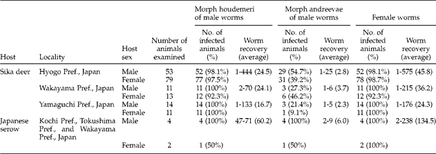
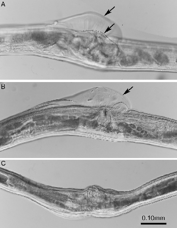
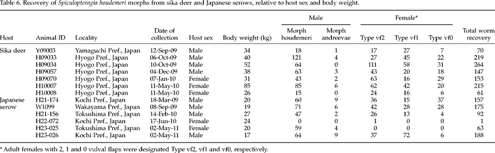
The recovery of male and female worms of S. houdemeri from 181 sika deer and six Japanese serows is summarized in table 5. Infection rate and recovery of female worms as well as morph houdemeri male worms were consistently high in both sika deer and Japanese serows, whereas those of morph andreevae male worms were lower or fewer. Adult females were divided into three morphotypes according to vulval ornamentation: Type vf2, with two flaps; Type vf1, with one flap; and Type vf0, flapless (fig. 1). Females of Types vf2 and vf1 were prominent, approximately 30–60% and 20–50% of classified females, respectively, regardless of host species. Type vf0 females were less common compared to Types vf2 and vf1, approximately 10–30%. Representative states of parasitism of 7 deer and 6 serows with different morphs are shown in table 6.
Recovery of Spiculopteragia houdemeri from sika deer and Japanese serows.
Three morphotypes of adult females of Spiculopteragia houdemeri with two vulval flaps (A), one vulval flap (B) and no vulval flap (C). Arrows denote the vulval flap.
Recovery of Spiculopteragia houdemeri morphs from sika deer and Japanese serows, relative to host sex and body weight.
* Adult females with 2, 1 and 0 vulval flaps were designated Type vf2, vf1 and vf0, respectively.
Genetic characterization of male and female morphotypes
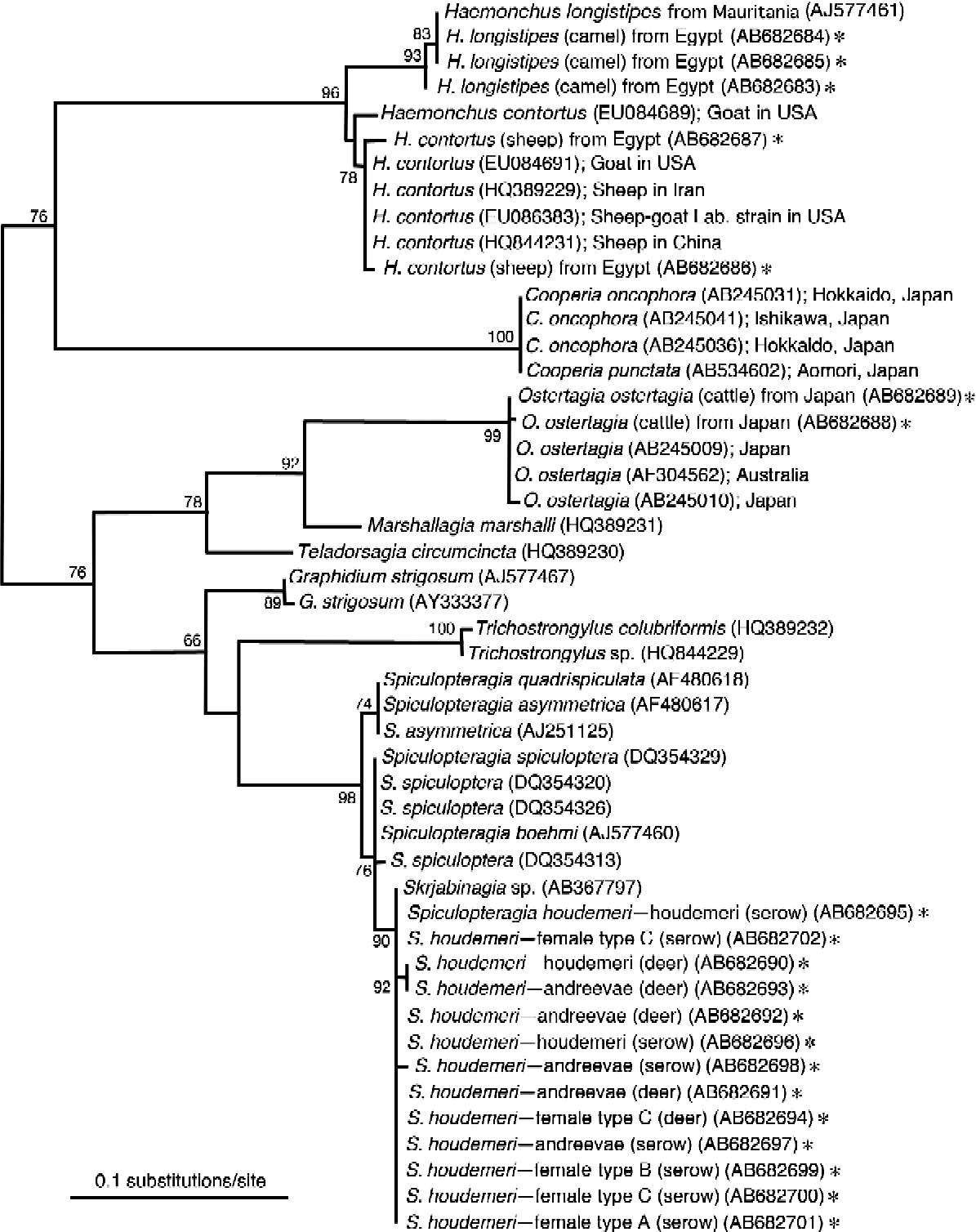
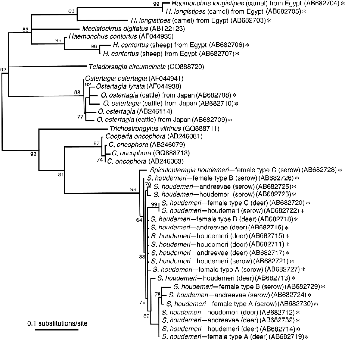
The ITS1, 5.8S and ITS2 sequences of all morphs of S. houdemeri were 383/385-bp, 152-bp and 241-bp long, respectively. Nucleotide substitutions in such regions were rather rare among the S. houdemeri morphs, including ‘Skrjabinagia sp. Iwate-01’ (syn. of S. houdemeri) which was collected from a Japanese serow in the northern part of Japan (DDBJ/EMBL/GenBank accession number AB367797). Due to higher nucleotide variations in ITS1 regions by different genera of the subfamily Ostertagiinae or the family Trichostrongylidae, only the ITS2 regions, which showed suitable variation for phylogenetic analysis, were used (fig. 2). All Spiculopteragia spp. formed a single clade, in which all morphs of S. houdemeri clustered in a single group, whereas S. quadrispiculata and S. asymmetrica, and S. spiculoptera and S. boehmi made two independent groups. Similarly, the partial COI region of mtDNA (423-bp long) showed almost identical nucleotide sequences (fig. 3), but with a few amino-acid substitutions. These changes, however, occurred irrespective of special morphotype or geographical origin.
An unrooted maximum likelihood (ML) phylogenetic tree based on the ITS2 region of rDNA sequence. Branch support scores calculated by aLRT (see text) and expressed after multiplication by 100 are given at nodes. Asterisks denote newly obtained sequences in the present study.
An unrooted ML phylogenetic tree based on the COI mtDNA sequence. Branch support scores calculated by aLRT and expressed after multiplication by 100 are given at nodes. Asterisks denote newly obtained sequences in the present study.
We simultaneously analysed H. contortus from a sheep in Egypt, H. longistipes from a camel in Egypt, and Ostertagia ostertagi morph ostertagi, all of which were morphologically characterized according to previous reports (Skrjabin et al., Reference Skrjabin, Shikhobalova, Shul'ts and Skrjabin1954; Lichtenfels & Hoberg, Reference Lichtenfels and Hoberg1993). All of these morphologically characterized specimens clustered with the specified species collected in countries other than Egypt in the phylogenetic tree based on either ITS2 or COI (figs 2 and 3).
Discussion
Certain taxa of the subfamily Ostertagiinae (Nematoda: Trichostrongylidae) have been shown to be polymorphic (Isenstein, Reference Isenstein1971; Morales & Cabaret, Reference Morales and Cabaret1985; Lichtenfels et al., Reference Lichtenfels, Pilitt and Lancaster1988, Reference Lichtenfels, Hoberg and Zarlenga1997; Andrews & Beveridge, Reference Andrews and Beveridge1990; Lichtenfels & Hoberg, Reference Lichtenfels and Hoberg1993), thus complicating trichostrongylid systematics. The present molecular genetic study confirms that ‘S. houdemeri’ and ‘S. andreevae’ are substantially conspecific, as indicated by Dróżdż (Reference Dróżdż1995) based solely on their co-occurrence in the same host individual and monomorphic females. This combination of Spiculopteragia spp. is the third genetically confirmed conspecific pair in the genus, coming after S. asymmetrica and S. quadrispiculata, and S. spiculoptera and S. mathevossiani (Santín-Durán et al., Reference Santín-Durán, de la Fuente, Alunda, Rosenthal and Hoberg2002; Liénard et al., Reference Liénard, Depaquit and Ferté2006).
The dorsal lobe and ray along with the morphology of the spicules clearly divide male S. houdemeri into morphs houdemeri and andreevae, whereas adult females exhibit uniform morphology except for vulval ornamentation, i.e. vulval flaps. We detected three types of females previously described by Ohbayashi (Reference Ohbayashi1966). As shown in table 6, there is no relationship between their occurrence and the occurrence of the different male morphs, i.e. houdemeri and andreevae. Michel (Reference Michel1967) and Michel et al. (Reference Michel, Lancaster and Hong1972) have shown that the development of vulval flaps of Ostertagia ostertagi is affected by an acquired immunity of the host to the parasite. Other researchers have also demonstrated that proportional changes of worms with different vulval flaps or ornamentations occur following deterioration of the niche for the parasite (Waller & Thomas, Reference Waller and Thomas1978; Lancaster & Hong, Reference Lancaster and Hong1981), while Le Jambre & Royal (Reference Le Jambre and Royal1977) suggested a genetic background for different vulval phenotypes. The synlophes of all male and female morphs have an identical pattern, 26–37 ridges, resembling that drawn for S. spiculoptera by Durette-Desset & Cabaret (Reference Durette-Desset and Cabaret1994).
Beyond the male polymorphism of several species of the subfamily Ostertagiinae, cryptic species of the T. circumcincta complex have been noted irrespective of morphotype using certain molecular markers, allozyme profiles, morphology and host preference (Gasnier & Cabaret, Reference Gasnier and Cabaret1996; Gasnier et al., Reference Gasnier, Cabaret and Durette-Desset1997; Leignel et al., Reference Leignel, Cabaret and Humbert2002). Our genetic examination presented here using a whole length of the ITS region of rDNA as well as a partial region of the COI mtDNA gene reveals no such cryptic species or special strains in our S. houdemeri samples.
Several parasitological and pathological examinations of Japanese serows have been conducted, and the following helminth species were recorded: Protostrongylus shiozawai, S. houdemeri, Okapinema japonica, Skrjabinema sp., Trichuris discolor, five Cercopithifilaria spp. (C. shohoi, C. multicauda, C. minuta, C. tumidicervicata and C. bulboidea), Dicrocoelium dendriticum, D. chinensis, Ogmocotyle capricorni and Moniezia monardi (Machida, Reference Machida1970; Machida et al., Reference Machida, Matsubura and Watanabe1974; Ohbayashi & Ueno, Reference Ohbayashi and Ueno1974; Wang et al., Reference Wang, Shiozawa, Akabane and Fujita1976; Suzuki et al., Reference Suzuki, Sugimura, Kamiya, Fujita, Yoshizawa, Kamiya and Ohbayashi1981; Yagi, Reference Yagi1982; Uni et al., Reference Uni, Suzuki, Baba, Mitani, Takaoka, Katsumi and Bain2001). Although Japanese serows have several unique helminth species, S. houdemeri (morphs houdemeri and andreevae) was found to have an identical genetic background to the S. houdemeri isolated from sika deer in Japan, with consistent growth and morphological variations in both host species.
Phylogenetic analysis of major taxa within the family Trichostrongylidae produced trees with different topologies, depending on the gene used for analysis, i.e. the ITS2 region of rDNA sequence (fig. 2) and COI mtDNA (fig. 3). Studies by other researchers have provided phylogenetic trees with the same topology as the present study when targeted genes are identical (Chilton et al., Reference Chilton, Newton, Beveridge and Gasser2001). The precise relationship of trichostrongylid genera is still not clear, but might be assessed by the addition of more sequences of congeners in the family in the future.
Acknowledgements
The authors are grateful to the Egyptian Government for the scholarship to K.S. and M.O., and the Japanese Government for the scholarship to P.M. This study was supported in part by Grants-in-Aid from the Japan Society for the Promotion of Science (nos. 19580355, 20570090, and 22580349).