Introduction
The roundworm Toxocara canis is an intestinal parasite of canids that can also infect paratenic hosts (rodents, but also humans) in its life cycle. In the intestine of the paratenic hosts, the larvae hatch from ingested eggs or are released from ingested tissue of paratenic hosts, actively penetrate intestinal walls and during hepato-pulmonary phase migrate through the bloodstream to the liver and then to the lungs. From there, larvae migrate to various tissues in whole body with different distribution according to species-dependent predilection sites (Strube et al. Reference Strube, Heuer and Janecek2013); however, nearly all organs may be affected with various amount of larvae. In the paratenic hosts, the parasite is either captured and eliminated by the immune system or the encapsulated larvae can survive for years (Maizels Reference Maizels2013). The lifecycle continues if the tissue of the paratenic host with larvae is ingested by the definitive host (Strube et al. Reference Strube, Heuer and Janecek2013). This mechanism enables the parasite to spread in space and time.
Long-term infections of humans can cause either symptomatic toxocarosis with four defined types – visceral larva migrans with organ-specific symptoms, ocular larva migrans when the eyes of the paratenic host are affected, covert toxocarosis with non-specific symptoms, and neurotoxocarosis/neural larva migrans connected with the invasion of larvae to the central nervous system (CNS) (Bardón et al. Reference Bardón, Cuéllar and Guillén1994; Fan et al. Reference Fan, Liao and Cheng2013; Chou et al. Reference Chou, Lee, Liao, Huang and Fan2017; Macpherson Reference Macpherson2013; Magnaval et al. Reference Magnaval, Glickman, Dorchies and Morassin2001; Strube et al. Reference Strube, Heuer and Janecek2013), or is widely considered asymptomatic, with no visible symptoms or health problems experienced by the patient.
The number of severe diagnoses is relatively low compared to the relatively high seroprevalence in the human population (19%) (Ma et al. Reference Ma, Holland, Wang, Hofmann, Fan, Maizels, Hotez and Gasser2018; Strube et al. Reference Strube, Waindok, Raulf and Springer2020). In general, higher seroprevalence is seen in tropical and subtropical countries, in rural settlements, and in populations with lower socioeconomic and hygiene standards (Ma et al. Reference Ma, Rostami, Wang, Hofmann, Hotez and Gasser2020; Rostami et al. Reference Rostami, Riahi, Holland, Taghipour, Khalili-Fomeshi, Fakhri, Omrani, Hotez and Gasser2019). When diagnosed, the severity of the infection is related to the location and number of larvae deposited (Hanh et al. Reference Hanh, Lee, Lin, Chou, Cheng, Quang and Fan2020; Pinelli et al. Reference Pinelli, Brandes, Dormans, Fonville, Hamilton and van der Giessen2007). Of all types of toxocarosis, the neural larva migrans is the scarcest (Deshayes et al. Reference Deshayes, Bonhomme and de La Blanchardière2016). However, some authors suggest possible connection between the presence of parasite in the CNS and development of neurodegenerative or neuropsychiatric diseases, such as multiple sclerosis or Alzheimer’s disease, supported by the detection of enhanced expression of associated biomarkers (e.g., glial fibrillary acidic protein, neurofilament light, or phosphorylated tau) in toxocara-infected mice (Liao et al. Reference Liao, Fan, Kao, Ji, Su, Lin and Cho2008; Söndergaard & Theorell Reference Söndergaard and Theorell2004).
Studies on animals experimentally infected with Toxocara eggs or larvae under defined conditions have helped to connect the impact of this infection on behaviour, cognition, and motor abilities of paratenic hosts (Cox & Holland Reference Cox and Holland2001a; Chou et al. Reference Chou, Lee, Liao, Huang and Fan2017; Janecek et al. Reference Janecek, Waindok, Bankstahl and Strube2017). In these studies, a wide range of T. canis infection doses (ID) of 5 to 3000 eggs/larvae per mouse were used by various authors (Cox & Holland Reference Cox and Holland2001b; Havasiová-Reiterová et al. Reference Havasiová-Reiterová, Tomašovicová and Dubinský1995; Holland & Hamilton Reference Holland and Hamilton2013; Kim et al. Reference Kim, Jin and Woo2024; Ollero et al. Reference Ollero, Fenoy, Cuéllar, Guillén and Del Aguila2008), with the most commonly used ID of 1000 T. canis eggs/larvae known for reliable neurological symptoms induction in mice (Dunsmore et al. Reference Dunsmore, Thompson and Bates1983; Holland & Hamilton Reference Holland and Hamilton2013; Janecek et al. Reference Janecek, Waindok, Bankstahl and Strube2017). Moderate or low ID infections are not that commonly used, although they are more similar to natural infections in wild mice (Holland & Hamilton Reference Holland and Hamilton2013). Furthermore, severe impact of lower IDs (250 eggs/mouse) was also detected, in combination with a longer period of 67 weeks of infection (Fan et al. Reference Fan, Lin, Du and Su2003; Fan et al. Reference Fan, Lin, Hung, Chang and Su2004a). The effect of infection over a longer time perspective has not been studied in detail. Therefore, our study focused on monitoring physical fitness, motor parameters and pathological changes in the brains of mice infected with low, medium and high ID T. canis for up to 100 weeks post infection (p.i.).
Material and methods
Material for infection and serological studies
Adults of Toxocara canis were obtained from the faeces of naturally infected shelter dogs previously treated with one tablet per 10 kilograms of praziquantel (Drontal, Bayer, Germany).
The unembryonated eggs were isolated from female worms and matured in vitro using the method by Kolbeková et al. (Reference Kolbeková, Kolářová, Větvička and Syrůček2011a). Afterwards, the L3 larvae obtained from fully embryonated eggs according to Fan et al. (Reference Fan, Lan, Hung, Chung, Liao, Du and Su2004b) were kept in RPMI 1640 medium (R8758, Sigma-Aldrich, Germany) with 100 IU/mL penicillin and 250 μg/mL streptomycin at 37 °C, with regular weekly change of the medium, until used in experimental infections or for the collection and preparation of larval excretory-secretory products (TcES).
The identity of the parasite was confirmed by polymerase chain reaction (PCR) technique, performed in the National Reference Laboratory for Tissue Helminthoses, General University Hospital in Prague. The sample of larvae in medium was mechanically ground and isolated using the QIAamp DNA Mini Kit (Qiagen, Hilden, Germany) according to the manufacturer’s instructions and stored at –20 °C until use. The internal transcribed spacer 2 was selected as a target for the amplification to distinguish T. canis and Toxascaris leonina (Jacobs et al. Reference Jacobs, Zhu, Gasser and Chilton1997; Li et al. Reference Li, Lin, Chen, Sani, Song and Zhu2007). For the PCR were used two forward primers specific for T. canis (5´-AGTATGATGGGCGCGCCAAT-3´) and T. leonina (5´-CGAACGCTCATATAACGGCATACTC-3´), and a common reverse primer for both species (5´-TTAGTTTCTTTTCCTCCGCT-3´) (de Moura et al. Reference de Moura, de Macedo, da Silva Terto, da Costa Avila, Leite, Scaini, Pinto, de Almeida Capella, Strothmann and Villela2018; Jacobs et al. Reference Jacobs, Zhu, Gasser and Chilton1997). The total volume of 25 μL reaction mixture contained 2.5 μL of extracted DNA as template, 0.5 μL of each primer, 0.5 μL of nucleotides, 0.25 μL of Taq polymerase (Qiagen), 2.5 μL of reaction buffer and distilled water. The reaction was initiated at 94 °C for 2 min followed by 30 cycles of denaturation at 94 °C for 30 s, annealing at 60 °C for 30 s and extension at 72 °C for 1 min, with the last extension at 72 °C for 7 min. Agarose gel electrophoresis was used for visualisation (Garibyan & Avashia Reference Garibyan and Avashia2013).
For the collection, preparation, and determination of protein concentration of TcES, the protocol by Novák et al. (Reference Novák, Panská, Macháček, Kolářová and Horák2017) was used. The antigen aliquots with a protein concentration of 0.0875 μg/mL were stored at –20 °C until used for enzyme-linked immunosorbent assay (ELISA; see the following section).
Experimental animals and infection
Altogether, 54 specific-pathogen-free inbred female BALB/c mice (Velaz Ltd., Prague, Czech Republic), aged 4 weeks, were used in this experiment for their susceptibility to infections and good comparison to up-to-date knowledge (Hamilton et al. Reference Hamilton, Stafford, Pinelli and Holland2006; Janecek et al. Reference Janecek, Beineke, Schnieder and Strube2014; Petit et al. Reference Petit, Diagne, Marechal, Owen, Taylor and Bain1992). Mice were divided into four experimental groups; three groups of 15 mice each were infected with various T. canis ID, the uninfected control group consisted of nine animals. The larvae were microscopically checked and adjusted to the decided quantity for the defined ID of 10 (low), 100 (moderate), or 1000 larvae (high) per mouse for the experimental infections of animals. Mice were administered orally with 8–30 μL of saline buffer per mouse containing the defined T. canis ID. Group G/10 was infected with 10 T. canis larvae per mouse, G/100 with 100 T. canis larvae per mouse, and G/1000 was infected with 1000 T. canis larvae per mouse. The remaining nine control mice (G/0) received 10 μL of tap water and were maintained under the same conditions as the infected groups.
Mice were housed in an animal facility under standard conditions in multiple cages, with commercial pellet food, water ad libitum, and a 12-h light/dark cycle. In each cage, five infected mice were kept together with one control mouse for check of equal housing conditions.
ELISA
Whole blood was collected from the tail tips of all experimental animals at week 16 p.i. and sera were stored at –20 °C until used. Additionally, the blood of five randomly selected mice from each group was collected on the following timeline: before infection and at weeks 1, 2, 3, 4, 12, 16, 24, 28, 32, 36, 41, 44, 48, 61, and 81 p.i. by the same process. The mouse infection was assessed by measuring specific anti-TcES immunoglobulin G (IgG) antibodies by indirect ELISA according to Novák et al. (Reference Novák, Panská, Macháček, Kolářová and Horák2017). Briefly, the wells of microplates (NUNC Maxi Sorp, Massachusetts, USA) were coated with 100 μL of bicarbonate coating buffer (pH 9.6) containing TcES (final protein concentration 0.0875 μg/mL; 1 h, 37 °C), followed by overnight incubation at 4 °C. Blocking of free binding sites was performed using of 1% bovine serum albumin (BSA) in phosphate-buffered saline (PBS) containing 0.02% Tween 20 (PBS/T/BSA) for 1 h at room temperature (RT). Afterward, all wells were washed in three cycles by PBS with 0.05% Tween 20 (PBS/T), 100 μL of mice sera diluted at 1:800 in PBS/T/BSA were incubated (1 h, RT) and washed. TcES-specific IgGs were detected by 100 μL peroxidase-conjugated goat anti-mouse IgG (ab6823, Abcam, UK) diluted at 1:10,000 in PBS/T/BSA (1 h, RT). Wells were washed 4× and visualized using 0.04% o-phenylenediamine dihydrochloride with 0.012% H2O2 in citrate buffer. The reaction was stopped after 25 min by adding 100 μL of 2M H2SO4. Optical density was measured at 492 nm and analysed in GraphPad Prism (version 8) using one-way ANOVA with Tukey´s multiple comparisons test for data on sera from week 16 p.i., and two-way ANOVA with mixed-effects analysis for data on sera from course of infection. The cut-off was stated as the mean optical density + 3 standard deviations, according to Frey et al. (Reference Frey, Di Canzio and Zurakowski1998).
Examination of animals and evaluation of general appearance and motor/balance skills
All mice were examined continuously starting from the day of initial infection until the end of the experiment (i.e., week 100 p.i.).
The weights of all mice were measured at 2-week intervals using analytic laboratory scales. The mean weight and standard deviation for each group were measured every examined week. Assessment of general appearance, activity, and motor/balance abilities, as well as testing on motor tests, were performed at weeks 4, 20, 33, 40, 44, 57, 60, 80, 92, 96, and 100 p.i. The animals were placed in a disinfected plastic container in an environment covered with a tight-fitting cloth to limit any visual stimuli. For 10 min, animals were observed and scored for the following parameters of general condition and obvious motor/balance abilities: (1) overt cachexia, defined as loss of body weight and muscle mass (Evans et al. Reference Evans, Morley, Argilés, Bales, Baracos, Guttridge, Jatoi, Kalantar-Zadeh, Lochs and Mantovani2008; Fok & Kassai Reference Fok and Kassai1998); (2) disheveled hair (Hermes et al. Reference Hermes, Ajioka, Kelly, Mui, Roberts, Kasza, Mayr, Kirisits, Wollmann and Ferguson2008; Janecek et al. Reference Janecek, Waindok, Bankstahl and Strube2017); (3) abnormal posture (kyphosis, reduced pelvic elevation) (Al-Hassnawi & Al-Quraishi Reference Al-Hassnawi and Al-Quraishi2014; Hermes et al. Reference Hermes, Ajioka, Kelly, Mui, Roberts, Kasza, Mayr, Kirisits, Wollmann and Ferguson2008; Janecek et al. Reference Janecek, Waindok, Bankstahl and Strube2017); (4) reduced general activity (Cox & Holland Reference Cox and Holland2001b; Janecek et al. Reference Janecek, Waindok, Bankstahl and Strube2017; Strube et al. Reference Strube, Waindok, Raulf and Springer2020); (5) stereotypical movements (Hermes et al. Reference Hermes, Ajioka, Kelly, Mui, Roberts, Kasza, Mayr, Kirisits, Wollmann and Ferguson2008; Heuer et al. Reference Heuer, Beyerbach, Lühder, Beineke and Strube2015; Janecek et al. Reference Janecek, Waindok, Bankstahl and Strube2017); (6) stomping; (7) tumbling movements (Janecek et al. Reference Janecek, Waindok, Bankstahl and Strube2017; Strube et al. Reference Strube, Waindok, Raulf and Springer2020); (8) impaired balance (Janecek et al. Reference Janecek, Waindok, Bankstahl and Strube2017); and (9) limb ataxia, partial or complete paralysis (Fok & Kassai Reference Fok and Kassai1998; Janecek et al. Reference Janecek, Wilk, Schughart, Geffers and Strube2015).
The subtle motor and balance abilities were examined by three tests in the same environment as general observation of animals:
-
(i) Turn-back test (Fox Reference Fox1965): The examined mouse was manipulated by the tail into a position on its back, then released. The righting reflex was tested in five consecutive trials. The regaining of natural body position by curling, spasmodic return to the side, or complete inability to regain natural body position was scored as a turn-back failure. Failure in 3 or more trials was counted as righting reflex impairment.
-
(ii) Tail-elevation test (Miedel et al. Reference Miedel, Patton, Miedel, Miedel and Levenson2017): The mouse was elevated by the tail approximately 30 cm above the surface of the testing plastic cage for 10 seconds and slowly returned to the surface. Inducible tremor or axial rotation, the connection of all paws (bat-like posture) to each other, and/or the loss of extension reflex were recorded.
-
(iii) The beam test (Deacon Reference Deacon2013) with modification consisted of two static horizontal beams: first, a thin metal beam with a diameter of 0.95 cm and a length of 46 cm was placed on a testing board 13 cm above the surface. The ability to hold on the beam was tested in three consecutive trials of 30 seconds. Inability to grasp the beam by forelimbs or falling off the beam within the first 10 seconds of all three trials was stated as motor impairment. Second, a thick beam covered by a rag with a diameter of 1.9 cm and a length of 30 cm was placed 33 cm above the surface of the testing board. The ability to stabilize on the beam and to move on the beam was evaluated for 30 seconds during a single trial. Paw slipping, visible balancing, or falling off the beam was assessed as motor impairment.
Larva recovery study and examination of pathologies
Euthanasia of all experimental mice was accomplished by cervical dislocation at the defined intervals at weeks 21, 41, 61, 81 and 100 p.i. or after the onset of severe infection symptoms to avoid suffering.
Brain, liver, spleen, and lungs of all mice were necropsied; brain was sagittally divided into cerebrum, cerebellum and medulla oblongata, the visceral organs were weighed and sampled for additional check of visceral migration of the larvae. More than half of the brain and 10 visceral tissue samples of 1 mm3 were compressed between two microscope slides (quantitative compressed biopsy technique) (Eid et al. Reference Eid, El-Kowrany, Othman, El Gendy and Saied2015; Springer et al. Reference Springer, Heuer, Janecek-Erfurth, Beineke and Strube2019) and microscopically examined for the presence, viability and number of larvae (Nikon Eclipse E200LED MV R, Nikon, Japan).
The remaining brain halves of mice dissected at weeks 21, 41, and 81 p.i. (G/10, G/100 and G/1000) and at weeks 27 and 81 of the experiment (G/0) were fixed in Bouin’s solution (HT101128, Sigma-Aldrich, Germany), histologically mounted to paraffin, and cut on an automated microtome (Shandon ME+ Finesse, Thermo Scientific, United States). Prepared histological sections were stained with hematoxylin-eosin (Cardiff et al. Reference Cardiff, Miller and Munn2014) using Gill’s hematoxylin solution (BaSo, Taiwan) and eosin Y (E4382, Sigma-Aldrich, Germany). The location of larvae and tissue damage extent was observed microscopically and compared with tissue of G/0 control mice. Regarding the euthanasia of mice by the cervical dislocation and evisceration of the brain from the medulla side, any pathology was carefully considered as a possible artefact and only hemorrhages detected further from the rim of the examined tissue were included.
Results
Validation of infection model
The amplified DNA was detected with use of primers specific for T. canis with the size of 380 base pairs (bp), as expected. No T. leonina DNA product (with expected size of 300 bp) was detected.
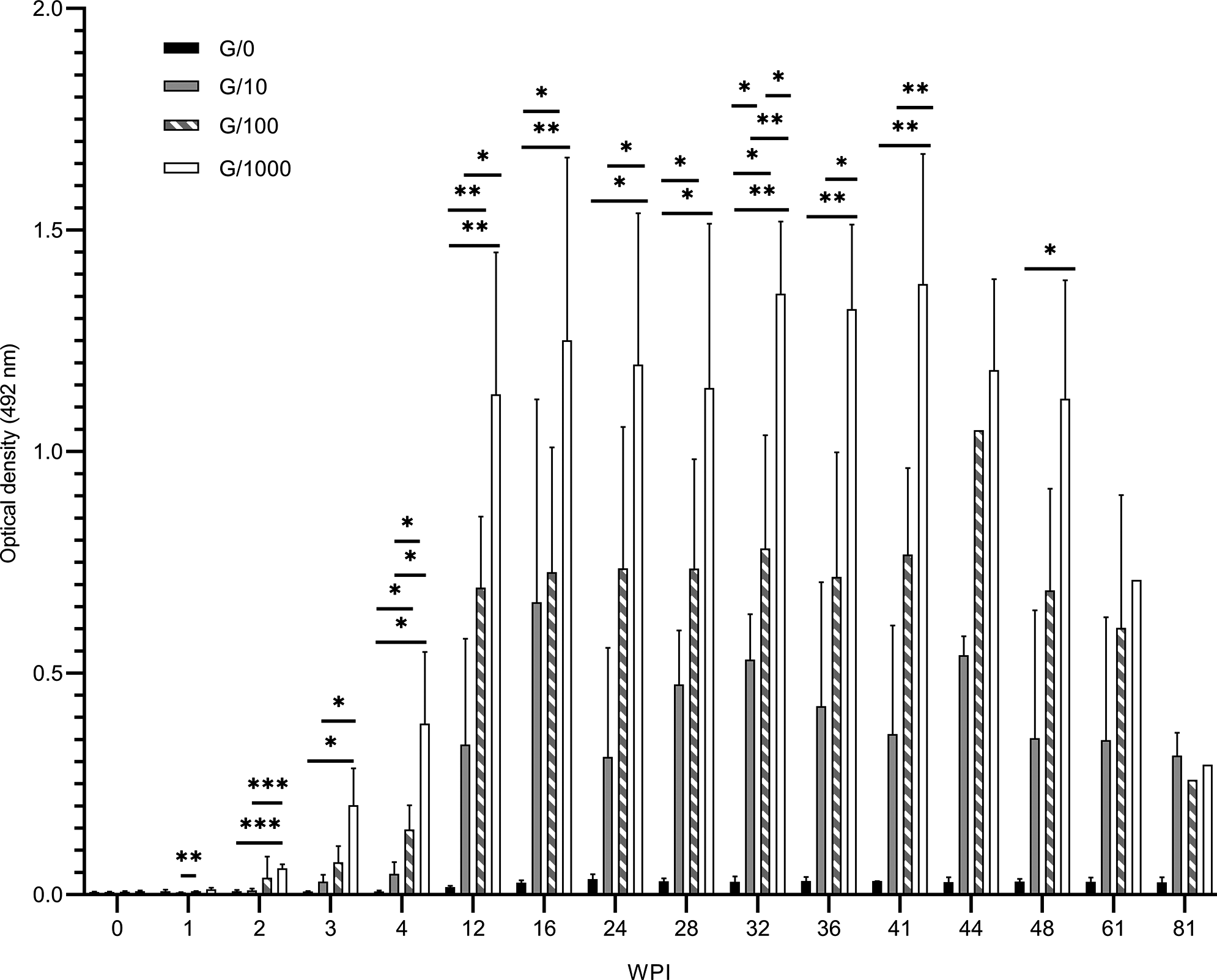
The control group G/0 showed no dynamics in the specific anti-TcES IgG antibody levels elevation, as expected. All infected groups showed an increase in antibody levels with ongoing infection. The antibody levels of all groups at week 16 p.i. are shown in Figure 1.
Levels of Toxocara canis excretory-secretory antigen-specific IgG antibodies in sera of mice infected by 10 (G/10), 100 (G/100) and 1000 (G/1000) T. canis larvae compared with controls (G/0) measured by ELISA at week 16 after infection. The antibody levels are displayed as the optical density (OD ± standard error of the mean) measured at 492 nm and analysed in GraphPad Prism (version 8) using one-way ANOVA with Tukey’s multiple comparisons test. Significant differences among groups are indicated by asterisks (*P<0.05, **P<0.01, ***P<0.001). The cut-off is stated as mean OD + 3 standard deviations.

As pictured in Figure 2, the elevation in antibody levels was detected from week 2 p.i., steadily increased to week 16 p.i., and showed a decreasing trend from week 48 p.i. in all infection groups. Starting from week 3 p.i., the higher the ID was, the higher the antibody levels detected.
Levels of Toxocara canis excretory-secretory antigen-specific IgG antibodies in sera of mice infected by 10 (G/10), 100 (G/100) and 1000 (G/1000) T. canis larvae compared with controls (G/0) measured by ELISA. This graph shows the development of antibody levels in course of infection to week 81 post infection (WPI) on 5 mice from each group. The antibody levels are displayed as the optical density (OD ± standard error of the mean) measured at 492 nm and analysed in GraphPad Prism (version 8) using two-way ANOVA with mixed-effect analysis. Significant differences among groups are indicated by asterisks (*P<0.05, **P<0.01, ***P<0.001).

General condition and motor and balance skills
The weights of all mice increased steadily to week 34 p.i. After this time point, a decreasing trend was observed; see Figure 3. Generally, when the general condition of mice rapidly worsened leading to euthanizing these animals immediately, this change was preceded by rapid weight loss in 2 weeks before reaching said condition. This occurred in all groups (data not shown).
Mean weights (in grams) of mice from three groups infected by 10 (G/10), 100 (G/100) and 1000 (G/1000) Toxocara canis larvae compared with controls (G/0) in the course of the infection. WPI, weeks post infection.

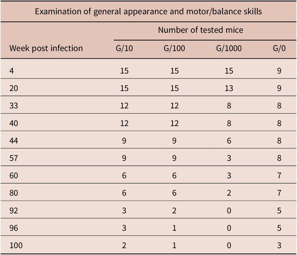
See Table 1 for the number of mice tested each week. The changes in general appearance, activity, and obvious motor changes were detected in G/1000 at week 20 p.i., in G/100 at week 40 p.i., and in G/10 at week 92 p.i., with higher incidence in the high ID group (see Figure 4). T. canis infection-specific stereotyped circular motion was detected in G/100 and G/1000 and disturbed balance, that clearly pointed out motor problem at week 20 p.i. (G/1000), week 80 p.i. (G/100) and week 92 p.i. (G/10).
Number of mice from three groups infected by 10 (G/10), 100 (G/100) and 1000 (G/1000) Toxocara canis larvae and control G/0 group that were regularly examined for any general appearance and motor/balance skills changes

The subtle motor/balance impairments detected by testing developed in G/1000 at week 4 p.i., in G/100 at week 20 p.i., and in G/10 at week 80 p.i. (Figure 4). In G/10, the first detected change was the loss of balance on the thin beam. The following detected changes were reduced overall activity, disturbed balance and paw slip on the thin beam. The bat-like posture and paw slip on the thin beam were the first detected changes in G/100 and G/1000. The paw slip on the thin beam and the loss of balance on the thick beam were apparent parameters of motor/balance impaired abilities, regarding the time of onset in every group, as well as number of presenting mice; see Figure 4. The changes in general appearance, activity, and obvious motor changes, as well as subtle motor/balance impairments, were detected also in G/0, with the first detection at weeks 33 and 44, respectively.
Changes in general appearance and activity, apparent impairments and subtle motor/balance impairments detected by testing of three groups infected by 10 (G/10), 100 (G/100) and 1000 (G/1000) Toxocara canis larvae and controls (G/0). Numbers of mice presenting each change in the week of testing are shown in the heat map. Abnormal post., Abnormal posture; Bat-like post., Bat-like posture; Dish. hair, Disheveled hair; Dist. balance, Disturbed balance; LoB/thin beam, Loss of balance on thin beam; LoB/thick beam, Loss of balance on thick beam; Loss of ext. ref., Loss of forelimb extension reflex; Overt cach., Overt cachexia; P. slip/thin beam, Paw slip on thin beam; Reduced o. act., Reduced overall activity; Right. ref. imp., Righting reflex impairment; Ster. move., Stereotyped movements; Tumbl. motion, Tumbling motion; WPI, weeks post infection.

The observation and testing results unequivocally prove the negative T. canis infection impact on mice in G/100 and G/1000. The results obtained in G/10 are more similar to those of the control G/0 group, with minimal differences.
Larvae in organs and brain pathologies
Larvae were detected microscopically in the brain and the liver of infected mice, but not in the lungs or spleen. Larvae were found in the liver of two G/1000 mice at weeks 14 and 61 p.i., one larva each time.
In the brains, larvae were found in eight of 15 examined mice G/10, in 13 of 14 examined mice G/100, and in all G/1000 mice in course of infection (i.e., from week 14 to week 100 p.i.). All larvae were intact and motile. In G/10, when detected, only one larva (i.e., 10 % of ID) was recovered in the brains of mice. In G/100 the highest detected number of larvae in one hemisphere was 12 (i.e., 12% of ID, week 61 p.i.) and the lowest was 1 larva (i.e., 1% of ID, weeks 41, 81 and 100 p.i.). The highest detected number of larvae in G/1000 was 115 (i.e., 11.5% of ID, week 14 p.i.) and the lowest was 4 larvae (i.e., 0.4 % of ID, week 81 p.i.). The detected numbers of larvae in G/10 and G/100 showed a stable trend in ongoing time of infection. In contrast, the numbers of detected larvae decreased in G/1000 with the ongoing infection (Table 2). One mouse from G/100 died unexpectedly at week 89 p.i., without examination of organs for larvae number and presence.
Mean percentage of ID ± standard deviation of recovered larvae from mouse brains of three groups infected by 10 (G/10), 100 (G/100) and 1000 (G/1000) Toxocara canis larvae

ID, infection dose; N/A, not available; WPI, weeks post infection.
Larvae were detected on hematoxylin-eosin sections in all infected groups (Table 3) without any surrounding pathology. Histological examination revealed the presence of foamy cells, hemorrhage, necrosis and infiltration of monocytes (Figure 5). Foamy cells were predominantly found in the corpus callosum with only one exception in cerebellum. Infiltrations were found in the cerebellum and corpus callosum. Hemorrhages were found in medulla oblongata/pons varoli, midbrain, cerebellum and cerebral cortex. Necroses were detected in midbrain, cerebellum, corpus callosum, optic tract in the hypothalamic area, anterior commissure and medulla oblongata/pons varoli, as described in Table 3.
The number of larvae and detected pathologies in hematoxylin/eosin-stained brain sections of mice infected by 10 (G/10), 100 (G/100) and 1000 (G/1000) Toxocara canis larvae

Brackets shows location of the larva/pathology and the number.
AntCom, Anterior commissure; BasFor, Basal forebrain; Cc, Corpus callosum; Crbl, Cerebellum; CrCt, Cerebral cortex; Fnx, Fornix; ID, Infection dose; Mb, Midbrain; Med, Medulla oblongata and pons varolli; No, larva/pathology in section not detected; Ot, Optic tract in hypothalamic area; WPI, Weeks post infection; Yes, pathology detected.
The brain pathologies in hematoxylin-eosin-stained sections of mice infected by 10 (G/10), 100 (G/100) and 1000 (G/1000) Toxocara canis larvae. The detected pathologies were (a) hemorrhage (arrows) in cortex close to cerebellum at week 21 post infection (p.i.) in brain of G/10 mouse, (b) necrosis (arrows) with debris of cells in midbrain at week 81 p.i. in G/100 brain and (c) huge mass of foamy cells (arrows) in corpus callosum at week 41 p.i. in G/1000 brain. The larva (d) in cerebellum (arrows) of G/1000 mouse at week 21 p.i. was not closely surrounded by any pathology.

Discussion
This study aimed to examine the progress and consequences of a 100 weeks infection of BALB/c mice with 10, 100 and 1000 larvae of Toxocara canis on motor/balance abilities. As T. leonina is broadly found in Eurasia (Bauer et al. Reference Bauer, Lider, Ussenbayev, Seitkamzina, Zhanabayev, Maksimov and Knaus2024; Rostami et al. Reference Rostami, Riahi, Fallah Omrani, Wang, Hofmann, Mirzapour, Foroutan, Fakhri, Macpherson and Gasser2020), morphological determination and, additionally, PCR were performed for confirmation of the parasite species. Similar dynamics of specific immune response to our study between G/10, G/100 and G/1000 (and in other infection schemes) were detected by other authors (Pinelli et al. Reference Pinelli, Brandes, Dormans, Fonville, Hamilton and van der Giessen2007).
Weight was regularly measured as one indicator of the general condition of the animals. Weight loss accompanied changes in the physical condition of mice and motor/balance abilities in infected mice. The weight loss in G/1000 starting from week 34 p.i. preceded the change in general appearance and activity, and apparent motor/balance impairment that onset at week 40 p.i. The weight loss of mice infected with 1000 T. canis eggs ID was already observed by Dolinsky et al. (Reference Dolinsky, Burright, Donovick, Glickman, Babish, Summers and Cypess1981), starting approximately after 12 weeks of infection. The weight loss was detected in G/100 at week 80 p.i., as either a consequence of infection and/or ageing of mice, as was previously stated by Miller et al. (Reference Miller, Harper, Galecki and Burke2002).
Another indicator of the general condition was their coat. A worsened state of coat could be considered as an effect of the unpredictable chronic mild stress-like stimuli to BALB/c mice (Farooq et al. Reference Farooq, Isingrini, Tanti, Le Guisquet, Arlicot, Minier, Leman, Chalon, Belzung and Camus2012; Isingrini et al. Reference Isingrini, Camus, Le Guisquet, Pingaud, Devers and Belzung2010; Mineur et al. Reference Mineur, Prasol, Belzung and Crusio2003). However, all experimental mice were kept in the same conditions and treated equally, which excludes dishevelled coat and reduced overall activity developed from week 33 of experiment as the consequence of any stress from housing or testing.
The neurodegenerative impairments, as well as other movement obstructions, may be also affected or caused by the process of ageing in long-time experiments. Age-associated osteoporosis, bone and joint disability, loss of muscle mass, poor vision causing reduced activity, and CNS impairment is known in BALB/c strain (Harkema et al. Reference Harkema, Youssef and de Bruin2016). Age-influenced behaviour and motor skills of the animals are impossible to exclude. Also, death of three G/0 mice at weeks 74, 90, and 100 of the experiment was tied to aging. We observed differences between all three infected groups, which are comparable with Toxocara infection consequences observed by other authors in mice (Corrêa et al. Reference Corrêa, Chieffi, Lescano and SVd2014; Hamilton et al. Reference Hamilton, Stafford, Pinelli and Holland2006; Janecek et al. Reference Janecek, Waindok, Bankstahl and Strube2017), as well as in gerbils (Akao et al. Reference Akao, Tomoda, Hayashi, Suzuki, Shimizu-Suganuma, Shichinohe and Fujita2003) and albino rats (Al-Hassnawi & Al-Quraishi Reference Al-Hassnawi and Al-Quraishi2014). However, G/10 and the control group G/0 demonstrated similar pattern of the onset of general condition and motor/balance abilities impairments, as described in Figure 4. In our case, symptoms in G/100 and G/1000 are clearly associated with the infection and ID, but it is not that clear for G/10, as some of the changes were also detected in the control group. Still, some impairments were detected only in G/10, but not in G/0 (i.e., disturbed balance or limb ataxia).
Larvae recovery was performed on sampled selected visceral organs and a randomly selected brain hemisphere using the quantitative compressed biopsy technique, which is advantageous for easy preparation and thorough screening of examined tissue (Eid et al. Reference Eid, El-Kowrany, Othman, El Gendy and Saied2015; Farjat et al. Reference Farjat, Minvielle, Pezzani and Niedfeld1995). From the visceral organs, larvae were found only in the liver of our infected mice. Corresponding to our study, Kolbeková et al. (Reference Kolbeková, Větvička, Svoboda, Skírnisson, Leissová, Syrůček, Marečková and Kolářová2011b) detected no larva in spleen of 1000 ID T. canis infected mice at 3 weeks’ examination, but detected 7.5% of ID in the brain after 4 months of infection. This is possibly caused by their cumulative effect in CNS (Bardón et al. Reference Bardón, Cuéllar and Guillén1994; Kolbeková et al. 2011A). Other studies on high ID infections of BALB/c mice overlap our time scheme minimally. However, based on available data, the number of larvae detected in the brains of G/1000 at week 14, 19 and 50 p.i. was higher than in study of Bardón et al. (Reference Bardón, Cuéllar and Guillén1994) and (Ollero et al. Reference Ollero, Fenoy, Cuéllar, Guillén and Del Aguila2008) at similar timepoints. The detected numbers were slightly higher in the cerebrum compared to cerebellum, which corresponds to the volume of the tissue and, therefore, is not considered as the affinity of the larvae. Our observation corresponds to other authors that also observed clustered larvae in higher numbers in the cerebrum (Kolbeková et al. 2011B). Larvae in brains were observed either single or in clusters with no visible preference for right or left half of the brain, which corresponds to observation of other authors (Janecek et al. Reference Janecek, Beineke, Schnieder and Strube2014; Kolbeková et al. 2011A; Kolbeková et al. 2011B). If we connect the presence of larvae in the brain and observed/tested impairments of mice, the early severe impairments in G/1000 were associated with the time when the highest numbers of larvae were detected in brains. However, we are convinced that the long-term presence of larvae in CNS actually causes changes in the tissue and subsequently leads to the development of inconspicuous impairments over a long time. Our hypothesis follows the proven presence of brain injury associated biomolecules and markers associated with neurodegenerative diseases, as β-amyloid precursor protein and amyloid β, transforming growth factor β1, or glial fibrillary acidic protein in T. canis–infected mice (Chou et al. Reference Chou, Lee, Liao, Huang and Fan2017; Liao et al. Reference Liao, Fan, Kao, Ji, Su, Lin and Cho2008; Springer et al. Reference Springer, Heuer, Janecek-Erfurth, Beineke and Strube2019; Waindok & Strube Reference Waindok and Strube2019).
The parasitic infections with a slowly developing impact on the physical state and the behaviour of the host are known to be caused by a wide range of migrating and non-migrating helminths (Blecharz-Klin et al. Reference Blecharz-Klin, Świerczyńska, Piechal, Wawer, Joniec-Maciejak, Pyrzanowska, Wojnar, Zawistowska-Deniziak, Sulima-Celińska and Młocicki2022; Finsterer & Auer Reference Finsterer and Auer2013; Macháček et al. Reference Macháček, Leontovyč, Šmídová, Majer, Vondráček, Vojtěchová, Petrásek and Horák2022; Othman et al. Reference Othman, Bruschi and Ganna2014; Wagner & Newton Reference Wagner and Newton2009). The mechanisms differ from feeding on nervous tissue, downregulation of neurophysiological pathways, neurotransmitter alterations, and epileptogenesis by calcified lesions or migrating larvae to discussed potential development of neurodegenerative diseases such as Alzheimer’s disease or multiple sclerosis, which still remains a question (Fan et al. Reference Fan, Holland, Loxton and Barghouth2015; Liao et al. Reference Liao, Fan, Kao, Ji, Su, Lin and Cho2008; Macháček et al. Reference Macháček, Leontovyč, Šmídová, Majer, Vondráček, Vojtěchová, Petrásek and Horák2022; Vojtechova et al. Reference Vojtechova, Machacek, Kristofikova, Stuchlik and Petrasek2022; Waindok et al. Reference Waindok, Janecek-Erfurth, Lindenwald, Wilk, Schughart, Geffers and Strube2022). As we proved in our study, T. canis infection is one of these infections with a severe impact on the host and therefore should not be underestimated.
Conclusions
All infectious doses resulted in infection measurable by the presence of specific antibodies and pathological findings in the brain. Only at medium (G/100) and high (G/1000) infection doses negative changes in physical condition and activity were observed. In these groups, deterioration of motor parameters was also observed at the same time.
Despite the fact that infection in the low-dose larvae-infected group (G/10) was demonstrated by the presence of antibodies serologically and histologically in the brain, there were no observable changes in animal condition or motor changes in this group compared to the uninfected control group. As an analogy to the high seroprevalence in the human population and the very low incidence of clinical signs, it can be assumed that in paratenic hosts, low-dose exposure leads to a detectable immune response and some pathological changes, but not to clinically manifested disease or symptoms or changes in physical condition and motor impairment.
Supplementary material
The supplementary material for this article can be found at http://doi.org/10.1017/S0022149X24000786.
Acknowledgements
The first author thanks Dr. Tomáš Macháček for consultations on manuscript and Mrs. Markéta Leissová for help with the preparation and implementation of the hatching, cultivation, and long-term maintenance of Toxocara canis larvae used in this study.
Author contribution
N.B. and M.C. conceived and designed the study. M.C. supervised the study. M.C. led an approved animal research project. N.B. conducted data gathering and analysis. N.B., J.N. and C.K.F. evaluated data. N.B. prepared the original draft. L.K., J.N., and C.K.F. consulted and reviewed the draft. All authors approved the final form of the manuscript before submission.
Financial support
This research was supported by the 1st Faculty of Medicine, Charles University, Prague (SVV 260 636); by Cooperatio – Infection and Immunity and Cooperatio – Biology, European Union project National Institute for Neurological Research (Program EXCELES, ID Project No. LX22NPO5107).
Competing interest
The authors declare no conflict of interest in this study.
Ethical standard
The authors assert that all procedures contributing to this work comply with the ethical standards of the relevant national and institutional guides on the care and use of laboratory animals.
Mice were housed under standard conditions in specifically accredited laboratories of the Institute of Immunology and Microbiology of the First Faculty of Medicine, Charles University and the General University Hospital in Prague (accreditation numbers 70030/2013-MZE-17214 and 8615/2019-MZE-17214). The certified person (CZ 02627) kept and euthanized the animals according to ethical standards, the animal welfare laws of the Czech Republic and European regulations for transport, housing and care of laboratory animals (Directive 2010/63/EU on the protection of animals used for scientific purposes). This project involving laboratory animals was approved by the Ministry of Education, Youth and Sports of the Czech Republic (approval numbers MSMT – 7063/2017-2 and MSMT – 11190/2020-4). The number of experimental animals was kept low for ethical reasons.