Introduction
The langbeinite group includes five isostructural cubic (space group P213) double sulfates: the widespread langbeinite K2Mg2(SO4)3 (Anthony et al., Reference Anthony, Bideaux, Bladh and Nichols2003) and four rare mineral species, namely efremovite (NH4)2Mg2(SO4)3 (Shcherbakova and Bazhenova, Reference Shcherbakova and Bazhenova1989; Shimobayashi et al., Reference Shimobayashi, Ohnishi and Miura2011), ferroefremovite (NH4)2Fe2+2(SO4)3 (Kasatkin et al., Reference Kasatkin, Plášil, Škoda, Campostrini, Chukanov, Agakhanov, Karpenko and Belakovskiy2021), calciolangbeinite K2Ca2(SO4)3 (Pekov et al., Reference Pekov, Zelenski, Zubkova, Yapaskurt, Chukanov, Belakovskiy and Pushcharovsky2012), and an insufficiently studied manganolangbeinite K2Mn2(SO4)3 (Zambonini and Carobbi, Reference Zambonini and Carobbi1924; Bellanca, Reference Bellanca1947). All these minerals are known in exhalations of volcanic fumaroles and in their genetic counterparts related to natural or anthropogene coal fires (see references above); moreover, all members of the group, except langbeinite, a typical evaporite mineral, have only been described from fumaroles or so called ‘pseudo-fumaroles’.
In this paper we characterise an orthorhombic (space group P212121) modification of K2Ca2(SO4)3, new for Nature and found in two regions, Kamchatka, Far-Eastern Region, Russia and Israel, in different geological environments. It is the first natural langbeinite-like sulfate with orthorhombic symmetry that differs clearly from cubic calciolangbeinite in the powder X-ray diffraction pattern, optical data and Raman spectrum. However, cubic and orthorhombic forms of K2Ca2(SO4)3 possess the same topology of crystal structure: the structure of the orthorhombic modification can be derived from the cubic one by distortion (see below). Such cases are regulated by the rules of the current mineral nomenclature: “…if the crystal structures of the polymorphs have essentially the same topology, differing only in terms of a structural distortion or in the order–disorder relationship of some of the atoms comprising the structure, such polymorphs are not regarded as separate species, and the names of such topologically-similar polymorphs can be distinguished by the addition of crystallographic suffixes to the mineral name” (Nickel and Grice, Reference Nickel and Grice1998). Thus, cubic and orthorhombic modifications of K2Ca2(SO4)3 are topologically-similar polymorphs and, from the nomenclature viewpoint, can be considered as structural varieties (or, maybe, the term ‘sub-species’ would be more correct) of the mineral species calciolangbeinite. Such varietal names should include the corresponding crystallographic suffixes, therefore, cubic calciolangbeinite described by Pekov et al. (Reference Pekov, Zelenski, Zubkova, Yapaskurt, Chukanov, Belakovskiy and Pushcharovsky2012) is now calciolangbeinite-C whereas its orthorhombic dimorph first described in the present paper is calciolangbeinite-O. Though no new mineral name has been introduced, and thus no official Commission on New Minerals, Nomenclature and Classification of the International Mineralogical Association (IMA) approval is necessary, we did discuss the naming of these varieties with the officers of the IMA and received approval.
In addition to the characterisation of calciolangbeinite-O, in this paper we give new data on calciolangbeinite-C, discuss the relationship between cubic and orthorhombic modifications of K2Ca2(SO4)3 and describe a novel solid-solution system between langbeinite and calciolangbeinite.
Occurrence and general appearance
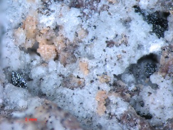
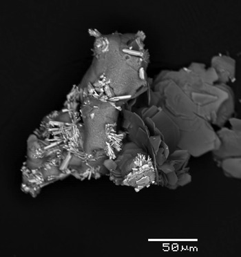
The Kamchatka samples of calciolangbeinite-O originate from the active Arsenatnaya fumarole located at the Second scoria cone of the Northern Breakthrough of the Great Tolbachik Fissure Eruption, Tolbachik volcano (55°41’N, 160°14’E). The Arsenatnaya fumarole is one of the mineralogically interesting fumaroles at the Tolbachik volcano. It was described by Pekov et al. (Reference Pekov, Zubkova, Yapaskurt, Belakovskiy, Lykova, Vigasina, Sidorov and Pushcharovsky2014, Reference Pekov, Koshlyakova, Zubkova, Lykova, Britvin, Yapaskurt, Agakhanov, Shchipalkina, Turchkova and Sidorov2018) and Shchipalkina et al. (Reference Shchipalkina, Pekov, Koshlyakova, Britvin, Zubkova, Varlamov and Sidorov2020). Calciolangbeinite-O occurs in the intermediate, so-called polymineralic zone of the fumarole. Temperatures measured by us using a chromel–alumel thermocouple in areas with this sulfate at the time of its collecting in 2013, 2014 and 2015 were 300–380°C. Calciolangbeinite-O forms interrupted porous crusts up to 1 cm × 1.5 cm in area and up to 0.5 mm thick on basalt scoria altered by fumarole gas, or on incrustations consisting mainly of sublimate silicates (sanidine and/or fluorophlogopite) and oxides (hematite and tenorite) overgrowing basalt scoria (Figs 1 and 2). Other associated minerals are aphthitalite, metathénardite, belomarinaite, langbeinite, calciolangbeinite-C, anhydrite, arcanite, krasheninnikovite, wulffite, fluoborite, pseudobrookite, gahnite, corundum, johillerite, nickenichite, bradaczekite, svabite, tilasite, lammerite, lammerite-β, urusovite, ericlaxmanite, kozyrevskite and popovite. Irregularly shaped single-crystal individuals of calciolangbeinite-O in the crusts are up to 0.3 mm across.
Porous interrupted crust of colourless to white calciolangbeinite-O with iron-black tenorite overgrowing beige sanidine and reddish-brown basalt scoria altered by fumarole gas. Arsenatnaya fumarole, Tolbachik volcano, Kamchatka, Russia. FOV width: 5.7 mm. Photo: I.V. Pekov & A.V. Kasatkin.

Calciolangbeinite-O crust overgrowing aggregates of lamellar crystals of fluorophlogopite and long-prismatic crystals of johillerite (white). Arsenatnaya fumarole, Tolbachik volcano. SEM back-scattered electron image.

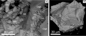
In Israel, calciolangbeinite-O has been described from two localities, as ‘calciolangbeinite', by Galuskina et al. (Reference Galuskina, Vapnik, Lazic, Armbruster, Murashko and Galuskin2014) and Galuskin et al. (Reference Galuskin, Galuskina, Gfeller, Krüger, Kusz, Vapnik, Dulski and Dzierżanowski2016). At Jabel Harmun, Judean Desert, West Bank, Palestinian Autonomy (31°46′N, 35°26′E) calciolangbeinite-O was identified in the specimen described as the holotype of harmunite (Galuskina et al., Reference Galuskina, Vapnik, Lazic, Armbruster, Murashko and Galuskin2014). This sample is a fragment of larnite pseudoconglomerate, a pyrometamorphic rock belonging to the well-known Hatrurim Complex (Gross, Reference Gross1977; Novikov et al., Reference Novikov, Ye and Safonova2013). Calciolangbeinite-O together with aphthitalite and minor thénardite fill small fractures on the boundary between porous ferrite aggregate (consisting of magnesioferrite, srebrodolskite and harmunite) and a rock mainly composed of larnite, fluorellestadite, ye'elimite, gehlenite, ternesite and fluormayenite–fluorkyuygenite and brownmillerite–srebrodolskite series minerals. Here calciolangbeinite-O forms interrupted granular incrustations up to 0.15 mm thick and up to 1 mm2 in area (Fig. 3a). The size of its single-crystal grains usually do not exceed 0.1 mm. The formation of görgeyite K2Ca5(SO4)6⋅H2O in the areas of supergene alteration of calciolangbeinite-O was observed (Fig. 3b). At Har Parsa, Hatrurim Basin, Negev Desert calciolangbeinite-O was detected in a pyrometamorphic rock which mainly consists of larnite and contains ternesite. Calciolangbeinite-O fills channels ~0.1 mm in diameter hosting gas bubbles and also occurs in intergranular cavities on the front of growth of ternesite poikilitic crystals (Galuskin et al., Reference Galuskin, Galuskina, Gfeller, Krüger, Kusz, Vapnik, Dulski and Dzierżanowski2016).
(a) Crust of calciolangbeinite-O (Clgb) and aphthitalite (Att) on magnesioferrite (Mfr) aggregate with larnite (Lrn) and srebrodolskite (Sre) from Jabel Harmun, Palestine Autonomy, Israel. (b) Grain of calciolangbeinite-O extracted from the aggregate shown in Fig. 3a, which after a few days was partially covered by tiny acicular görgeyite crystals.

In the present paper, calciolangbeinite-O from Tolbachik and Jabel Harmun is characterised.
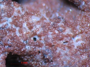
Calciolangbeinite-C was described originally from the Yadovitaya (Poisonous) fumarole at the same Second scoria cone of the Northern Breakthrough of the Great Tolbachik Fissure Eruption, (as ‘calciolangbeinite’: Pekov et al., Reference Pekov, Zelenski, Zubkova, Yapaskurt, Chukanov, Belakovskiy and Pushcharovsky2012). Later, in 2013 we also found it in the Arsenatnaya fumarole, in the same mineral association as calciolangbeinite-O. In the majority of samples, these varieties of calciolangbeinite are visually indistinguishable from one another. A common specimen of calciolangbeinite-C from the Arsenatnaya fumarole is shown in Fig. 4. However, in some samples well-shaped crystals of calciolangbeinite-C occur (Fig. 5), unlike calciolangbeinite-O which was observed only as irregularly shaped individuals (Figs 1 and 2). Langbeinite with different contents of admixed Ca is widespread in sublimates of both Arsenatnaya and Yadovitaya fumaroles. The most common variety of Tolbachik fumarolic langbeinite is Ca-poor (<1 wt.% CaO).
Numerous reddish-brown (impregnated with hematite micro-inclusions) aggregates of calciolangbeinite-C overgrowing a white sanidine crust. Arsenatnaya fumarole, Tolbachik volcano. FOV width: 3.2 mm. Photo: I.V. Pekov & A.V. Kasatkin.

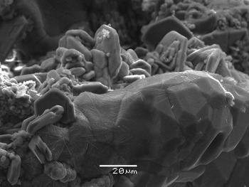
Compact aggregate of well-shaped calciolangbeinite-C crystals with numerous hexagonal flattened corundum crystals. Arsenatnaya fumarole, Tolbachik volcano. SEM (SE) image.

Physical properties and optical data of calciolangbeinite-O
Calciolangbeinite-O is transparent or, in aggregates, translucent, with vitreous lustre. It is colourless to white; some specimens from Tolbachik are orange-reddish to reddish-brown or greenish due to the presence of scattered micro-inclusions of hematite or copper arsenates, respectively. It is brittle, cleavage or parting was not observed. The fracture is uneven or conchoidal. The Mohs hardness is ca. 3. Density calculated for the sample from Tolbachik using the empirical formula and unit-cell volume determined from the single-crystal X-ray diffraction (XRD) data is 2.685 g cm–3.
In plane polarised transmitted light, calciolangbeinite-O from Tolbachik is colourless and non-pleochroic. It is optically biaxial (+), α = 1.475(3), β = 1.490(2), γ = 1.508(3) (589 nm); 2Vmeas. = 85(5)°; and 2Vcalc. = 86°. Dispersion of optical axes was not observed.
Raman spectroscopy
The data on analytical technique and conditions are given as supplementary information.
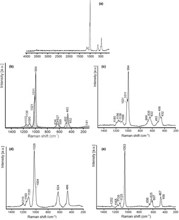
The Raman spectra of calciolangbeinite-O from Jabel Harmun and from Tolbachik (Figs 6a–c) are similar to one another and clearly differ from the spectra of calciolangbeinite-C (Fig. 6d) and langbeinite (Fig. 6e).
The Raman spectra of (a, b) calciolangbeinite-O from Jabel Harmun, Israel (a – spectrum in the range 100–4000 cm–1, b – in the range 100–1500 cm–1); (c) calciolangbeinite-O from Arsenatnaya fumarole, Tolbachik, Kamchatka, Russia; (d) calciolangbeinite-C from Arsenatnaya fumarole; (e) langbeinite from Stebnik potassium salt deposit, Ukraine.

The assignments of Raman bands for calciolangbeinite-O made in accordance with Prieto-Taboada et al. (Reference Prieto-Taboada, Fdez-Ortiz de Vallejuelo, Veneranda, Lama, Castro, Arana, Larrañag and Madariaga2019) are given in Table 1. All bands with Raman shifts above 300 cm–1 correspond to vibrations in (SO4)2–: ν3 is 1090–1180 cm–1; ν1 is 996, 1011 and 1021 cm–1; ν4 is 700–550 cm–1; and ν2 is 550–400 cm–1. Very weak unresolved bands below 200 cm–1 (in the spectrum of calciolangbeinite-O from Jabel Harmun) correspond to external modes involving K⋅⋅⋅O and Ca⋅⋅⋅O stretching vibrations (‘⋅⋅⋅’ denotes weak non-covalent bonds) as well as libration and translational vibrations of SO42– as a whole (Table 1). The very weak band at 3520 cm–1 in the Raman spectrum of calciolangbeinite-O from Jabel Harmun is probably caused by a minor impurity of görgeyite (see Fig. 6b).
Raman shifts (cm–1) of bands in Raman spectra of calciolangbeinite-O, calciolangbeinite-C and langbeinite and their assignment, in accordance with Nakamoto (Reference Nakamoto2008).

Characteristics of spectral bands: s – strong, w – weak, vw – very weak, sh – shoulder, br – broad.
In the Raman spectrum of Ca-free langbeinite from the Stebnik potassium salt deposit, Ukraine (Fig. 3e), the total number of bands is less than in the spectra of calciolangbeinite-O because the SO4 tetrahedron occupies only one site in the structure of the former. This is confirmed by the presence of a single band at 1053 cm–1 corresponding to the nondegenerate symmetric S–O stretching mode. This band is shifted towards higher frequencies as compared to analogous bands of calciolangbeinite which confirms the involvement of Mg cations in S–O stretching vibrations. The shoulder at 1004 cm–1 and a poor resolution of Raman bands in the range 600–670 cm–1 in the spectrum of calciolangbeinite-C from the Arsenatnaya fumarole (Fig. 3d) may be due to a different local environment around the SO42– anion (presumably, without Mg and including Mg).
Thus, Raman spectroscopy seems a good, express tool to distinguish calciolangbeinite-C and calciolangbeinite-O, especially in micro-samples.
Chemical composition
The chemical composition of the langbeinite-group minerals studied from the Arsenatnaya fumarole was determined in the Laboratory of Analytical Techniques of High Spatial Resolution, Dept. of Petrology, Moscow State University (MSU), using a Jeol JSM–6480LV scanning electron microscope (SEM) equipped with an INCA-Wave 500 wavelength-dispersive spectrometer. The conditions were: an acceleration voltage of 20 kV, a beam current of 20 nA and a 5 μm beam diameter. Сalciolangbeinite-O from Jabel Harmun was studied using a Philips XL30 scanning electron microscope in EDS mode (Laboratory of Electron Microscopy, Faculty of Earth Sciences, University of Silesia (UoS), Sosnowiec) with an acceleration voltage of 15 kV, a beam current of 1 nA, and a ~5 μm beam diameter.
The following standards were used (MSU / UoS): Na (albite / albite), K (microcline / orthoclase), Rb (Rb2Nb4O11 / –), Mg (diopside / diopside), Ca (CaSiO3 / anhydrite), and S (SrSO4 / anhydrite). Contents of other elements with atomic numbers higher than carbon are below detection limits.
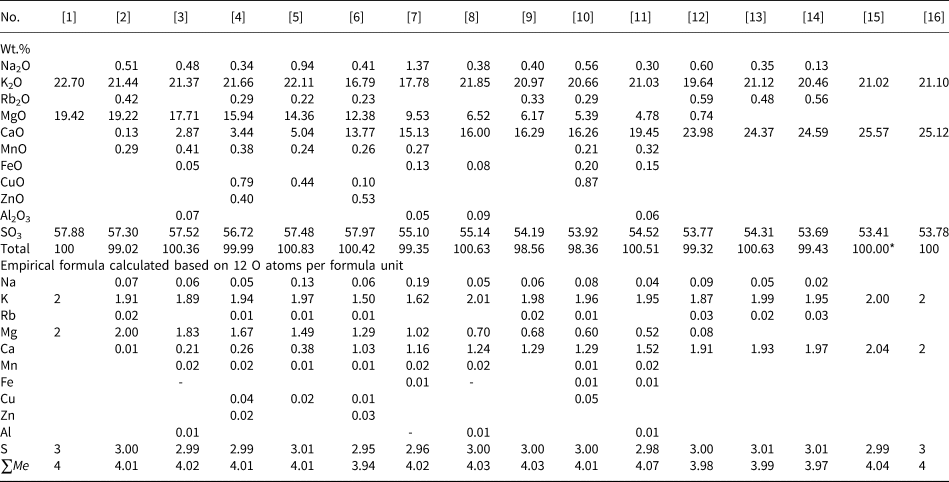
The chemical composition of the samples studied is given in Table 2. The empirical formulae were calculated on the basis of 12 O atoms per formula unit (apfu). The ratios of Ca and Mg, the major octahedrally coordinated cations M are shown in Fig. 7.
Ratios of the major M cations: Mg and Ca in minerals of the langbeinite–calciolangbeinite solid-solution series from fumaroles of the Tolbachik volcano, Kamchatka, Russia: 1 – langbeinite; 2 – calciolangbeinite-C; 3 – calciolangbeinite-O.

Chemical composition (wt.%) and empirical formulae of minerals of the langbeinite–calciolangbeinite solid-solution system.

[1] – K2Mg2(SO4)3; [2–6] – langbeinite; [7–11] – calciolangbeinite-C; [12–15] – calciolangbeinite-O; [16] – K2Ca2(SO4)3; [2–14] – Tolbachik volcano, Kamchatka, Russia [3, 7, 8 and 11] – Yadovitaya fumarole, others – Arsenatnaya fumarole); [15] – Jabel Harmun, Israel; [3, 7, 8 and 11] – after Pekov et al. (Reference Pekov, Zelenski, Zubkova, Yapaskurt, Chukanov, Belakovskiy and Pushcharovsky2012), others – this work. Analyses [8] and [14] represent the composition of samples on which the crystal structures of calciolangbeinite-C and calciolangbeinite-O, respectively, were solved. *Analytical total was normalized for 100 wt.%. ΣMe is the sum of all metal cations. Empty cell means that the content of a constituent is below detection limit and dash means 0.00 apfu (for minor admixtures).
In general, the samples studied correspond well to the formula A +2M 2+2(SO4)3 in which the major cations M = Mg or Ca with minor admixtures (≤ 0.08 apfu together) of Mn, Fe, Cu, Zn and/or Al, and A = K >> Na, Rb. Two samples (an. ## 6 and 7 in Table 2) contain less K (1.5–1.6 apfu) and larger amounts of bivalent cations: Ca + Mg = 2.2–2.3 apfu.
X-ray crystallography and crystal structure of calciolangbeinite-O
The data on analytical technique and conditions for both powder and single-crystal XRD studies are given in the Supplementary material (see below).
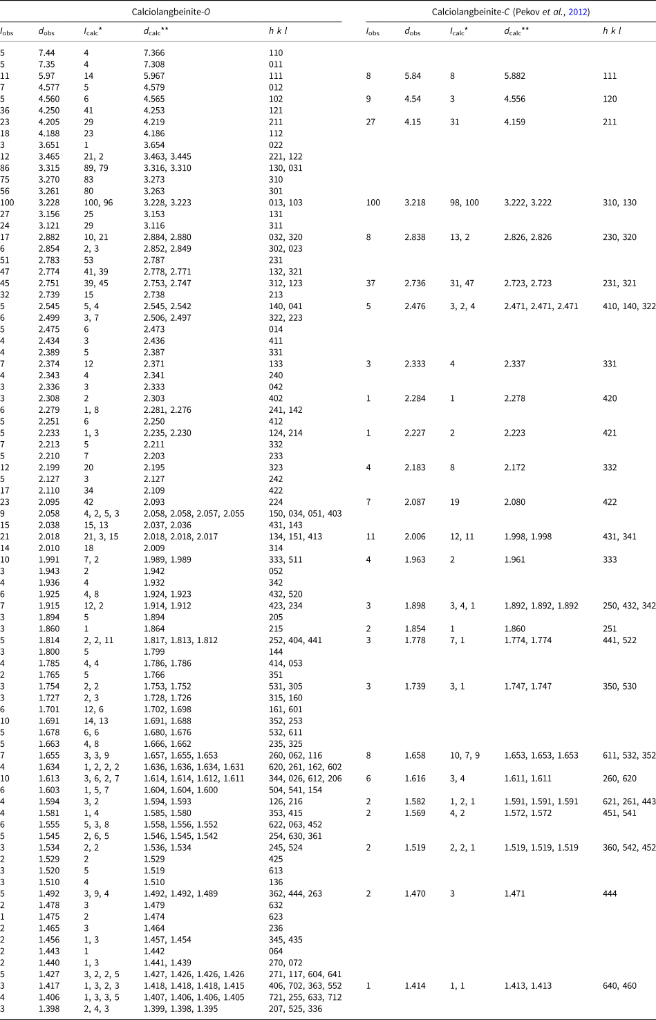
The powder XRD data of calciolangbeinite-O from Tolbachik are given in Table 3. Parameters of the orthorhombic unit cell calculated from the powder data are: a = 10.341(2), b = 10.509(3), c = 10.175(2) Å and V = 1105.8(8) Å3.
Powder X-ray diffraction data (d in Å) of calciolangbeinite-O and calciolangbeinite-C from the Tolbachik volcano.

* For the calculated pattern, only reflections with intensities ≥1 are given; ** for the unit-cell parameters calculated from single-crystal data.
The single-crystal XRD study of calciolangbeinite-O from Jabel Harmun gave the following parameters of the orthorhombic unit cell: a = 10.2828(2), b = 10.4675(2), c = 10.0996(3) Å and V = 1087.07(4) Å3.
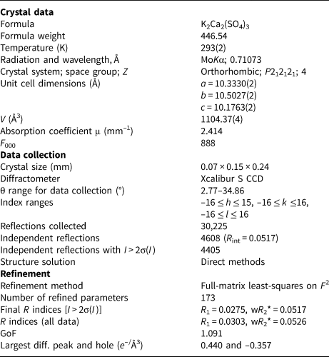
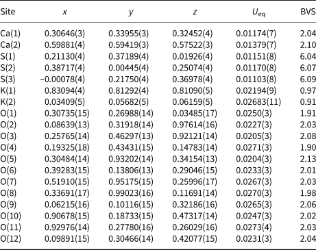
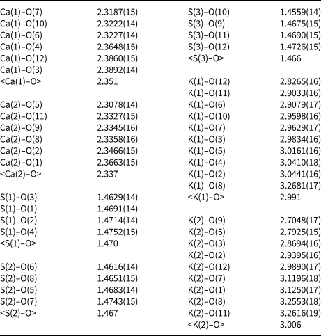
Single-crystal XRD studies of calciolangbeinite-O from Tolbachik were carried out using an Xcalibur S diffractometer equipped with a CCD detector. A full sphere of three-dimensional data was collected. Crystal data, data collection information and structure refinement details are given in Table 4. Data reduction was performed using CrysAlisPro Version 1.171.35.21 (Agilent Technologies, 2012). The XRD data were corrected for Lorentz factor and polarisation effects. The structure was solved by direct methods and refined with the use of SHELX software package (Sheldrick, Reference Sheldrick2008) on the basis of 4405 unique reflections with I > 2σ(I) to the final R = 0.0275. Atom coordinates, their displacement parameters and bond-valence sums are given in Table 5 and selected interatomic distances in Table 6. The crystallographic information files have been deposited with the Principal Editor of Mineralogical Magazine and are available as Supplementary material (see below).
Crystal data, data collection information and structure refinement details for calciolangbeinite-O from Tolbachik.

* w = 1/[σ2(Fo 2) + (0.0176P)2 + 0.2728P]; P = ([max of (0 or Fo 2)] + 2Fc 2)/3
Сoordinates, equivalent displacement parameters (U eq, in Å2) of atoms in and bond-valence sums (BVS) calciolangbeinite-O from Tolbachik. Parameters for BVS were taken from Gagné and Hawthorne (Reference Gagné and Hawthorne2015).

Selected interatomic distances (Å) in the crystal structure of calciolangbeinite-O from Tolbachik.

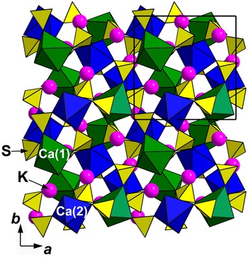
As crystal structure data show, calciolangbeinite-O is a natural analogue of synthetic orthorhombic K2Ca2(SO4)3 (Speer and Salje, Reference Speer and Salje1986), a representative of the structure type of K2Cd2(SO4)3 which is a distorted derivative of the cubic langbeinite-type structure (Abrahams and Bernstein, Reference Abrahams and Bernstein1977). The crystal structure of calciolangbeinite-O (Fig. 8) is based upon the three-dimensional complex framework formed by Ca(1)O6 and Ca(2)O6 octahedra and SO4 tetrahedra. Two crystallographically non-equivalent Ca sites centre M octahedra with mean Ca–O distances of 2.351 [Ca(1)] and 2.337 Å [Ca(2)]. There are three crystallographically non-equivalent S sites centring tetrahedra with mean S–O distances of 1.470 [S(1)], 1.467 [S(2)] and 1.466 Å [S(3)] (Table 6). Each CaO6 octahedron shares all oxygen vertices with SO4 tetrahedra and thus there is no linkage between CaO6 polyhedra. Potassium atoms occupy two crystallographically non-equivalent sites in the voids of the framework: K(1) cations are located in ten-fold polyhedra while K(2) cations centre nine-fold polyhedra. The K–O distance limit was 3.3 Å.
The crystal structure of calciolangbeinite-O in the ab projection. The unit cell is outlined.

Discussion
Solid-solution series between langbeinite and calciolangbeinite
As our data on Tolbachik samples show (Fig. 7; Table 2), a solid-solution system between langbeinite and calciolangbeinite exists, and the samples studied occupy the whole compositional range from K2Mg2(SO4)3 to K2Ca2(SO4)3. In the langbeinite part of the series, most analyses lie in the region (Mg2.0–1.6Ca0.0–0.4) and in the calciolangbeinite part – in the region (Ca1.2–2.0Mg0.8–0.0), whereas in the middle part, with (Mg1.6–0.8Ca0.4–1.2), only a few samples were found (Fig. 7). We suggest that it could be caused by the significant difference in ionic radii of Mg2+ and Ca2+ which hampers their occurrence in comparable amounts in the same M sites in the structure. For samples which contain fewer univalent cations (first of all, K) and more bivalent cations (Ca + Mg) in comparison with the idealised general formula A +2M 2+2(SO4)3 (see, e.g. an. ## 6 and 7 in Table 2), we suggest partial substitution of K by Ca.
Scanning electron microscopic, XRD and Raman spectroscopic data confirm that the samples studied represent a homogeneous solid solution and not micro-intergrowths of Mg- and Ca-rich phases formed as a result of its exsolution. The Raman spectroscopy turned out to be both a reliable and express tool for checking the homogeneity of the langbeinite-type minerals and the identification of the calciolangbeinite form as cubic or orthorhombic (Fig. 6). The Raman spectra obtained for samples studied by electron microprobe showed that the border between calciolangbeinite-C and calciolangbeinite-O under room-temperature conditions lies near the following ratio of M cations: Ca1.9Mg0.1: the Mg-poorest samples belong to calciolangbeinite-O while the samples with Mg > 0.1 apfu correspond to calciolangbeinite-C (Fig. 7).
It is noteworthy that the continuous solid-solution series between synthetic K2Mg2(SO4)3 and K2Ca2(SO4)3 has been known since 1935. It was synthesised and studied using powder XRD by Ramsdell (Reference Ramsdell1935) who reported that the calcium end-member of the series demonstrates a poor-quality (with diffuse reflections) langbeinite-type diffraction pattern and is optically birefringent, unlike K2Mg2(SO4)3 which is a full analogue of langbeinite. The synthetic solid solution K2Mg2(SO4)3–K2Ca2(SO4)3 was later studied in more detail and it was found that members of the series with >73.5 wt.% K2Ca2(SO4)3 are optically biaxial at room temperature whereas at 200°C all members of the series are isometric (Morey et al., Reference Morey, Rowe and Fournier1964). However, the structural study of K2Ca2(SO4)3 was carried out only in the 1980s (Speer and Salje, Reference Speer and Salje1986).
Relationship between calciolangbeinite-O, calciolangbeinite-C and synthetic langbeinite-like sulfates with the general formula K2M2 + 2(SO4)3 in which M = Ca, Cd, Zn, Co, Ni or Mg
Orthorhombic [space group P212121, structure type of K2Cd2(SO4)3] and cubic [space group P213, structure type of langbeinite] structural varieties (‘sub-species’) of calciolangbeinite, a mineral with the idealised, end-member formula K2Ca2(SO4)3, are dimorphous. The structural difference between cubic and orthorhombic modifications of this sulfate is in significant tilting of SO4 tetrahedra, shifting of Ca and K sites and thus the distortion of the heteropolyhedral, octahedral–tetrahedral framework in calciolangbeinite-O in comparison with calciolangbeinite-C.
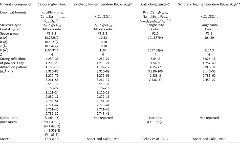
Dimorphism of synthetic K2Ca2(SO4)3 (Table 7) was studied by Speer and Salje (Reference Speer and Salje1986) who solved crystal structures of both modifications of this sulfate and found that the temperature of the phase transition from high-temperature cubic langbeinite-type modification (P213, the structure was studied at 260°C) to the orthorhombic low-temperature modification (P212121, the structure was studied at room temperature) is 184°C. Orthorhombic K2Ca2(SO4)3 belongs to the structure type of K2Cd2(SO4)3 attracting research attention due to ferroelastic properties (Abrahams and Bernstein, Reference Abrahams and Bernstein1977). K2Cd2(SO4)3 demonstrates the same dimorphism as K2Ca2(SO4)3 with the temperature of transition from high-temperature cubic modification to low-temperature (room-temperature) orthorhombic form equal to 159°C. Unlike these two compounds, sulfates K2M 2+2(SO4)3 with M = Mg, Ni, Co and Zn belong to the langbeinite structure type (cubic, P213) at room temperature. In general, the temperature of transition from a cubic, langbeinite-type modification to the orthorhombic, K2Cd2(SO4)3-type modification for sulfates K2M 2+2(SO4)3 clearly depends on the ionic radius of M 2+ [radius of M 2+ for octahedral oxygen coordination, after Shannon (Reference Shannon1976), is given below in square brackets]: 184°C for K2Ca2(SO4)3 [Ca: 1.00 Å], 159°C for K2Cd2(SO4)3 [Cd: 0.95 Å], –135°C for K2Zn2(SO4)3 [Zn: 0.74 Å] and –148°C for K2Co2(SO4)3 [Co: 0.745 Å]. Orthorhombic modifications are unknown for K2Mg2(SO4)3 [Mg: 0.72 Å] and K2Ni2(SO4)3 [Ni: 0.69 Å] (Speer and Salje, Reference Speer and Salje1986).

Note also that both cubic and orthorhombic modifications of langbeinite-type K2Mn2(SO4)3 (Oelkrug et al., Reference Oelkrug, Brückel, Hohlwein, Hoser and Prandl1988) and cubic K2Fe2(SO4)3 (Lander et al., Reference Lander, Rousse, Batuk, Colin, Corte and Tarascon2017) are known among synthetic compounds. Synthetic sulfates K2Cu2(SO4)3 and Rb2Cu2(SO4)3, as well as so-called ‘hydrolangbeinite’ Rb2Cu2(SO4)3(H2O), having the langbeinite-type stoichiometry, differ from langbeinite-type compounds in both structure and unit-cell metrics (Lander et al., Reference Lander, Rousse, Batuk, Colin, Corte and Tarascon2017; Siidra et al., Reference Siidra, Nekrasova, Charkin, Zaitsev, Borisov, Colmont, Mentré and Spiridonova2021), due to the Jahn-Teller distortion of the Cu2+-centred polyhedra. It is also noteworthy that such Cu-bearing sulfates are described as synthetic analogues of fumarolic minerals (Siidra et al., Reference Siidra, Nekrasova, Charkin, Zaitsev, Borisov, Colmont, Mentré and Spiridonova2021).
In terms of crystal structure, the difference between calciolangbeinite-C and calciolangbeinite-O is mainly in the configuration of the langbeinite-type 3D heteropolyhedral M–S–O framework: it is distorted in calciolangbeinite-O in comparison with the cubic modification (see above) (Fig. 9). Such distortion was reported for synthetic K2Ca2(SO4)3 by Speer and Salje (Reference Speer and Salje1986) who noticed, in particular, that in the orthorhombic modification the average Ca–O distances become longer in heavily distorted octahedra in comparison with the cubic form whereas the average K–O distances become shorter and SO4 tetrahedra shift and rotate during the phase transition. In the discussed natural pair, we see the same situation: the average M–O distances in calciolangbeinite-O are longer but it is not possible to compare these values correctly due to the presence of a significant Mg admixture in both M sites in calciolangbeinite-C whereas the M sites in calciolangbeinite-O are occupied only or almost only by Ca cations. Considering the distance ranges in MO6 octahedra, the orthorhombic mineral demonstrates a greater range of values: from 2.319 to 2.389 Å and from 2.308 to 2.366 Å in calciolangbeinite-O, whereas in calciolangbeinite-C these values vary from 2.202 to 2.204 Å and from 2.194 to 2.198 Å, respectively. The values of the distortion index of the M-centred octahedra (calculated using the Vesta program: Momma and Izumi, Reference Momma and Izumi2011) in calciolangbeinite-O are 0.013 and 0.005 whereas in calciolangbeinite-C these values are 0.000 and 0.001. At the same time, SO4 tetrahedra are more distorted in calciolangbeinite-C, with the S–O distances in the range from 1.430 to 1.458 Å and distortion index 0.006, whereas in calciolangbeinite-O three crystallographically non-equivalent tetrahedra have S–O distances in the ranges from 1.463 to 1.475 Å, from 1.462 to 1.474 Å and from 1.456 to 1.473 Å with distortion indices 0.003, 0.003 and 0.004, respectively. The M–O–S angles in calciolangbeinite-C are 3 × 142.3 and 3 × 157.8° [M1] and 3 × 129.9 and 3 x 161.5° [M2]. In calciolangbeinite-O six different values of M–O–S angles vary from 115.1 to 163.7° [M1] and from 116.2 to 157.3° [M2]. The change of K-centred polyhedra is close in the pairs calciolangbeinite-O–calciolangbeinite-C and orthorhombic–cubic synthetic K2Ca2(SO4)3. In both natural and synthetic orthorhombic sulfates, the K(1) site centres a ten-fold polyhedron while the K(2) site is coordinated by nine O atoms with average K–O distances 2.991 [K(1)] and 3.006 Å [K(2)] in calciolangbeinite-O and 2.986 [K(1)] and 3.000 [K(2)] in synthetic K2Ca2(SO4)3. In cubic sulfates, both K sites centre nine-fold polyhedra [in calciolangbeinite-C the K(2) polyhedra can be also considered as distorted octahedra, not taking into account strongly elongated K(2)–O bonds: 3.273(6) Å]. The average K–O distances in cubic forms are longer than in orthorhombic ones: in calciolangbeinite-C they are 3.012 [K(1)] and 3.034 Å [K(2)] (Pekov et al., Reference Pekov, Zelenski, Zubkova, Yapaskurt, Chukanov, Belakovskiy and Pushcharovsky2012) and in synthetic high-temperature K2Ca2(SO4)3 they are 3.073 [K(1)] and 3.091 Å [K(2)] (Speer and Salje, Reference Speer and Salje1986).
The comparison of crystal structures of calciolangbeinite-O (a) and calciolangbeinite-C (b), in simplified form.

It is noteworthy that, regardless of the topological similarity of the crystal structures and similar unit-cell dimensions between calciolangbeinite-C [a = 10.1887(4) Å, V = 1057.68(4) Å3] and calciolangbeinite-O [a = 10.3330(2), b = 10.5027(2), c = 10.1763(2) Å, V = 1104.37(4) Å3] (Table 7), these varieties of calciolangbeinite differ strongly from one another in powder XRD patterns and some physical properties. They can be distinguished easily using powder XRD diagrams, Raman or IR spectra and optical data. Due to the difference in systematic absences and unequal lattice parameters a, b and c between space groups P212121, and P213, the powder XRD pattern of calciolangbeinite-O contains many more reflections in comparison with that of calciolangbeinite-C, including the most informative low-angle region (d > 2.5 Å). In particular, six strong, well-resolved reflections present in the powder XRD pattern of calciolangbeinite-O instead of a narrow singlet in the powder XRD pattern of calciolangbeinite-C in the region of 3.0 < d < 3.5 Å and four strong reflections instead of a singlet in the region of 2.7 < d < 2.8 Å (Table 3). The Raman spectra of orthorhombic and cubic varieties of calciolangbeinite are different, first of all, in the number of bands (Table 1 and Fig. 6). Calciolangbeinite-C, a cubic form, is optically isotropic and has a refractive index distinctly higher in comparison with calciolangbeinite-O which is optically anisotropic and demonstrates significant birefringence: 0.033 (Table 7).
For the natural cubic modification described as calciolangbeinite, the idealised, end-member formula K2Ca2(SO4)3 was suggested originally (Pekov et al., Reference Pekov, Zelenski, Zubkova, Yapaskurt, Chukanov, Belakovskiy and Pushcharovsky2012), by analogy with synthetic cubic (P213) high-temperature langbeinite-type modification of K2Ca2(SO4)3, reported by Speer and Salje (Reference Speer and Salje1986). However, all the studied samples of cubic calciolangbeinite (now calciolangbeinite-C) from its type locality, the Yadovitaya fumarole, contain a significant admixture of Mg: 4.8–9.5 wt.% MgO = 0.5–1.0 apfu Mg (Pekov et al., Reference Pekov, Zelenski, Zubkova, Yapaskurt, Chukanov, Belakovskiy and Pushcharovsky2012). As the present work has shown, calciolangbeinite-C from the Arsenatnaya fumarole contains not less than ca. 1 wt.% MgO = 0.1 apfu Mg, unlike calciolangbeinite-O in which Mg ≤ 0.1 apfu (Fig. 7); most typically, the Mg content in calciolangbeinite-O is below the detection limit and, thus, this modification is chemically close to the end-member K2Ca2(SO4)3 (an. 13–15 in Table 2). In some specimens from the Arsenatnaya fumarole, the association of Mg-bearing calciolangbeinite-C with Mg-free calciolangbeinite-O is observed and both structural varieties of calciolangbeinite in all cases demonstrate these individual chemical features.
We believe that a significant admixture of Mg (> 0.1 apfu) in calciolangbeinite (Tables 2 and 7) stabilises its cubic structure at room temperature while Mg-free or Mg-poor (with Mg ≤ 0.1 apfu: Fig. 7) varieties of the mineral can only exist at room temperature in orthorhombic form. This suggestion is confirmed by the above-discussed clear correlation between the temperature of the transition of the cubic form of a langbeinite-type sulfate to the orthorhombic one and ionic radius of the M cation reported by Speer and Salje (Reference Speer and Salje1986). Based on this, the simplified formula of Tolbachik calciolangbeinite-C existing under room-temperature conditions could be written as K2(Ca,Mg)2(SO4)3. We cannot exclude the existence of calciolangbeinite-C varieties stabilised by other cations smaller than Ca. In any case, the idealised, end-member formula of calciolangbeinite-C remains K2Ca2(SO4)3.
Notes on genesis of calciolangbeinite
At the Tolbachik volcano, calciolangbeinite is a fumarolic mineral deposited directly from the gas phase as a volcanic sublimate or, more probably, formed as a result of the interaction between volcanic gas and basalt scoria. The latter could be a source of Ca which has a very low volatility in fumarolic systems at temperatures up to 400–500°C (Symonds and Reed, Reference Symonds and Reed1993). The temperature of formation of both varieties of calciolangbeinite in the Arsenatnaya fumarole was not lower than 380–400°C. In the Yadovitaya fumarole, we estimate, based on direct temperature measurements, that calciolangbeinite-C was formed at temperatures not lower than 300°C (Pekov et al., Reference Pekov, Zelenski, Zubkova, Yapaskurt, Chukanov, Belakovskiy and Pushcharovsky2012).
In pyrometamorphic rocks of the Hatrurim Complex, calciolangbeinite was presumably formed at temperatures ~800–900°C. We believe that it could crystallise from small portions of a specific Na–Ca–K-rich sulfate melt, one of the products of pyrometamorphism, which can be liquated to Na–K and K–Ca melts (Galuskina et al., Reference Galuskina, Vapnik, Lazic, Armbruster, Murashko and Galuskin2014). The temperature of the melt could be not lower than 1000–1020°C, i.e. not lower than the estimated melting point of K2Ca2(SO4)3 (Morey et al., Reference Morey, Rowe and Fournier1964; Tesfaye et al., Reference Tesfaye, Lindberg, Moroz and Hupa2020 and references therein).
Thus, in all known cases in Nature the temperature of crystallisation of calciolangbeinite is higher than the point of phase transition found even for pure K2Ca2(SO4)3, 184°C (Speer and Salje, Reference Speer and Salje1986). We can conclude that calciolangbeinite crystallises in cubic modification with the langbeinite-type structure and during cooling its ‘fate’ depends on the composition of M 2+ cations. The chemical variety close to the calcium end-member K2Ca2(SO4)3 undergoes phase transition to the K2Cd2(SO4)3-type compound and becomes calciolangbeinite-O, whereas the Mg-enriched chemical varieties keep the langbeinite-type structure (stabilised in this case by Mg) and remain calciolangbeinite-C, a calciolangbeinite variety (‘sub-species’) with the most correct, for room-temperature conditions, simplified formula K2(Ca,Mg)2(SO4)3.
The high-temperature crystallisation favours Ca–Mg isomorphism (disorder) in calciolangbeinite-C. It is not excluded that the variety of calciolangbeinite-C richest in Ca existing at room temperature is in fact a quenched form of the cubic modification.
Acknowledgements
We thank Peter Leverett and anonymous referees for valuable comments. This study was supported by the Russian Science Foundation, grant no. 19-17-00050. The technical support by the SPbSU X-Ray Diffraction Resource Center in the powder XRD study is acknowledged.
Supplementary material
To view supplementary material for this article, please visit https://doi.org/10.1180/mgm.2021.95