The pulmonary arterial sling is a rare congenital condition, in which the left pulmonary artery originates extrapericardially from the posterior aspect of the right pulmonary artery, passing leftward around the right bronchus and between the lower trachea and oesophagus, thereby indenting these structures and displacing the trachea to the left. The main stem of the right bronchus, and the oesophagus, are both compressed anteriorly. The arterial duct, if patient, takes origin from the pulmonary trunk, passing anteriorly and superiorly to the main stem of the left bronchus as it joins the descending thoracic aorta, thus completing the ring.Reference Bové, Demanet, Casimir, Viart, Goldstein and Deuvaert1, Reference Fiore, Brown, Weber and Turrentine2 Its association with tetralogy of Fallot is even rarer. Successful single-stage correction, with extensive reconstruction of the pulmonary artery, has been reported only occasionally.Reference Hanafusa, Uemura, Yagihara, Kagisaki, Takahashi and Kitamura3, Reference Takeda, Asou, Fakhri, Rahayoe, Yoshimura and Rachmat4 We present an 18 month-old girl who underwent single-stage total correction for both pathologies.
Case report
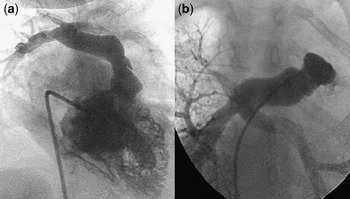
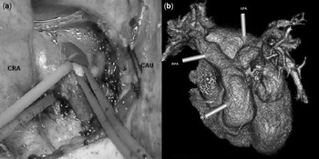
An 18 month-old girl was referred to our clinic with tetralogy of Fallot and an associated pulmonary arterial sling. She had undergone construction of 2 palliative modified Blalock-Taussig shunts, through left and right thoracotomies, following the initial echocardiographic diagnosis of tetralogy of Fallot. An angiography was performed following failure of both shunts within 3 months of the procedure to demonstrate the anatomy. Selective angiography of the pulmonary arteries demonstrated the anomalous course of the left pulmonary artery (Fig. 1a and b). We performed a median sternotomy to evaluate the pathology, and provide relief of the tracheobronchial symptoms resulting from recurrent infections of the upper and lower respiratory tract, as well as cyanotic and hypoxic spells. We confirmed the presence of the pulmonary arterial sling (Fig. 2a), and found that the left pulmonary artery was 4 millimeters in diameter as it reached the left hilum. Both Blalock-Taussig shunts were occluded, this contributing on the left side to the small diameter of the pulmonary artery near the left hilum. On standard cardiopulmonary bypass, we liberated the left pulmonary artery from its origin on the right, transferring it to the left behind the trachea and the aorta following extensive liberation from adhesions with blunt and sharp dissections. Cold cardioplegic arrest was achieved for intracardiac repair of the ventricular septal defect, and a transjunctional patch of autologous pericardium was placed up to the bifurcation of the pulmonary trunk. Division of the occluded shunt from the left pulmonary artery yielded a small luminal area, permitting augmentation with a pericardial patch and anastomosis to the reconstructed pulmonary trunk, thus achieving an acceptable perioperative ratio of right ventricular to left ventricular pressures (Fig. 2b). The patient was weaned off cardiopulmonary bypass on moderate doses of dopamine, and was extubated at the 6th postoperative hour. She was discharged to the ward on the second day, did not experience any complications postoperatively, and was discharged on the 8th day.
The anatomy is revealed by a right ventricular injection (a). A selective injection in the pulmonary artery (b) shows the left pulmonary artery to be originating from the inferoposterior aspect of the right pulmonary artery.

The intraoperative view shows the left pulmonary artery originating posteroinferiorly from the right pulmonary artery (a). Rightward, cranial and caudal sides are shown as R, CRA and CAU, respectively. The postoperative computed tomographic scan (b) shows the satisfactory anatomical correction of both the right ventricular outflow tract and the pulmonary arterial branches subsequent to the extensive reconstruction.

Discussion
The principal areas of controversy involving patients who present with pulmonary arteral sling and congenital tracheal stenosis include the optimal methods to establish the diagnosis, the relative merits of pulmonary arterial re-implantation versus translocation, the need for cardiopulmonary bypass, and the most advantageous technique to repair extensive segments of congenital tracheal stenosis.Reference Fiore, Brown, Weber and Turrentine2 Bronchoscopic investigation eliminated the latter complication in our patient. Irrespective of the associated intracardiac lesions, the optimal method to repair the sling is sternotomy with cardiopulmonary bypass. This technique permits easy dissection of the left pulmonary artery, and provides a safe uncompromised environment to re-implant the left pulmonary artery into the pulmonary trunk after adequate resection of all residual ductal tissue with deflation of the lungs.Reference Fiore, Brown, Weber and Turrentine2
Its association other cardiac defects, mostly atrial or ventricular septal defects, has been reported. Single stage correction of the sling associated with tetralogy of Fallot with extensive reconstruction of the pulmonary arteries and trunk, however, has been reported only rarely.Reference Hanafusa, Uemura, Yagihara, Kagisaki, Takahashi and Kitamura3, Reference Takeda, Asou, Fakhri, Rahayoe, Yoshimura and Rachmat4 Others have preferred a staged approach, with total correction after the repair of the pulmonary sling and an additional palliative shunt.Reference Fiore, Brown, Weber and Turrentine2 The approach may depend on the timing of diagnosis of the sling, the size of the left pulmonary artery, and the feasibility of repairing as opposed to palliating the tetralogy of Fallot. In our patient, a later diagnosis precluded early repair of the sling during initial palliation. Repair of the sling during palliation avoids subsequent compression as the left pulmonary artery starts to grow.
Our experience shows that extensive reconstruction of the pulmonary arteries, particularly of the left, with an adequate relief of the right ventricular outflow tract and the pulmonary trunk, does permit single-stage correction of both pathologies in selected patients, resulting in favourable physiology and anatomy.