Non-ischemic idiopathic dilated cardiomyopathy is the most common cause of advanced heart failure in children albeit it is more frequently seen in adults. Reference Towbin, Lowe and Colan1,Reference Lipshultz, Sleeper and Towbin2 Despite the natural history and long-term progression of dilated cardiomyopathy in children not being fully characterised, limited available data from international registries suggest that up to 50% of such patients either die or undergo heart transplantation within 5 years of presentation. Reference Towbin, Lowe and Colan1 Complex interplay of genetic factors influenced by environmental triggers predates the pathophysiology of left ventricle dilatation and remodelling, myocyte apoptosis, collagen accumulation, and fibrosis that leads to the replacement of the viable myocardium. Reference Schultheiss, Fairweather and Caforio3 Also, there is a finite risk of sudden death in the paediatric age group that is not well defined, although population-based studies have described certain risk factors like age < 14 years, familial dilated cardiomyopathy, higher left ventricular dilation, and lower posterior wall thickness and fractional shortening. Reference Pahl, Sleeper and Canter4–Reference Bharucha, Lee and Daubeney6
Myocardial fibrosis assessed by late gadolinium enhancement on cardiac MRI highlights the focal pathology of the disease in the affected myocardial segments. Myocardial fibrosis is known to be associated with adverse events in dilated cardiomyopathy. Reference Assomul, Prasad and Lyne7,Reference Al-Wakeel-Marquard, Degener and Herbst8 A native T1 mapping on cardiac MRI (combined proton signal from myocytes and interstitium before gadolinium contrast administration) is a surrogate of extracellular volume. The histopathologic correlation with myocardial fibrosis provides an attractive imaging alternative in the evaluation of end-stage heart failure. Reference Nakamori, Dohi and Ishida9 It is particularly useful to understand the diffuse nature of involvement in dilated cardiomyopathy, especially early onset of fibrosis that may not be apparent on late gadolinium enhancement. Reference Nakamori, Dohi and Ishida9
T1 mapping is reported to be independently predictive of all-cause mortality and heart failure-related events in adults with non-ischemic dilated cardiomyopathy. Reference Puntmann, Carr-White and Jabbour10 We sought to assess the conditional survival of children and adolescents with advanced heart failure due to idiopathic dilated cardiomyopathy based on native T1 mapping as a means to assess early myocardial fibrosis.
Methods
Following approval from the institutional review board (reference approval number NHH/AEC-CL-2019-421), this retrospective case–cohort study was done to evaluate all consecutive patients aged ≤ 21 years with newly diagnosed non-ischemic dilated cardiomyopathy and advanced heart failure (NYHA class ≥ 2) and underwent a cardiac MRI between 1 January, 2017 and 30 June, 2019. Data on follow-up were tracked till 31 December, 2019. We found 69 records of eligible patients. Only 57 patient’s records were included (Supplemental Text 1 & 2). We obtained a waiver of obtaining a written informed consent, owing to the nature of the study; a retrospective data review and capturing de-identified patient records. An attempt was made to contact patients/relatives of patients, who did not have a follow-up record. This consent was obtained orally from the parents to do a telephonic follow-up.
All patients had an echocardiographic diagnosis of dilated cardiomyopathy (m-mode left ventricular internal dimension in diastole z score ≥ 2, left ventricular ejection fraction ≤ 45%, and the absence of structural heart disease) prior to cardiac MRI. Patients with heart failure and left ventricular dysfunction due to other cardiomyopathies (hypertrophic cardiomyopathy, restrictive cardiomyopathy, and tachycardia-induced cardiomyopathy) or structural heart disease were excluded. Our primary objective was to evaluate the conditional survival (defined as follow-up ≥ 6 months after cardiac MRI) and its association with native T1 mapping. Our secondary objective was to study the effect of potential co-variates including NYHA functional class and imaging data on survival.
The cardiac MRI studies were performed (3-T scanner Ingenia, Philips Healthcare, the Netherlands) in accordance with the standardised protocols. Reference Kramer, Barkhausen, Flamm, Kim and Nagel11 (Please see Supplemental text 3 for the detailed institutional protocol of cardiac MRI acquisition and analysis.) A steady-state free precession, single breath-hold shortened modified Look-Locker inversion recovery sequence was used for T1 mapping, performed in a mid-cavity short-axis slice before and at 15 minutes after contrast administration. Cardiac MRI analysis was performed using commercially available software (Philips Intellispace, Philips Healthcare). The volumetric indices were adjusted to body surface area. Region of interests were placed within the septal myocardium to reduce substantial segmental variation in T1 values, in lateral and the septal segments. Reference Piechnik, Ferreira and Dall’ Armellina12 Normative data for native T1 values in children have been described to be similar to those in normal adult subjects. Reference Barczuk-Falecka, Malek, Werys, Roik, Adamus and Brzewski13 The average normal native T1 values derived as per our institutional protocol is 1250 ms in males and 1280 ms in females.
Statistical analysis was performed using R version 3.6.2. Normally distributed continuous variables were expressed as mean ± SD (median and interquartile range when applicable). For data showing a non-Gaussian distribution, comparisons were made using the Mann–Whitney U test, with a significance level of p = .05 (two-tailed). Correlations between cardiac MRI characteristics of late gadolinium enhancement and native T1 values with survival events were calculated using the two-tailed Spearman coefficient for non-parametric correlations. Multiple binary logistic regression was used to calculate the effect of multiple predictor variables (e.g., NYHA class, left ventricle ejection fraction, mitral regurgitation, pulmonary hypertension, late gadolinium enhancement, and native T1 duration) on the dichotomous outcome of survival versus death or heart transplantation.
Results
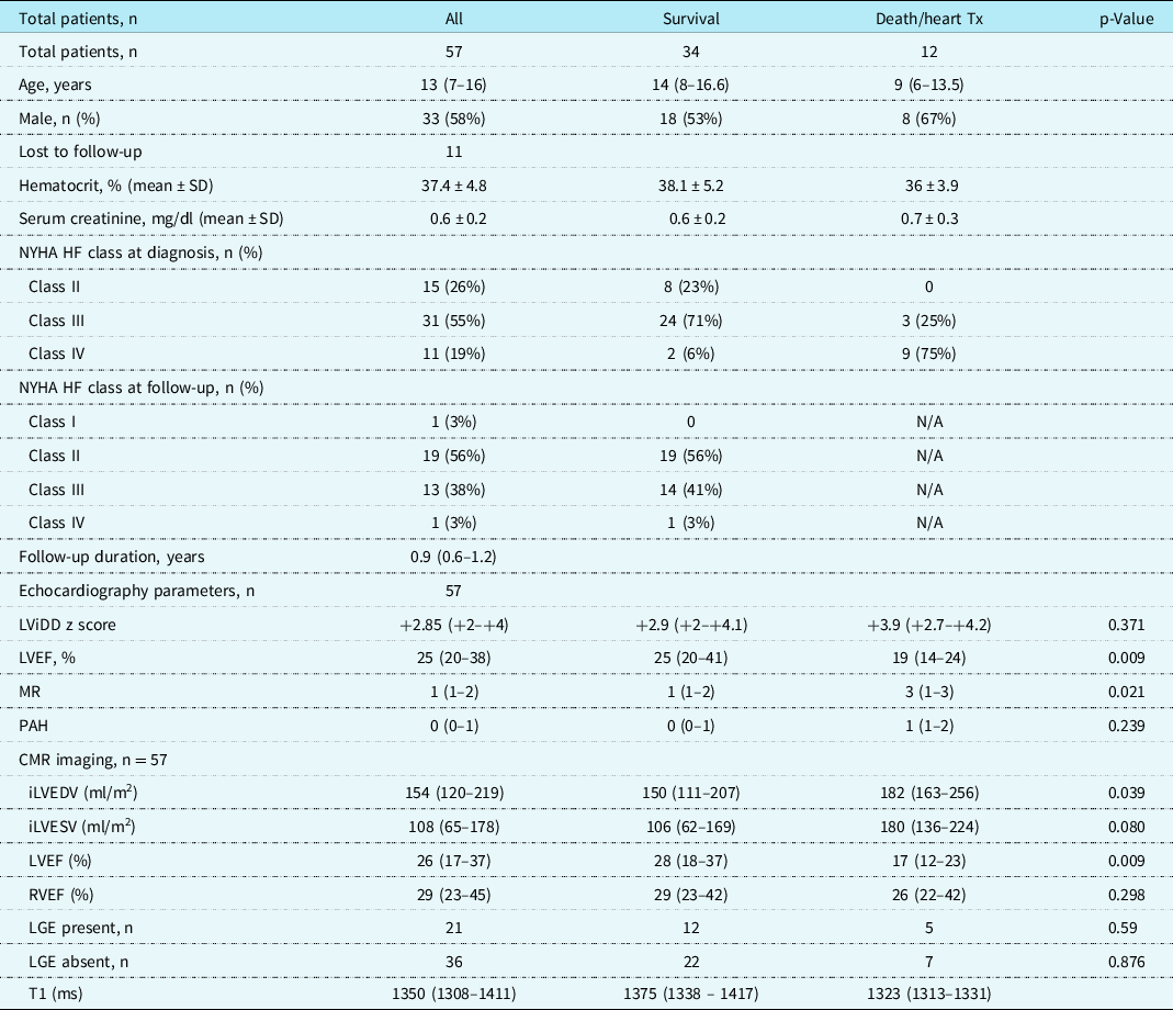
A total of 57 patients with dilated cardiomyopathy and advanced heart failure were included in the study (Table 1) [median age 13 years (range 9 months–21 years); median NYHA Class III; median duration of follow-up 0.9 years (IQR 0.6–1.2)]. Follow-up records were not available for 11patients (Supplemental Table 1). We obtained 6-month conditional survival data for 81% patients (46/57). The mortality rate was 24% (11/46), and one child had a heart transplantation. None of the study patients (n = 57) received mechanical circulatory support. Among the surviving patients, 56% patients (19/34) were in NYHA Class II and 41% patients (15/34) in either NYHA Class III or IV. The median echocardiographic left ventricle ejection fraction and left ventricle internal dimension in diastole z score were 25% (IQR 20–38) and + 2.85 (IQR + 2–+4), respectively.
Demographic, echocardiography, and CMR variables of patients with DCM and advanced HF

All values are median (interquartile range) unless otherwise specified.
CMR – cardiac magnetic resonance; HF – heart failure; iLVEDV – indexed left ventricular end-diastolic volume; iLVESV – indexed left ventricular end-systolic volume; LGE – late gadolinium enhancement; LVEF – left ventricular ejection fraction;
LViDD – left ventricular internal dimension in diastole; MR – mitral regurgitation; PAH – pulmonary artery hypertension; RVEF – right ventricular ejection fraction.
The group who survived had significantly higher left ventricle ejection fraction (p = 0.009) and lower grade of mitral regurgitation (p = 0.021) but had no difference in left ventricle internal dimension in diastole z score or pulmonary artery hypertension when compared to those who died or had heart transplant. The median left ventricle ejection fraction and right ventricle ejection fraction by cardiac MRI were 26% (IQR 17–37) and 29% (IQR 23–45), respectively. The left ventricle ejection fraction by echocardiography and cardiac MRI had a strong correlation (correlation coefficient of 0.8). The group of patients who died/underwent heart transplant had a significantly higher indexed left ventricular end-diastolic volume (p = 0.039) and lower left ventricle ejection fraction (p = 0.009) when compared to those who survived.
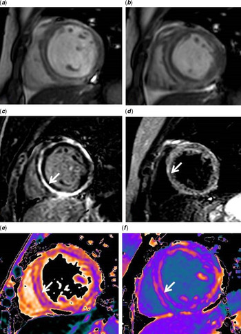
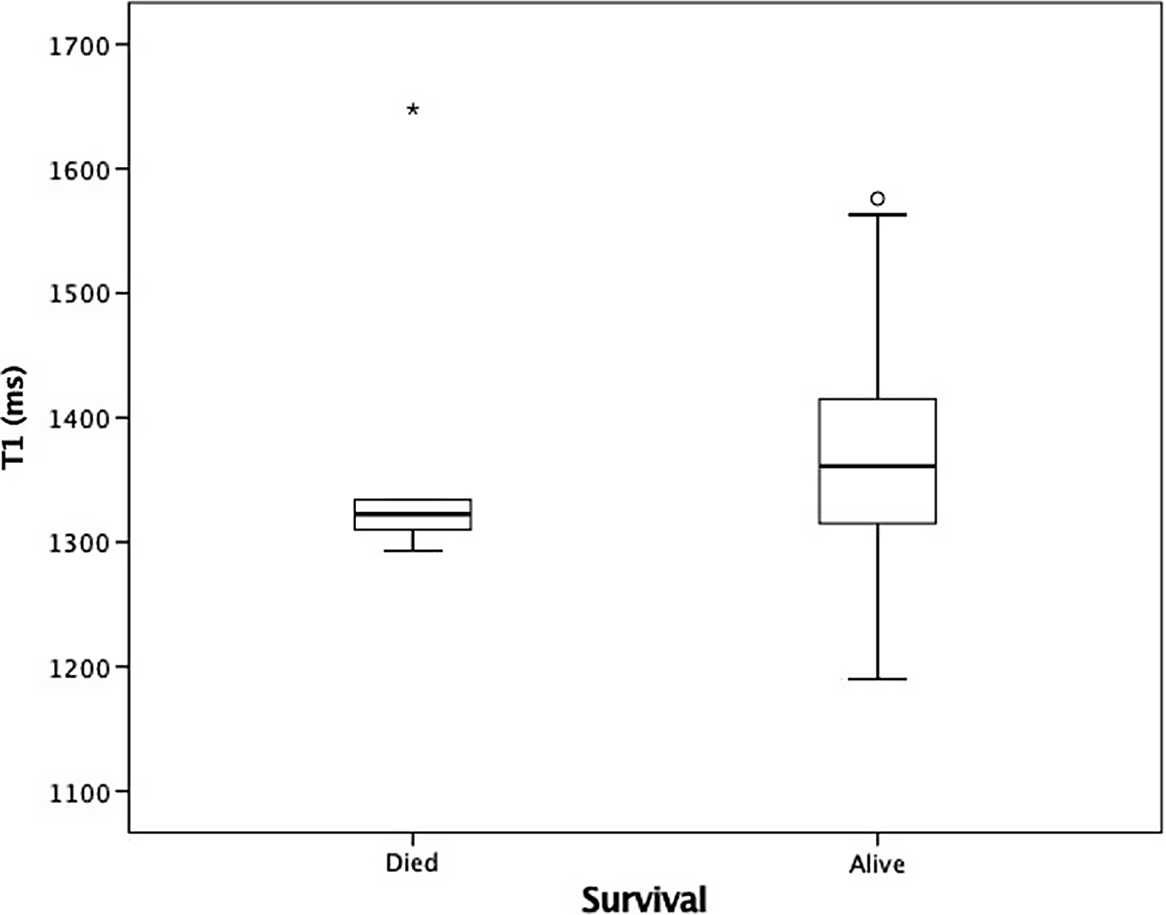
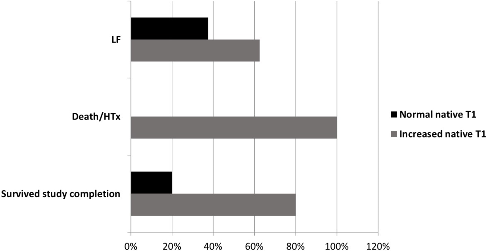
Late gadolinium enhancement on cardiac MRI was seen in 37% patients (21/57) with predominantly diffuse transmural and epicardial involvement (10/22) (Fig 1). All patients with late gadolinium enhancement had increased native T1 values, while 53% (19/36) patients without late gadolinium enhancement had increased native T1 values. The median native T1 value for the entire cohort was 1351 ms (range 1180–1648) available in 77% patients (44/57) (Fig 2). None of the patients in death group and six patients in survival group had normal T1 values. Overall, increased native T1 values were seen in 79.5% patients (35/44) (95% confidence interval, 1332–1394 ms). There was no association between absolute native T1 values with late gadolinium enhancement. On univariate analysis of native T1 values, there was no association with NYHA class at diagnosis or at follow-up, left ventricle ejection fraction by echocardiography and cardiac MRI, and severity of mitral regurgitation and pulmonary artery hypertension. On multivariate regression analysis when controlled for other co-variates (age, gender, imaging characteristics of echocardiography, and cardiac MRI) elevated native T1 values did not predict survival events (Fig 3).
(a and b) steady-state free precession short-axis end-diastolic and end-systolic images. (c) Short-axis LGE image shows mid-myocardial linear enhancement (white arrow) with further involvement of the epicardial surface along lateral and inferior wall. (d) Corresponding linear mid-myocardial oedema (white arrow) seen on short-T1 inversion recovery imaging. (e and f) Pre- and post-contrast (white arrow) T1 maps show corresponding linear pattern with high native T1 value of 1563 ms with ECV of 46%.

Boxplot depicting native T1 values of patients with DCM and advanced heart failure who died/underwent heart transplant versus those who survived study completion.

Increased versus normal native T1 values in the three groups (lost to follow-up (LOF), study survival, and death/heart transplant.

Discussion
The present study shows that native T1 values on cardiac MRI are elevated in children and adolescents with advanced heart failure due to dilated cardiomyopathy, and native T1 values do not predict 6-month conditional survival events in children and adolescents with dilated cardiomyopathy despite controlling for disease co-variates like left ventricle ejection fraction and diastolic dimension, severity of mitral regurgitation and pulmonary artery hypertension, and late gadolinium enhancement. To our knowledge, this is the first study to evaluate short-term conditional survival in children and adolescents in this cohort of heart failure in relation to 3-T cardiac MRI-derived native T1 mapping.
Although multiple complex histopathologic changes at the myocardial cellular level are known to occur in heart failure due to non-ischemic dilated cardiomyopathy, replacement of the interstitium with fibrosis predates cell death. Reference Li, Liu, Villarreal and Garcia14 Hence, recently several researchers have reported on the utility of native T1 mapping in the assessment of extracellular volume as a surrogate of histologic collagen volume. Reference Nakamori, Dohi and Ishida9 Also noteworthy is the limited ability of late gadolinium enhancement on cardiac MRI to demonstrate diffuse fibrosis in dilated cardiomyopathy due to its inherent need for spatial heterogeneity at the extracellular level of the myocytes. Reference Nakamori, Dohi and Ishida9 The findings of late gadolinium enhancement in our patients did not correlate with absolute native T1 values. This was not contrary to the fact that histological collagen volume has shown not to have strong relationship with quantitative late gadolinium enhancement. Reference Nakamori, Dohi and Ishida9 A small retrospective study in adults with dilated cardiomyopathy showed strong correlation of native T1 values of septal myocardium with late gadolinium enhancement when compared to those without late gadolinium enhancement and of normal controls. Reference Yanagisawa, Amano, Tachi and Inui15 However, lack of follow-up and unknown survival events limit the application of these results in the prognostication of young patients with advanced heart failure.
The predictive ability of native T1 does not improve when combined with left ventricle ejection fraction or late gadolinium enhancement, highlighting the differences between diffuse and focal nature of myocardial involvement and the independent disease processes in dilated cardiomyopathy (DCM). Reference Puntmann, Carr-White and Jabbour10 Majority of our patients of dilated cardiomyopathy were in NYHA Class III or IV (74%) with likely advanced disease progression. Also, the influence of goal-directed medical therapy in the clinical management of heart failure on left ventricle remodelling and consequent effect on native T1 values cannot be overemphasised.
Our study has some other limitations. First, being a retrospective study, we could not account for all the biochemical (e.g., brain natriuretic peptide or N-terminal pro-brain natriuretic peptide) and imaging attributes (e.g., extracellular volume on cardiac MRI) of advanced heart failure in the prognostic value of native T1 mapping. Second, the effect of heteroscedasticity in terms of age, body size, and gender on native T1 values in heart failure in children is not completely understood, and our study findings were limited to evaluate its association. Also, some patients with advanced heart failure may not have undergone cardiac MRI due to variation in clinical practice amongst providers contributing to the inherent selection bias. In the era of ever-increasing cost of healthcare delivery, the risk/benefits of using cardiac MRI especially in younger children is quite challenging, and a larger study with longer follow-up may give clarity on its utility in the care of such patients.
To conclude, in children and adolescents with advanced heart failure due to idiopathic dilated cardiomyopathy, diffuse myocardial fibrosis can be depicted by native T1 mapping on cardiac MRI. Although, all patients with poor outcome had elevated native T1, it did not predict 6-month conditional survival in our retrospective cohort. Finally, in contrast to our smaller retrospective study, a larger multicentre prospective study with standardised protocol of clinical management of advanced heart failure is warranted to assess the true influence of the disease process on cardiac MRI characteristics of native T1 and its potential prognostic utility.
Supplementary material
To view supplementary material for this article, please visit https://doi.org/10.1017/S1047951121001268
Acknowledgements
None.
Financial support
This research received no specific grant from any funding agency, commercial or not-for-profit sectors.
Conflict of interest
None.
Ethical standards
The authors assert that all procedures contributing to this work comply with the ethical standards of the Joint Commission International, The Government of India (National Organ and Tissue Transplant Organization under The Directorate General of Health Services), The State of Karnataka, ISHLT ethical guidelines, the 2008 Declaration of Istanbul, and the Helsinki Declaration of 1975 as revised in 2008. This research work has been approved by the institutional ethics committees (Narayana Health academic ethics committee approval number – NHH/AEC-CL-2019-421).