INTRODUCTION
Dientamoeba fragilis is a trichomonad-like protozoan parasite with a worldwide distribution commonly found in the gastrointestinal tract of humans (Stark et al. Reference Stark, Beebe, Marriott, Ellis and Harkness2006; Cepicka et al. Reference Cepicka, Hampl and Kulda2010; Stark et al. Reference Stark, Barratt, Roberts, Marriott, Harkness and Ellis2010a,Reference Stark, Barratt, Roberts, Marriott, Harkness and Ellisb; Barratt et al. Reference Barratt, Harkness, Marriott, Ellis and Stark2011a). Despite widespread belief to the contrary, numerous reports document that D. fragilis is a common cause of gastrointestinal disease in both developed and developing regions of the world and has the propensity to exist as a chronic infection with associated clinical signs of disease (Grendon et al. Reference Grendon, Digiacomo and Frost1991, Reference Grendon, Digiacomo and Frost1995; Dickinson et al. Reference Dickinson, Cohen and Schlenker2002; Norberg et al. Reference Norberg, Nord and Evengård2003; Stark et al. Reference Stark, Beebe, Marriott, Ellis and Harkness2006, Reference Stark, Barratt, Roberts, Marriott, Harkness and Ellis2010b; Barratt et al. Reference Barratt, Harkness, Marriott, Ellis and Stark2011a). It is found in all patient groups studied so far including the general population, travellers and HIV-infected individuals (Stark et al. Reference Stark, Beebe, Marriott, Ellis and Harkness2005, Reference Stark, Beebe, Marriott, Ellis and Harkness2007a, Reference Stark, Fotedar, Van Hal, Beebe, Marriott, Ellis and Harknessb; Barratt et al. Reference Barratt, Harkness, Marriott, Ellis and Stark2011a).
Several studies have reported that children are susceptible to infection with D. fragilis and present with clinical symptoms at higher rates than adults (Preiss et al. Reference Preiss, Ockert, Broemme and Otto1991; Ayadi and Bahri, Reference Ayadi and Bahri1999; Crotti et al. Reference Crotti, D'annibale, Fonzo, Lalle, Caccio and Pozio2005). Consequently, D. fragilis should be considered in the differential diagnosis of gastrointestinal infections in children (Spencer et al. Reference Spencer, Garcia and Chapin1979; Keystone et al. Reference Keystone, Yang, Grisdale, Harrington, Pillon and Andreychuk1984; Preiss et al. Reference Preiss, Ockert, Brömme and Otto1990; Stark et al. Reference Stark, Barratt, Ellis, Harkness and Marriott2009) but it is generally ignored as a cause of disease. Clinical symptoms reported include acute and chronic diarrhoea, lower abdominal pain, nausea, flatulence and constipation (Spencer et al. Reference Spencer, Millet, Garcia, Rhee and Masterson1983; Cuffari et al. Reference Cuffari, Oligny and Seidman1998). Transmission of D. fragilis is believed to be via the faecal-oral route, but the mechanism and whether it includes a helminth or a cyst-like stage, are still unclear (Stark et al. Reference Stark, Beebe, Marriott, Ellis and Harkness2006; Barratt et al. Reference Barratt, Harkness, Marriott, Ellis and Stark2011b).
Here we review existing knowledge on dientamoebiasis in children. A case-controlled study was also conducted to document the extent of D. fragilis infections in children presenting to a major Sydney Hospital.
MATERIALS AND METHODS
The study was performed at St Vincent's Hospital, Sydney. Laboratory and clinical records of D. fragilis-infected children were collected from August 2004 to July 2010. The following criteria were used for inclusion into this study: children were aged 15 years or less, full clinical history, confirmed laboratory diagnosis of D. fragilis as the sole pathogen, treatment history, follow-up stool samples were analysed to evaluate treatment regimes, clinical follow up and bacteriological cultures for enteric pathogens and virological screening for the presence of rotavirus and enteric adenoviruses was performed. A total of 41 children were included in the study along with a control group. A control group of children (established using the same criteria) free from infection of D. fragilis and confirmed by PCR (as described below), were included in the study. Where possible control subjects were age and sex matched with D. fragilis-infected children. However, this was not possible for 5 children. In the majority of these cases (90%, 37/41) only a single stool sample was collected and analysed.
Microbiological analysis
Laboratory diagnosis was performed by microscopy of permanently stained fixed faecal smears and by real-time PCR as previously described (Stark et al. Reference Stark, Barratt, Roberts, Marriott, Harkness and Ellis2010a). Bacterial cultures were performed using standard microbiological techniques to rule out the following infections; Salmonella spp., Shigella spp., Campylobacter spp., Yersinia enterocolitica, Vibrio spp., Plesiomonas spp., Aeromonas and Clostridium difficile. Virology testing was performed by an immunochromatographic screening test (Adeno/Rota STAT-PAK; Chembio Diagnostic Systems Inc., Sydney) for the detection of adenovirus and rotavirus antigen in faeces according to the manufacturer's recommendations.
Statistical methods
Differences between means of gastrointestinal symptoms of control subjects and D. fragilis-infected cases were evaluated by using a Chi-Square (χ 2) Test of Association.
RESULTS
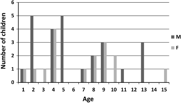
In total, 41 children were identified from laboratory and hospital records as meeting the inclusion criteria for the study. All 41 children had laboratory confirmed D. fragilis infection along with clinical notes detailing symptoms, anti-microbial treatment regime and follow-up stool samples to check for parasite clearance following treatment. The study group ranged in age from 1 to 15 years with 88% (n=36) being less than 10 years of age (Fig. 1). None of the children were immunosuppressed. No bacterial pathogens were isolated from the stools of the 41 D. fragilis-infected children. All children's stools were also negative for enteric adenoviruses and rotaviruses. The control group was comprised of 41 children, in which no D. fragilis was detected by microscopy and PCR. No bacterial or viral pathogens were detected in this group.
Age of children with dientamoebiasis, (M, Male; F, Female).

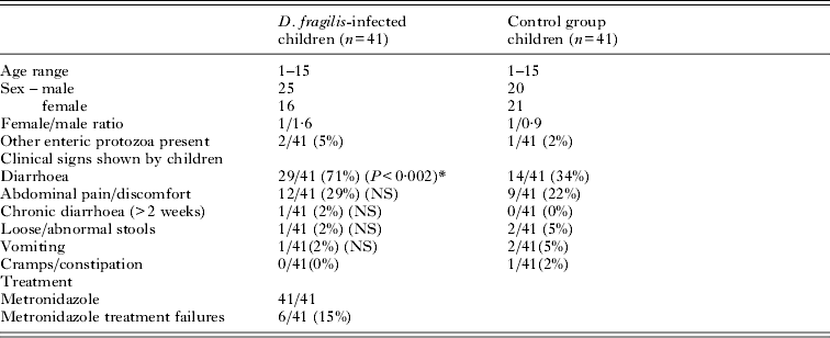
Initially, 47 D. fragilis-infected children were identified from records; among them 10 were infected with other enteric parasites; 4 with Blastocystis hominis, 2 with Cryptosporidium spp. and B. hominis, 2 with Endolimax nana, 1 with Entamoeba coli, and 1 with Enteromonas hominis. As Cryptosporidium spp. is considered to be pathogenic and B. hominis is potentially pathogenic and capable of causing gastrointestinal symptoms (Yakoob et al. Reference Yakoob, Jafri, Beg, Abbas, Naz, Islam and Khan2010), these 6 children were excluded from the data dealing with symptoms and treatment. Of the children with D. fragilis infection, 98% (40/41) presented with at least 1 or more gastrointestinal symptoms. Diarrhoea (71%) was found to be the most common symptom followed by abdominal pain (29%) (Table 1). Persistent or chronic infection was reported in 1 child.
Summary of results from children with Dientamoeba fragilis infection
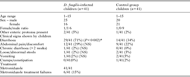
* P⩽0·05, (NS, not significant).
When compared to the control group diarrhoea was significantly higher (P<0·002) in children with dientamoebiasis. Other enteric protozoa were present in 2% (1/41) of control children. Originally, 44 control patients were identified; however, 3 were removed because they harboured B. hominis and had symptoms of diarrhoea.
In total, 41 children were treated with metronidazole and 35/41 (85%) reported complete resolution of symptoms. Follow-up stool samples collected between 1 and 4 weeks following treatment were collected from these children and all showed clearance of the parasite. However, in 6 children, gastrointestinal symptoms did not resolve following initial metronidazole treatment. Four of those children were subsequently treated with a repeat course of metronidazole for either 10 or 14 days and the other 2 children were treated with iodoquinol. Following this additional anti-microbial therapy the children reported improved clinical symptoms and follow-up stool samples collected 2 weeks post-treatment were negative for D. fragilis.
DISCUSSION
This study, conducted over a 6-year period, highlights the association of D. fragilis with clinical signs of disease: 98% of the children infected with D. fragilis studied presented with gastrointestinal symptoms. Diarrhoea was found in 29/41 (71%) of the children and abdominal pain in 12/41 (29%). The control group represents a group of symptomatic children free of D. fragilis infection. Diarrhoea was, however, more common in children with D. fragilis infection compared to the control group.
There are other reports from various parts of the world that also describe an association between D. fragilis infection in children and various clinical symptoms, most commonly diarrhoea and abdominal pain. A large study comprising over 43 029 children first reported a correlation between D. fragilis infection and symptoms of diarrhoea, abdominal pain and loose stools (Yang and Scholten, Reference Yang and Scholten1977). In that study, chronic infections were found in 2% of children. Spencer et al. (Reference Spencer, Millet, Garcia, Rhee and Masterson1983) reported a study from 104 children, in which diarrhoea and abdominal pain were the most common symptoms in those with D. fragilis infection. Preiss et al. (Reference Preiss, Ockert, Brömme and Otto1990) reported that among 102 children, 7 had acute diarrhoea, 39 had relapsing diarrhoea, 7 had bloody stools and 29 had abdominal pain. A retrospective study of 87 Swedish children diagnosed with D. fragilis found the majority of children had symptoms of diarrhoea, abdominal pain and flatus (Norberg et al. Reference Norberg, Nord and Evengård2003). Previous reports have also highlighted the propensity of the parasite to cause prolonged infection (Stark et al. Reference Stark, Beebe, Marriott, Ellis and Harkness2005; Crotti and D'Annibale, Reference Crotti and D'annibale2007) with chronic infections reported in the literature to last as long as 2 years (Wenrich, Reference Wenrich1944).
Dientamoeba fragilis is a commonly encountered enteric protozoan parasite in children that should be considered in any differential diagnosis of gastrointestinal disease. Several studies have shown D. fragilis to be more prevalent than Giardia intestinalis in paediatric populations (Preiss et al. Reference Preiss, Ockert, Brömme and Otto1990; Crotti and D'Annibale, Reference Crotti and D'annibale2007; Rayan et al. Reference Rayan, Ismail and El Gayar2007). One serological study carried out in children reported a D. fragilis seroprevalence of 91% (Chan et al. Reference Chan, Stewart, Guan, Robb, Fuite, Chan, Diaz-Mitoma, King, MacDonald and Mackenzie1996). When compared to the seroprevalence of Giardia and Cryptosporidium this study suggests that childhood contact with Dientamoeba is common (Chan et al. Reference Chan, Stewart, Guan, Robb, Fuite, Chan, Diaz-Mitoma, King, MacDonald and Mackenzie1996). Yang and Scholten (Reference Yang and Scholten1977) found D. fragilis in 4·2% of individuals who submitted stools for parasitological examination during 1970–1974 in Ontario, Canada. Infections were found to be more common in children, with nearly half of the infections occurring in individuals under 20 years of age (Yang and Scholten, Reference Yang and Scholten1977). A recent study from the Netherlands found that D. fragilis infection was most common in children aged between 5 and 14 years (de Wit et al. Reference De Wit, Koopmans, Kortbeek, van Leeuwen, Vinjé and van Duynhoven.2001).
Interestingly, an association was observed between D. fragilis infection and carriage of other enteric protozoa normally transmitted via the faecal-oral route. This association has previously been observed (Stark et al. Reference Stark, Barratt, Roberts, Marriott, Harkness and Ellis2010b) and suggests that transmission of D. fragilis also occurs in the same way (by the faecal-oral route). There was no evidence for the presence of helminths in this study, which have previously been suggested to be a host for D. fragilis (Stark et al. Reference Stark, Beebe, Marriott, Ellis and Harkness2006). Recent studies also suggest that helminths such as Enterobius vermicularis appear to play no role in transmission of D. fragilis (Stark et al. Reference Stark, Barratt, Roberts, Marriott, Harkness and Ellis2010b, Barratt et al. Reference Barratt, Harkness, Marriott, Ellis and Stark2011b).
Anti-microbial treatment most commonly used for treatment of D. fragilis infection in children includes metronidazole and idoquinol (diidohydroxyquin) (Stark et al. Reference Stark, Barratt, Roberts, Marriott, Harkness and Ellis2010b). Therapy with metronidazole was effective for most of the children in this study. Forty-one children were administered the drug, with the duration of treatment varying from 5 to 10 days. Of the children treated with metronidazole 85% (35/41) resulted in clearance of D. fragilis as determined by analyses of follow-up stools and complete resolution of gastrointestinal symptoms. However, 6/41 (15%) children who underwent metronidazole treatment failed to clear the infection parasitologically or clinically. There was no correlation between the dose received, the duration of treatment and treatment failure associated with metronidazole use. These 6 children underwent further treatment, 4 were put on a repeat course of metronidazole for 2 weeks duration and the other 2 children were treated with iodoquinol. On follow-up examination all children reported marked clinical improvement and clearance of parasite from stool samples. Iodoquinol has been widely used to treat D. fragilis infections (Butler, Reference Butler1996). In a recent study, 27/33 children were successfully treated with clioquinol, a member of the same drug family as iodoquinol (Bosman et al. Reference Bosman, Benninga, van De Berg, Kooijman and van Gool2004).
There are varying reports on the efficacy of metronidazole treatment for D. fragilis infections in children. Spencer et al. (Reference Spencer, Garcia and Chapin1979) reported that therapy with metronidazole was effective in 35 children. Similarly, in New Zealand, metronidazole was successfully used in the treatment of dientamoebiasis in 3 children (Oxner et al. Reference Oxner, Paltridge, Chapman, Cook and Sheppard1987). Preiss et al. (Reference Preiss, Ockert, Brömme and Otto1990) studied 123 paediatric children with D. fragilis infections. They found metronidazole to be effective, with 70% of children eliminating the parasite and symptoms after a single treatment. A second treatment was required for 21 other children with another drug. Ten children were treated a third time in order to eliminate D. fragilis and accompanying abdominal complaints. They recommended a 10-day treatment with metronidazole for D. fragilis infections. Cuffari et al. (Reference Cuffari, Oligny and Seidman1998) showed that metronidazole was effective in the treatment of 5 children. A study from Sweden included 32 children whom were treated with metronidazole. The drug was given at various doses for various lengths of time, and they found that only 4 children responded to the metronidazole treatment (Norberg et al. Reference Norberg, Nord and Evengård2003). Recently, it was also documented that 2 children presented with D. fragilis infection over prolonged periods (Stark et al. Reference Stark, Barratt, Ellis, Harkness and Marriott2009). In that study, metronidazole was used initially, but subsequently treatment was done with paramomycin for 10 days to effect clearance of the parasite. At this point in time, it is not clear why some cases of dientamoebiasis do not respond to metronidazole treatment; such observations may be the result of metronidazole resistance or failure to comply with medication (Stark et al. Reference Stark, Barratt, Roberts, Marriott, Harkness and Ellis2010b).
In conclusion, this study serves to highlight the fact that children infected with D. fragilis typically have diarrhoea. Studies such as those reported here, and elsewhere, strongly implicate D. fragilis as a common cause of gastrointestinal disease in children and other patient populations (Reference Stark, Beebe, Marriott, Ellis and HarknessStark et al. Reference Stark, Beebe, Marriott, Ellis and Harkness2006, Reference Stark, Barratt, Roberts, Marriott, Harkness and Ellis2010b; Barratt et al. Reference Barratt, Harkness, Marriott, Ellis and Stark2011a). Dientamoeba fragilis was not detected in the control group. We therefore recommend that all laboratories must routinely test for D. fragilis because treatment that eliminates the parasite usually results in the resolution of symptoms. It is essential that a correct clinical and laboratory diagnosis is made in order that treatment can be initiated.
ACKNOWLEDGMENTS
We acknowledge the help of the staff at St Vincent's Hospital Microbiology Department in the collection and processing of stool samples positive for D. fragilis. Part of this work was presented at the XIIth International Congress of Parasitology (ICOPA 2010), Melbourne, Australia (16 August, 2010). This research was supported by a grant from the St Vincent's Hospital, Darlinghurst, Australia, University of Technology, Sydney and Australian Research Council. Ms S. Fletcher provided advice on statistics.