Implications
Nutrient sensing by free fatty acid receptors (FFARs) and hydroxycarboxylic acid receptors (HCARs) links energy substrates with cellular response in different tissues or cells. Sensing by these receptors allows reactions on a cellular level, which are independent of the responses that depend on the function of their ligands as energy substrates. The receptors of both families are expressed in different organs including intestine, where the receptor ligands are produced by the intestinal microbiota. Compared with the human or rodent models, less data are available regarding farm animal species. The available data are primarily linked to the distribution of mRNA compared with proteins.
Introduction
Nutrient sensing represents the ability of the organism to recognise and respond to macronutrients linked to energy metabolism and biomass production (Efeyan et al., Reference Efeyan, Comb and Sabatini2015). Nutrient sensing involves sensory mechanisms at different organism localisations (e.g. tongue, stomach and other parts of the intestinal tract) (Steinert et al., 2011; Kokrashvili et al., Reference Kokrashvili, Yee, Ilegems, Iwatsuki, Li, Mosinger and Margolskee2014; Vancleef et al., Reference van der Wielen, van Avesaat, de Wit, Vogels, Troost, Masclee, Koopmans, van der Meulen, Boekschoten, Muller, Hendriks, Witkamp and Meijerink2015) and also on different organs or cells (e.g. pancreatic β-cells that affect insulin secretion) (Kristinsson et al., Reference Kristinsson, Bergsten and Sargsyan2015), or adipose tissue, thereby affecting insulin sensitivity (Kimura et al., Reference Kimura, Ozawa, Inoue, Imamura, Kimura, Maeda, Terasawa, Kashihara, Hirano, Tani, Takahashi, Miyauchi, Shioi, Inoue and Tsujimoto2013). Carbohydrates, amino acids, fatty acids, β-hydroxybutyric acid (BHBA) and lactate are involved in these nutrient-sensing mechanisms. All of the above mentioned substrates directly affect the metabolism because of their function as energy sources and as receptor ligands (Offermanns et al., 2011; Janssen and Depoortere, Reference Janssen and Depoortere2013). Therefore, they act as metabolic regulators. Nuclear receptor binding (e.g. long-chain fatty acids (LCFA)) (Kliewer et al., 1997) and membrane-bound receptor binding are involved in these sensing mechanisms. The latter is the focus of this paper and refers to short-chain fatty acids (SCFA), medium-chain fatty acids (MCFA), LCFA (Briscoe et al., 2003; Brown et al., 2003), BHBA (Taggart et al., 2005) and lactate (Cai et al., 2008).
Fatty acids sense (e.g. caloric intake) via the gut-brain dopamine axis as demonstrated in mice (Ferreira et al., 2012) and are important fuels during fasting (e.g. for skeletal muscles) (de Lange et al., 2007). The nuclear receptor peroxisome proliferator-activated receptor δ, which binds LCFA, is the important component that switches glucose oxidation to fatty acid oxidation during starvation in skeletal muscle (Tanaka et al., 2003). Ketone bodies are the energy sources during starvation. The ketone body BHBA reduces lipolysis in men (Senior and Loridan, 1968) and is, therefore, part of a classical feedback mechanism in the millimolar range, as discussed by Offermanns et al. (2011). The inhibitory effects of the ketone body BHBA on lipolysis were demonstrated in the bovine in the 1970 of last century (Metz and van den Bergh, 1972). The authors reported the reduction of lipolysis by BHBA using subcutaneous adipose tissue explants in vitro. Moreover, a comparable observation was performed by these authors using SCFA butyrate.
As discussed by Efeyan et al. (Reference Efeyan, Comb and Sabatini2015), effective nutrient-sensing mechanisms might have evolved due to nutrient scarcity during the evolution. These mechanisms should help control the intestinal energy uptake, as well as switching between energy storage and energy release. Limited knowledge is available on direct nutrient-sensing mechanisms in different species, as stated by Efeyan et al. (Reference Efeyan, Comb and Sabatini2015). This statement includes farm animal species. In these species, some data on receptor abundances, mostly the level of RNA, are available. However, the information on functional importance is scarce.
Family of free fatty acid receptors
General aspects of free fatty acid receptor signalling and metabolic consequences
SCFA, MCFA and LCFA are endogenous ligands for the receptor family of FFARs that belong to the group of seven transmembrane-spanning G-protein-coupled receptors (GPR) (Brown et al., 2005). Originally, the group consisted of GPR40, GPR41, GPR42 and GPR43 (Sawzdargo et al., 1997). After deorphanisation they were named FFAR1 (GPR40), FFAR2 (GPR43) and FFAR3 (GPR41) (Stoddart et al., 2008). Recently, after the identification of specific ligands of receptor GPR120 by Hirasawa et al. (2005), the receptor was integrated into the family as FFAR4. The human receptor GPR42, which is expressed only in primates and does not respond to SCFA, was characterised as a pseudogene (Brown et al., 2003). However, the additional six amino acids and R/W exchange at position 174, which is essential for functionality but is found only in parts of the studied human population, exhibit polymorphisms (Liaw and Connolly, 2009). In addition, recently, it was shown that the receptor sequence of GPR42 is highly heterogeneous in humans, and its variant was functional and comparable with FFAR3. Therefore, the authors suggested that the importance of GPR42 should be revaluated (Puhl et al., Reference Puhl, Won, Lu and Ikeda2015).
The receptor FFAR1, which is stimulated by MCFA and LCFA, is coupled to G-protein subunit Gα q11 and Gα i/o signalling that is comparable with FFAR2, which is activated by SCFA such as FFAR3; the latter is also coupled to Gα i/o signalling (Stoddart et al., 2008). The receptor FFAR4 differs from FFAR1 and is preferentially activated by LCFA (Hirasawa et al., 2005). For the Gα q11-coupled receptor FFAR4 (Hirasawa et al., 2005; Oh et al., 2010), which is similar to FFAR1 and FFAR2, the β-arrestin pathway signalling was demonstrated (Hudson et al., 2012; Watson et al., 2012; Mancini et al., Reference Mancini, Bertrand, Vivot, Carpentier, Tremblay, Ghislain, Bouvier and Poitout2015).
The activation of FFAR1 increases glucose-stimulated insulin secretion (Doshi et al., 2009; Nagasumi et al., 2009) and glucagon secretion (Flodgren et al., 2007). The activation of FFAR1 improves hepatic insulin sensitivity, as demonstrated in vitro (Wu et al., 2012), and steatosis in mice in vivo (Ou et al., Reference Ou, Wu, Lu, Su, Hung, Wu, Yang, Wu and Chang2014). The receptor FFAR2 shows antilipolytic activity. It is linked to adipogenesis and reduces isoproterenol-stimulated lipolysis (Hong et al., 2005). Notwithstanding the functions of FFAR2, the activation of FFAR3 increases leptin secretion from adipose tissue (Xiong et al., 2004). Both, FFAR2 and FFAR3 affect ghrelin secretion. However, compared with FFAR3, the activation of FFAR2 is mainly responsible for a reduction of ghrelin secretin from gastric cells (Engelstoft et al., Reference Engelstoft, Park, Sakata, Kristensen, Husted, Osborne-Lawrence, Piper, Walker, Pedersen, Nohr, Pan, Sinz, Carrington, Akiyama, Jones, Tang, Ahmed, Offermanns, Egerod, Zigman and Schwartz2013). Recently, it was demonstrated in the FFAR2 and FFAR3 knockout mice that SCFA acetate reduces glucose-stimulated insulin secretion (Tang et al., Reference Tang, Ahmed, Gille, Lu, Grone, Tunaru and Offermanns2015). In contrast, it was recognised in the 1960 of the last century that SCFA propionate, butyrate and valerate, in contrast to acetate, stimulate insulin secretion in ruminants but not in monogastrics (Horino et al., 1968).
The activation of FFAR4 increases adipogenesis (Gotoh et al., 2007), lipogenesis (Ichimura et al., 2012), improves insulin sensitivity and shows anti-inflammatory properties (Oh et al., 2010).
All receptors of the family are expressed on the innate immune system cells but show different expression patterns on different immune cells (Briscoe et al., 2003; Brown et al., 2003; Konno et al., Reference Konno, Ueki, Takeda, Kobayashi, Tamaki, Moritoki, Oyamada, Itoga, Kayaba, Omokawa and Hirokawa2015).
The fatty acid receptors are expressed on enteroendocrine cells with differences in their distribution between different cell types. All receptors are found on L-cells, which are responsible for the secretion of glucagon-like peptide-1 (GLP-1) and peptide yy (PYY). In FFAR3 knockout mice it was shown that this receptor is linked to the PYY release (Samuel et al., 2008), which is associated with reduced intestinal motility and reduced appetite; the latter is also relevant for GLP-1 release (De Silva and Bloom, 2012). Recently, it was proven that FFAR2 was associated with PYY secretion (Psichas et al., Reference Psichas, Sleeth, Murphy, Brooks, Bewick, Hanyaloglu, Ghatei, Bloom and Frost2015). In knockout mice it was also demonstrated that FFAR2 was linked with the GLP-1 release, though to a higher extend than FFAR3 (Tolhurst et al., 2012). In addition, the receptor FFAR2 is found on leukocytes located within the lamina propria in contrast with FFAR3, which is detected on cell bodies of submucosal and myenteric ganglia of mice (Nohr et al., Reference Nohr, Pedersen, Gille, Egerod, Engelstoft, Husted, Sichlau, Grunddal, Poulsen, Han, Jones, Offermanns and Schwartz2013).
The colocalisation of FFAR1 in association with different gastrointestinal hormones in different parts of the gastrointestinal tract was evaluated (Edfalk et al., 2008). As discussed by Hara et al. (Reference Hara, Ichimura and Hirasawa2014), based on the expression of gastrointestinal hormones, FFAR1 and FFAR4 were found on L-cells. In addition, the receptor FFAR1 was described on I-cells (responsible for glucose-dependent insulinotropic peptide secretion) and K-cells (associated with cholecystokinin (CCK) secretion), comparable with FFAR4 (Liou et al., 2011). However, using the murine enteroendocrine cell line STC-1, it was shown that the effect on GLP-1 secretion (Hirasawa et al., 2005) and CCK secretion (Tanaka et al., 2008) was linked to FFAR4 and not to FFAR1.
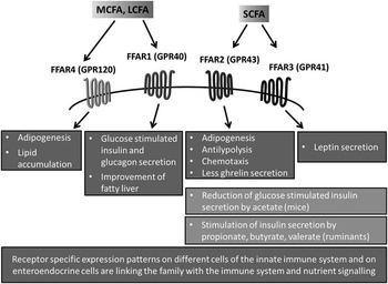
Based on recent studies, it is becoming increasingly clear that proximal and distal L-cells differ regarding the gastrointestinal hormone cosecretion. This can signify that different L-cell types exist (Svendsen et al., Reference Svendsen, Pedersen, Albrechtsen, Hartmann, Torang, Rehfeld, Poulsen and Holst2015) and FFAR1 activation by linoleic acid may affect GLP-1 release only by vascular and not luminal stimulation (Christensen et al., Reference Christensen, Kuhre, Janus, Svendsen and Holst2015). Therefore, the data on cosecretion solely from in vitro models should be scrutinised with care. General functions of FFARs are shown in Figure 1.
General functions of free fatty acid receptors (FFARs) 1 to 4 in different tissues or cells. The information came mainly from studies in rodents or humans. The receptors are also found on immune cells and are involved in different tasks of the immune system (Alvarez-Curto and Milligan, Reference Alvarez-Curto and Milligan2016) in addition to the release of several gastrointestinal hormones, besides the down-regulation of gastric ghrelin secretion. Activated FFAR2 and FFAR3 reduce ghrelin secretion, but the impact of FFAR3 is of minor importance (Engelstoft et al., Reference Engelstoft, Park, Sakata, Kristensen, Husted, Osborne-Lawrence, Piper, Walker, Pedersen, Nohr, Pan, Sinz, Carrington, Akiyama, Jones, Tang, Ahmed, Offermanns, Egerod, Zigman and Schwartz2013). The stimulation of insulin secretion by short-chain fatty acids (SCFA) in ruminants is well known but an association with FFAR2 or FFAR3 was not researched yet. Other references are part of the manuscript. LCFA=long-chain fatty acids; MCFA=medium-chain fatty acids; GPR=G-protein-coupled receptors.

Free fatty acid receptors in farm animal species
Pig
The messenger RNAs (mRNAs) expression patterns of FFAR2 and FFAR3 were researched at first in different segments of the porcine intestine by Haenen et al. (Reference Haenen, Zhang, Souza da Silva, Bosch, van der Meer, van Arkel, van den Borne, Perez Gutierrez, Smidt, Kemp, Muller and Hooiveld2013). In 2014, the porcine FFAR2 was cloned by two groups (Li et al., Reference Li, Su, Zhou and Yao2014a; Zhang et al., Reference Zhang, Cheng, Wang, Yu and Li2014). In parallel, the receptor FFAR3 was cloned by Li et al. (Reference Li, Su, Zhou and Yao2014a) and mRNA (Li et al., Reference Li, Su, Zhou and Yao2014a; Zhang et al., Reference Zhang, Cheng, Wang, Yu and Li2014), and also the protein data (Li et al., Reference Li, Su, Zhou and Yao2014a) became available from other tissues. Because of mRNA expression patterns with the highest abundance of FFAR2 in spleen and FFAR3 in ileum, it was suggested that FFAR2 in spleen may indicate an important interaction with the immune response, and FFAR3 may be linked with PYY secretion. However, the available data on protein abundances illustrate that protein may be not representative of the quantitative mRNA data.
Based on a study by Haenen et al. (Reference Haenen, Zhang, Souza da Silva, Bosch, van der Meer, van Arkel, van den Borne, Perez Gutierrez, Smidt, Kemp, Muller and Hooiveld2013) in multiparous female pigs, it became clear that the highest occurrences of both receptor mRNAs, which were found in the distal part of the small intestine, are not consistent with the abundance of produced SCFA, which is highest in caecum and colon. Nevertheless, there was an overlap with the mRNAs of glucagon, angiopoietin-like protein 4, that linked gut microbiota with energy storage in adipose tissue (Zhu et al., 2012), sodium-coupled monocarboxylate transporter 5A8, responsible for SCFA, lactate transport (Coady et al., 2004) and PYY; the latter showed a higher abundance in small intestine and colon.
Colocalisation of FFAR2 and FFAR3 with PYY, GLP-1 and 5-hydroxytryptamine was demonstrated in pig colon using immunohistochemistry (Hamilton Weatherburn, Reference Hamilton Weatherburn2015). An ontogenetic pattern of the receptor mRNA abundance in different tissues was recorded by Li et al. (Reference Li, Yao and Jiang2014b) from day 1 postpartum to day 160. In most tissues, such as in liver, the expression was down regulated in growing and finishing pigs. However, based on the mRNA abundance in adipose tissue, the authors concluded that both receptors might have a critical role in adipogenesis between day 25 and day 70. An increase in adipogenesis by SCFA was demonstrated by Li et al. (Reference Li, Su, Zhou and Yao2014a) in vitro. However, the effects were rather independent of FFAR2 and FFAR3 because these receptor mRNAs were not detected during differentiation of porcine adipocytes using the stromal vascular cell fraction.
The SCFA binding to porcine FFAR2 was characterised by Zhang et al. (Reference Zhang, Cheng, Wang, Yu and Li2014). In addition, this group also identified an orthologue named FFAR2-like (FFAR2L) that was found in 9 out of 42 analysed mammalian species, including elephants and cats. The mRNA of FFAR2L was primarily observed in the gastrointestinal tract in addition to a very weak signal found in liver and spleen (Zhang et al., Reference Zhang, Cheng, Wang, Yu and Li2014). In contrast to FFAR2, the activation of FFAR2L by butyrate was not found.
Yan and Ajuwon (Reference Yan and Ajuwon2015) detected that butyrate inhibits basal and isoproterenol-stimulated lipolysis in pigs. The authors suggested that this effect could be an indication of transmission by FFAR3 rather than by FFAR2 because of its higher affinity for butyrate found in other monogastric species in addition to the higher abundance of FFAR3 mRNA compared with FFAR2 mRNA after stimulation with butyrate. Indeed, the maximal effective concentration (EC50) of FFAR3 is 1.05 mM, and the EC50 of FFAR2 is 16.2 mM, as shown in a thesis by Hamilton Weatherburn (Reference Hamilton Weatherburn2015).
First data on FFAR1 and FFAR4 mRNA came from 42-day-old pigs. In different segments of the intestine, FFAR4 mRNA was found with a higher abundance in colon compared with small intestine (Colombo et al., 2012). This is comparable with observations by van der Wielen et al. (Reference van der Krieken, Popeijus, Mensink and Plat2014) in 10-week-old piglets in which the abundance was also higher in the distal parts of the intestine. In contrast, FFAR1 mRNA was not detected within the intestine (Colombo et al., 2012), and further data on this receptor is not yet available in the pig.
The porcine FFAR4 was cloned and characterised by Song et al. (Reference Song, Peng, Ren, Wei and Peng2015). In total, three splice variants, comparable with the human receptor, were identified. The variant 3 did not have the 4th and 5th transmembrane domains. The wild-type receptor mRNA was detected in adipose tissue, jejunum, duodenum and spleen but not in other tissues; colon was not analysed. The variant 2 was abundantly expressed in spleen. As calculated by the authors, the EC50 in case of the wild-type receptor was between 1 and 10 µM and depended on the fatty acid ligand. The potential of activation by the analysed n-3 polyunsaturated fatty acids was higher compared with tested n-6 polyunsaturated fatty acids. The variant 2 with a shortened 4th transmembrane domain could not be stimulated by LCFA and did not act as a dominant-negative isoform. Based on the number of only three small nuclear polymorphisms found by Fontanesi et al. (Reference Fontanesi, Bertolini, Scotti, Schiavo, Colombo, Trevisi, Ribani, Buttazzoni, Russo and Dall’Olio2015), it was suggested that the FFAR4 gene was conserved in this species.
Recently, it was demonstrated that the expression of FFAR4 in porcine adipose tissue was driven by the transcription factor CCAAT/enhancer binding protein β (Chen et al., Reference Chen, Zhou, Zhang, Zhang, Zhang, Zhan, Tang, Deng, Lei and Xiong2016), which is involved in metabolic regulation, adipogenesis and inflammation (van der Krieken et al., Reference Vancleef, Van Den Broeck, Thijs, Steensels, Briand, Tack and Depoortere2015). An association with back fat thickness was not found (Fontanesi et al., Reference Fontanesi, Bertolini, Scotti, Schiavo, Colombo, Trevisi, Ribani, Buttazzoni, Russo and Dall’Olio2015).
Chicken
Compared with pig and ruminant species, fewer data are available on FFARs in chicken, and some of the data may be controversial. In 2008, it was demonstrated that linoleic acid-stimulated gluconeogenesis in chicken primary hepatocytes by activating FFAR1 (Suh et al., 2008). The specificity of this observation was approved by the stimulation of gluconeogenesis using linoleic acid, which decreased to basal levels by transfection with FFAR1 small interfering RNA. However, genomic analysis revealed the absence of FFAR1 and FFAR3 genes in the chicken genome (Meslin et al., Reference Meslin, Desert, Callebaut, Djari, Klopp, Pitel, Leroux, Martin, Froment, Guilbert, Gondret, Lagarrigue and Monget2015). Using tBLASTn on NCBI genome and Expressed Sequence Tags databases, no link was found. As the small interfering RNA used by Suh et al. (2008) was not published, the authors suggested that chicken FFAR1 might be rather FFAR2.
More than 20 FFAR2 paralogs within the chicken genome were identified by Meslin et al. (Reference Meslin, Desert, Callebaut, Djari, Klopp, Pitel, Leroux, Martin, Froment, Guilbert, Gondret, Lagarrigue and Monget2015), and their mRNA abundances were quantified. The highest expression of the total FFAR2 mRNA was observed in testes followed by spleen, peripheral blood mononuclear cells and adipose tissue. In the latter, it was demonstrated that more FFAR2 mRNA was found in a line selected for its lower abdominal adipose tissue. However, as discussed by the authors, the functionality of different paralogs remains unknown.
Ruminants
First data on the existence and possible functionality of FFARs in cows was provided by Yonezawa et al. (2008 and 2009). These researchers demonstrated mRNAs of FFAR1, FFAR2 and FFAR3 in bovine mammary gland epithelial cells. Both SCFA and LCFA were capable of stimulating the cytosolic Ca2+ release (Yonezawa et al., 2008 and 2009). In parallel, bovine FFAR2 and FFAR3 were cloned, and SCFA dose response curves using the luciferase reporter assay were generated by Wang et al. (2009).
Effects of non-esterified fatty acids on oxidative burst activity in bovine polymporphonuclear cells (Scalia et al., 2006), matrix metalloproteinase-9 release and adhesion, granule release and chemotaxis in bovine neutrophils were demonstrated (Hidalgo et al., 2011; Mena et al., Reference Mena, Manosalva, Ramirez, Chandia, Carroza, Loaiza, Burgos and Hidalgo2013).
After cloning the bovine FFAR1 by Manosalva et al. (Reference Manosalva, Mena, Velasquez, Colenso, Brauchi, Burgos and Hidalgo2015), the dose-dependent increase in intracellular Ca2+ mobilisation was shown in transfected Chinese hamster ovary cells (CHO-K1) stimulated with LCFA or with the agonist GW9508. Using GW9508, an increase of reactive oxygen species production by FFAR1 was observed, which was decreased using the FFAR1 antagonist GW1100 (Manosalva et al., Reference Manosalva, Mena, Velasquez, Colenso, Brauchi, Burgos and Hidalgo2015).
Recently, it was demonstrated by Loaiza et al. (Reference Loaiza, Carretta, Taubert, Hermosilla, Hidalgo and Burgos2016) that several LCFA activate different signalling pathways in primary bovine umbilical vein endothelial cells in vitro. They showed that linoleic acid in contrast to other LCFA increases nitric oxide production, which was independent of FFAR1. In contrast the linoleic acid-stimulated expression of interleukin-8 and intercellular adhesion molecule 1 was dependent on FFAR1 signalling. Because of the variable effects of the different non-esterified fatty acids they suggested that the function of non-esterified fatty acids on pro-inflammatory processes, for example, during the transition period, should be reconsidered.
We observed FFAR1 mRNA in different adipose tissue depots, muscle and liver of multiparous cows; the latter showed the highest abundance (Friedrichs et al., Reference Friedrichs, Saremi, Winand, Rehage, Danicke, Sauerwein and Mielenz2014). In this tissue, the mRNA of FFAR1 decreased during the transition period (Friedrichs et al., Reference Friedrichs, Saremi, Winand, Rehage, Danicke, Sauerwein and Mielenz2014).
Comparable with LCFA, the effects of SCFA on bovine neutrophils were demonstrated. Propionate induces the release of matrix metalloproteinase-9, myeloperoxidase and lactoferrin from neutrophils (Carretta et al., 2013). There is a broad distribution of FFAR2 and FFAR3 mRNA in bovine tissues. A relatively high mRNA abundance was found for both receptors in spleen and intestine and no expression in adipose tissue (Wang et al., 2009); the latter was different to our results. We analysed the FFAR3 mRNA abundance in different adipose tissue depots of sheep and detected more FFAR3 mRNA in visceral adipose tissue depots compared with subcutaneous adipose tissue (Lemor et al., 2010). Relating the abundance of both receptor mRNAs in mammary epithel cells in vitro, the mRNA of FFAR3 was more abundant (Yonezawa et al., 2009).
The cloned bovine FFAR2 was used to develop an anti-bovine FFAR2 antibody, which allowed to demonstrate the FFAR2 protein in ruminal mucosa; however, the receptor was not detected in pancreatic islets (Wang et al., 2012). The pharmacological characterisation of bovine FFAR2 and FFAR3 compared with the human counterparts revealed different affinities for SCFA with a shift to a longer carbon backbone in bovine, that is, the highest affinity of FFAR2 for hexanoic acid in cows compared with propionate or acetate in humans (Hudson et al., 2012).
The infusion of propionate increases leptin mRNA and the genes involved in lipogenesis and lipid metabolism such as peroxisome proliferator-activated receptor gamma lipoprotein lipase and acetyl CoA carboxylase mRNA in ovine subcutaneous adipose tissue (Lee and Hossner, 2002). The effects of propionate are tissue dependent, as demonstrated in goats in vivo (Mielenz et al., 2008) and in cattle in vitro (Hosseini et al., 2012). The infusion of propionate enhances the expression of FFAR3 mRNA in subcutaneous but not in perirenal adipose tissue of goats (Mielenz et al., 2008). However, these in vivo effects in sheep and goat can be linked to insulin (Horino et al., 1968). Nevertheless, we could not detect the effect of insulin at least on FFAR3 mRNA in bovine adipose tissue explants (Hosseini et al., 2009).
Comparable with the in vivo experiment in goats, also in the explant model, the differential effects of propionate on subcutaneous fat compared with kidney fat were observed. Most changes of mRNA abundances (i.e. on the increase of adiponectin receptor 1 mRNA) were linked to subcutaneous fat, but a decrease of insulin receptor substrate 1 was only observed in kidney fat (Hosseini et al., 2009).
In contrast to FFAR2 mRNA, we detected an increase of FFAR3 mRNA in back fat of multiparous dairy cows during the transition period (Lemor et al., 2009). However, this increase was not confirmed by Friedrichs et al. (Reference Friedrichs, Saremi, Winand, Rehage, Danicke, Sauerwein and Mielenz2014) in subcutaneous adipose tissue from the tail head. Nevertheless, in this study, we detected a decrease of FFAR2 mRNA from day −30 up to day 182 postpartum. In addition, we found the highest abundance of FFAR2 and FFAR3 mRNA in visceral adipose tissue depots compared with subcutaneous fat depots and compared with liver of primiparous cows (Friedrichs et al., Reference Friedrichs, Saremi, Winand, Rehage, Danicke, Sauerwein and Mielenz2014).
In a recent study, we analysed FFAR1, FFAR2 and FFAR3 mRNA in subcutaneous adipose tissue of dairy cows from the tail head compared with retroperitoneal kidney (RPAT) fat during the transition period (Friedrichs et al., Reference Friedrichs, Sauerwein, Huber, Locher, Rehage, Meyer, Danicke, Kuhla and Mielenz2016). We detected an increase in FFAR3 mRNA from day −21 to day 1 postpartum in both adipose tissues of multiparous dairy cows. The mRNA of FFAR2 decreased during the same time period but only in RPAT in which FFAR3 mRNA was more abundant antepartum. In parallel, we analysed the effects of a 60 : 40 concentrate-to-roughage ration compared with a 30 : 70 concentrate-to-roughage ration starting day 1 postpartum, leading to differences in the energy balance postpartum (−15.3 MJ/day v.−33.7 MJ/day, respectively (Locher et al., 2011)). Due to the diet, FFAR2 mRNA in RPAT of the low-concentrate group was enhanced at day 21 postpartum, and FFAR3 mRNA was lower in subcutaneous adipose tissue of the same feeding group. Overall, these results indicated that sensing by both receptors might be more important during a more severe negative energy balance (Friedrichs et al., Reference Friedrichs, Sauerwein, Huber, Locher, Rehage, Meyer, Danicke, Kuhla and Mielenz2016).
Recently, we demonstrated the FFAR1 and FFAR2 protein abundance in bovine liver in cows, which were grouped by their plasma BHBA concentrations postpartum (Aguinaga Casanas et al., Reference Aguinaga Casañas, Schäff, Albrecht, Hammon, Kuhla, Röntgen, Nürnberg and Mielenzsubmitted). In contrast to FFAR1, which was higher over the entire time in cows with lower BHBA concentrations postpartum, the FFAR2 protein was higher in cows with higher BHBA concentrations as a trend, and an increase in FFAR2 over time was observed.
Family of hydroxycarboxylic acid binding receptors
General aspects on hydroxycarboxylic acid receptor signalling and metabolic consequences
The family of HCAR is composed by three GPR, named HCAR1, HACR2, HACR3; the latter is found only in higher primates. The signalling by these receptors is linked to Gα i/o (Offermanns et al., 2011). Most information is available on HCAR2. The ligands of this receptor, formerly named PUMA-G in mice or HM74a, also called GPR109A, in humans, are nicotinic acid in pharmacological doses and endogenously the ketone body BHBA (Tunaru et al., 2003 and 2005; Taggart et al., 2005). It was demonstrated by Thangaraju et al. (2009) that HCAR2 is also a receptor for butyrate, produced by intestinal fermentation.
Taggart et al. (2005) showed for the first time that BHBA inhibits lipolysis by activating HCAR2 in mice. The highest expression of this receptor in humans was observed in adipose tissue and spleen. In contrast, the abundance of HCAR2 mRNA in human liver is very low (Wise et al., 2003). A feedback mechanism between the hepatic ketone body synthesis and regulation of lipolysis during starvation, which involves ligand–receptor interaction, was suggested by Gille et al. (2008). The activation of receptor induces the inhibition of adenylate cyclase, which results in reduced protein kinase A activity and therefore leads to reduced lipolysis.
Anti-inflammatory effects due to the HCAR2 activation were discussed (Graff et al., Reference Graff, Fang, Wanders and Judd2016). Intestinal binding of the fermentation product butyrate to HCAR2 may be perceived as the missing link between the intestinal microbiota and the gut health (Jobin, Reference Jobin2014). Butyrate or niacin induces anti-inflammatory interleukin-10 mRNA expression by colonic macrophages or dendritic cells in an HCAR2-dependent manner as well as interleukin-18 mRNA expression by colon epithel cells, as demonstrated in HCAR2 knockout mice (Singh et al., Reference Singh, Gurav, Sivaprakasam, Brady, Padia, Shi, Thangaraju, Prasad, Manicassamy, Munn, Lee, Offermanns and Ganapathy2014). The function of interleukin-18 in colitis might be a matter of debate. Nonetheless, recently, it was shown that interleukin-18 was essential for the regulatory T cell-dependent adjustment of intestinal inflammation and that interleukin-18 restricted the T helper 17 cell differentiation (Harrison et al., Reference Harrison, Srinivasan, Pott, Schiering, Krausgruber, Ilott and Maloy2015).
The distribution of HCAR3 in higher primates, formerly known as GPR109b, is comparable with HCAR2 (Offermanns et al., 2011). In addition, HCAR3 has an antilipolytic function but is stimulated by the β-oxidation intermediate 3-hydroxy-octanoate (Ahmed et al., 2009). Both receptors are found on immune cells such as macrophages and neutrophil granulocytes.
The receptor HCAR1, formerly called GPR81, is activated by l-lactate. It is primarily found on adipocytes and is also involved in antilipolytic function (Cai et al., 2008; Liu et al., 2009). In brain, lactate is suggested as a volume transmitter, and HCAR1 may be involved (Morland et al., Reference Morland, Lauritzen, Puchades, Holm-Hansen, Andersson, Gjedde, Attramadal, Storm-Mathisen and Bergersen2015). Depending on HCAR1, lactate reduces the toll-like receptor-mediated inflammatory signalling and may reduce liver and pancreatic injury (Hoque et al., Reference Hoque, Farooq, Ghani, Gorelick and Mehal2014). However, the receptor abundance is down-regulated by the inflammatory stimuli at least in adipose tissue of mice (Feingold et al., 2011).
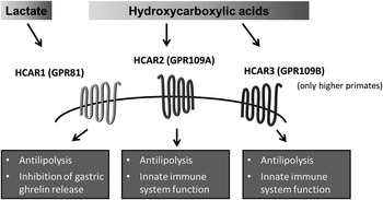
Antilipolysis mediated by lactate and HCAR1 may involve auto and paracrine effects (Ahmed et al., 2010; Offermanns et al., 2011). However, the function of lactate as an inhibitor of norepinephrine-stimulated lipolysis, as demonstrated in vitro in rats by Bjorntorp (1965), may be a matter of debate in vivo. In dogs, a decrease of plasma-free fatty acid concentration after infusion of lactate was detected (Gold et al., 1963); however, no effect of lactate on lipolysis in subcutaneous adipose tissue was observed in male humans during the exercise (Trudeau et al., 1999). Nevertheless, using HCAR1 knockout mice, the negative effect of l-lactate on lipolysis was demonstrated and approved using a specific HCAR1 agonist in vivo (Sakurai et al., Reference Sakurai, Davenport, Stafford, Grosse, Ogawa, Cameron, Parton, Sykes, Mack, Bousba, Parmar, Harrison, Dickson, Leveridge, Matsui and Barnes2014). An inhibitory effect of lactate on ghrelin secretion was also found. This observation was verified by a specific HCAR1 agonist (Engelstoft et al., Reference Engelstoft, Park, Sakata, Kristensen, Husted, Osborne-Lawrence, Piper, Walker, Pedersen, Nohr, Pan, Sinz, Carrington, Akiyama, Jones, Tang, Ahmed, Offermanns, Egerod, Zigman and Schwartz2013). General functions of HCARs are shown in Figure 2.
General functions of hydroxycarboxylic acid receptors (HCARs) in different tissues or cells, based on data from humans or rodents. Moreover, butyrate is a ligand for HCAR2. References are part of the manuscript. GPR=G-protein-coupled receptors.

Hydroxycarboxylic acid receptors in farm animal species
Pig
Data on porcine HCARs is scarce. It is well established that BHBA, the potential ligand for HCAR2, has some growth promoting effects directly after weaning (Piva et al., 2002) and affects intestinal morphology in piglets. In addition, it was shown that niacin induced the skeletal muscle fibre switch from type II to the oxidative type I in growing pigs (Khan et al., Reference Khan, Couturier, Kubens, Most, Mooren, Kruger, Ringseis and Eder2013a). However, these processes could be linked to butyrate as an energy source or its function as a histone deacetylase inhibitor as discussed by Hamer et al. (2008). The studies showing specific data linked to HCAR2 activity in pigs are not available. Nonetheless, an HCAR2-like sequence is predicted (National Institutes of Health (NIH) GenBank, XM_005670607). The porcine receptor HCAR1 was cloned, and the EC50 value for l-lactate of 5.62 mM was established (Liu et al., 2009). However, no further information is available.
Chicken
Currently, no data on HCAR1 or HCAR2 are available in chicken. However, the data on niacin and butyrate effects on lipid or insulin metabolism, which can be linked to epigenetic effects, are known. For example, niacin reduces plasma non-esterified fatty acids and abdominal fat in females of two broiler breads (Jiang et al., Reference Jiang, Zhao, Zhao, Chen, Zheng, Liu and Wen2014), and recently, it was shown that butyrate reduced insulin sensitivity in chicken, which differed from mammals (Matis et al., Reference Matis, Kulcsar, Turowski, Febel, Neogrady and Huber2015).
Ruminants
Comparable with other species, the epigenetic effects of butyrate were found in bovine kidney MDBK cells in vitro (Li et al., 2010). In sheep, data on the predicted ovine HCAR1 and HCAR2 mRNAs are available from the NIH GenBank (XM_012097734.2 and XM_004023416.3, respectively). However, the data on niacin effects are not directly linked to the receptor HCAR2 in this species. It was shown that niacin-induced skeletal muscle fibre switch from type II to the oxidative type I (Khan et al., Reference Khan, Ringseis, Mooren, Kruger, Most and Eder2013b), which was comparable with pig (Khan et al., Reference Khan, Couturier, Kubens, Most, Mooren, Kruger, Ringseis and Eder2013a).
Ketosis in association with fatty liver are the commonly observed metabolic disorders during the transition period in dairy cows (Grummer, 1993). Therefore, the knowledge of BHBA effects in dairy cows is important. In the 1970s, it was shown in bovine AT in vitro that BHBA and butyrate were efficient in inhibiting lipolysis, which was in contrast to acetate and propionate (Metz and van den Bergh, 1972; Metz et al., 1974).
The abundance of HCAR2 mRNA is higher in visceral compared with subcutaneous adipose tissue as demonstrated in primiparous dairy cows (Friedrichs et al., Reference Friedrichs, Saremi, Winand, Rehage, Danicke, Sauerwein and Mielenz2014). The bovine HCAR2 mRNA was up-regulated in tendency during the transition period in subcutaneous adipose tissue of dairy cows but not in leukocytes (Lemor et al., 2009). Recently, we extended this observation during the transition period to retroperitoneal adipose tissue in which more HCAR2 mRNA was detected and in which a decrease between day 42 antepartum and day 1 postpartum was found. However, beside an increase of the HCAR2 protein in subcutaneous adipose tissue over time, the protein abundance was higher in subcutaneous adipose tissue at days 1, 21 and 100 compared with retroperitoneal adipose, which was opposite to the abundance of mRNA (Weber et al., Reference Weber, Locher, Huber, Kenez, Rehage, Tienken, Meyer, Danicke, Sauerwein and Mielenz2016a). First data on the HCAR2 protein were published by Titgemeyer et al. (2011) in steers. The data showed the highest abundance of HCAR2 mRNA and high protein amount in liver compared with adipose tissue, which is different from humans and differs from the low mRNA abundance found in adipose tissue by Friedrichs et al. (Reference Friedrichs, Saremi, Winand, Rehage, Danicke, Sauerwein and Mielenz2014) in primiparous cows. The HCAR2 protein was not regulated by the nicotinic acid supplementation in vivo (Titgemeyer et al., 2011; Weber et al., Reference Weber, Locher, Huber, Kenez, Rehage, Tienken, Meyer, Danicke, Sauerwein and Mielenz2016a) and showed different abundance in different adipose depots such as kidney fat, tail head fat and back fat. The lowest values were in the back fat in steers (Titgemeyer et al., 2011).
Kenez et al. (Reference Kenez, Locher, Rehage, Danicke and Huber2014) found more HCAR2 protein in subcutaneous adipose tissue from tail head compared with kidney fat in multiparous dairy cows. In addition, they demonstrated in vitro that the reduction of lipolysis by niacin and BHBA involves the dose dependent increase of phosphorylation of hormone sensitive lipase.
In differentiated bovine preadipocytes in vitro we showed an increase of HCAR2 mRNA by niacin, which was not blocked by pertussis toxin in contrast to adiponectin secretion and the amount of adenosine monophosphate (AMP)-activated protein kinase. The abundance of adiponectin and AMP-activated protein kinase were distinctly reduced but not totally blocked to basal concentrations. Therefore, the reduction of adiponectin in the medium and AMP-activated protein kinase in cell extracts could be at least partly linked to HCAR2 (Kopp et al., Reference Kopp, Hosseini, Singh, Regenhard, Khalilvandi-Behroozyar, Sauerwein and Mielenz2014). This link needs to be confirmed in detail after the bovine receptor cloning, which is still missing. In addition, the confounding effects between the in vitro and in vivo situation may occur because Weber et al. (Reference Weber, Locher, Huber, Kenez, Rehage, Tienken, Meyer, Danicke, Sauerwein and Mielenz2016a) detected no niacin effect on adiponectin secretion as well as on HCAR2 mRNA and HCAR2 protein in multiparous dairy cows in vivo (Weber et al., Reference Weber, Locher, Huber, Rehage, Tienken, Meyer, Danicke, Webb, Sauerwein and Mielenz2016b).
The bovine receptor HCAR1 was cloned, the binding of l-lactate was analysed, and the EC50 value of 4.95 mM was calculated (Liu et al., 2009). However, the protein data on this receptor in cows regarding its abundance and regulation are not yet available.
The first data on mRNA was provided by Ji et al. (Reference Ji, Drackley, Khan and Loor2014). The HCAR1 mRNA was down-regulated in non-pregnant, non-lactating cows by high-energy diet. However, we could not confirm the regulation of mRNA in subcutaneous and retroperitoneal adipose tissue by the dietary energy supply in multiparous cows (Weber et al., Reference Weber, Locher, Huber, Rehage, Tienken, Meyer, Danicke, Webb, Sauerwein and Mielenz2016b). Based on our data, we suggested that there might be a tissue-specific lipolytic regulation and a higher importance of the receptor in retroperitoneal adipose tissue during the transition period. However, plasma concentrations of lactate to activate the receptor were reached only in exercising cows (Davidson and Beede, 2009) or in cows with abomasal disorders (Figueiredo et al., 2006). In general, the lactate concentration in plasma was 4- to 10-fold lower compared with the EC50 value. However, paracrine or autocrine effects should be taken into account, as discussed by Ahmed et al. (2010).
Conclusions
The number of data on FFARs and HCARs are increasing in human or rodent models. The importance of these receptor families regarding to the communication between the intestinal microbiota and the host becomes clearer. In addition, in farm animals, the available data from intestine and other tissues increase. However, currently, those data are mostly linked to the distribution of receptor mRNAs and proteins. The functional data on the importance of these receptors in farm animal species is rather scarce. Considering that the understanding of microbiota–host interaction in animal science is of significant research interest, both receptor groups may come increasingly into focus in animal physiology in the future.
Supplementary material
To view supplementary material for this article, please visit https://doi.org/10.1017/S175173111600238X