If coronary artery sequelae are the only suggestive signs of previous Kawasaki disease, the diagnosis may easily be missed. These patients are usually diagnosed and cared for by adult cardiologists. We present a unique case of a child presenting with severe occlusive coronary disease most likely owing to missed Kawasaki disease. Paediatric cardiologists should consider this rare diagnosis in children presenting with coronary symptoms or cardiomyopathy as timely diagnosis can lead to successful treatment and prevention of sudden death.
Case report
A previously healthy 12-year-old Asian-American boy was referred to the paediatric heart failure clinic at our centre for dilated cardiomyopathy. He presented 2 months earlier with exertional chest pain. His evaluation at an outside hospital included an echocardiogram with severely decreased left ventricular function, with an ejection fraction of 34.3% by 5/6×area−length method. An electrocardiogram showed sinus rhythm, a left axis, and ST and T-wave changes. His troponin-I (6.46 ng/ml) and NT-pro-BNP (4554 pg/ml) levels were elevated. Cardiac MRI showed severely decreased left ventricular ejection fraction (35%), thinning and dyskinesia of the apical cap, but no evidence of infarction with late gadolinium enhancement. An extensive laboratory evaluation for myocarditis was unrevealing. He was diagnosed with dilated cardiomyopathy and treated with ibuprofen, furosemide, digoxin, and lisinopril with slight improvement in his function. He was discharged with follow-up at our centre.
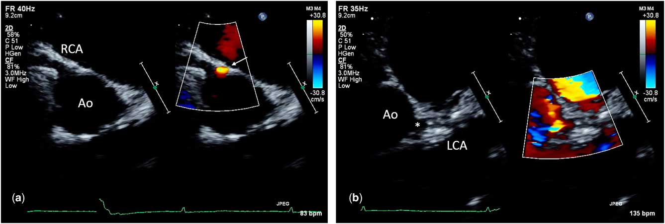
At our centre, his echocardiogram demonstrated diffuse dilation of the right coronary artery (Fig 1a). The left coronary artery was visualised on two-dimensional imaging, but no flow was seen on color Doppler (Fig 1b; Supplementary video 1). There was a separate turbulent flow jet seen on color Doppler arising from the right coronary cusp coursing leftwards and inter-arterially, raising suspicion for a possible anomalous origin of the left coronary artery (Fig 1a; Supplementary video 2). The left ventricle was severely dilated (end diastolic volume z-score +7) with severely depressed function (ejection fraction 31%).
Two-dimensional and color Doppler echocardiogram images of the right ( a ) and left ( b ) coronary arteries. ( a ) The dilated right coronary artery (RCA) is seen originating from the right coronary cusp of the aorta (Ao). Additionally, there is flow seen next to the origin of RCA coursing leftward (arrow), raising suspicion for an anomalous origin of the left coronary artery (LCA). ( b ) The possibility of the origin of the LCA from the left coronary cusp is raised (*), but there is no flow visualised in the LCA on color Doppler.

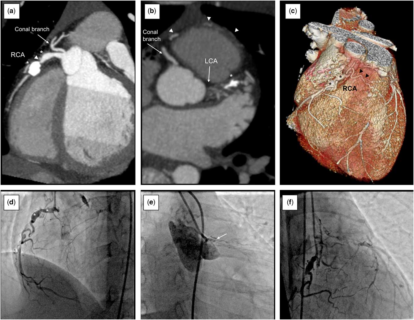
A low-radiation, electrocardiogram-gated CT angiogram (dose-length product 27.9 mGy×cm) was performed to further assess the coronary anatomy (Fig 2). The CT angiogram showed that the right coronary artery originated normally from the right coronary cusp but was dilated and ectatic (Fig 2a). A prominent conal branch originating near the right coronary artery gave off small branches to the anterior right ventricular free wall, the right ventricular outflow tract and the left anterior descending artery territory. The vessel raising concern for an anomalous left coronary artery origin was a collateral arising from the right coronary cusp and coursing inter-arterially. There were two large saccular aneurysms within the proximal-to-mid portion of the right coronary artery measuring 6×5 and 8×9 mm in diameter with>90% luminal stenosis between them. The distal aneurysm was heavily calcified and nearly occluded with thrombus. The left main coronary artery originated normally from the left coronary cusp, but was nearly completely occluded just distal to its origin from the aorta (Fig 2b). The proximal left anterior descending artery was occluded with a 9-mm-long, 6×6-mm-diameter calcified and thrombosed aneurysm. The myocardial territory normally supplied by the left anterior descending artery was fed by multiple small collaterals arising from the conal artery (Fig 2c).
Low-radiation coronary CT scan ( a – c ) and coronary angiograms ( d – f ). ( a ) Dilated and ectatic proximal right coronary artery (RCA) containing two large saccular aneurysms (*). The distal aneurysm was calcified and nearly occluded with thrombus. There was>90% luminal stenosis of the RCA between the aneurysms (arrowhead). A prominent conal artery gave off collaterals to the left. ( b ) Axial view showing normal origins of the right and left coronary arteries with near-total occlusion of the left coronary artery (LCA) just distal to its origin. A large aneurysm in the proximal left anterior descending was calcified and thrombosed (*). Collaterals from the right traverse the right ventricular (RV) free wall (arrowheads). ( c ) A three-dimensional reconstruction showing the RCA with two large saccular aneurysms (*) and collaterals passing leftwards across the RV outflow tract (arrowheads). ( d ) Angiogram of the RCA showing two large saccular aneurysms within the proximal-to-mid RCA (*). ( e ) Angiogram of the LCA showing 100% chronic total occlusion of the left main coronary artery (arrow). ( f ) Angiogram of the RCA showing collateral flow to the LCA.

A presumptive diagnosis of missed Kawasaki disease leading to ischaemic cardiomyopathy was made. Even though the family could not recall any significant febrile childhood illnesses that could suggest a history of Kawasaki disease, he did not have any risk factors for coronary artery disease and there were no alternative diagnoses that were more likely. The patient was otherwise healthy, non-syndromic, had normal cardiac anatomy without evidence of coronary fistulae, and did not have a history of trauma, connective tissue, or autoimmune disease. The patient was admitted to the hospital, started on a heparin drip, and a cardiac catheterisation was performed. Angiograms confirmed the findings seen on CT angiogram of 100% chronic total occlusion of the left main and left anterior descending coronary arteries with collaterals from the right coronary system and 100% occlusion of the right coronary artery aneurysm with right-to-right collaterals (Fig 2d–f).
The patient underwent bilateral internal mammary artery bypass grafting without ligation of the proximal coronary arteries, given the severe obstruction. His postoperative course was uneventful. He is maintained on aspirin, enalapril, and metoprolol. At his most recent follow-up, 5 months post bypass grafting, his function had normalised, with an ejection fraction of 58%, with improved dilation (end diastolic volume z-score +3.8). His troponin and NT-pro-BNP normalised and he was asymptomatic without recurrent chest pain. He will continue to be followed up with serial echocardiograms, as well as periodic consideration of angiography and stress imaging, to assess graft and distal coronary patency and to look for inducible ischaemia.
Discussion
Kawasaki disease is an acute, self-limited vasculitis that can be easily mistaken for other childhood illnesses leading to misdiagnosis and late presentation with potentially catastrophic coronary artery sequelae, usually in adulthood.Reference Gordon, Kahn and Burns 1 Burns et alReference Burns, Shike and Gordon 2 reported that the mean age for coronary artery sequelae was 24.7±8.4 years. A recent case series of adults, aged 30–40 years, with presumed missed Kawasaki disease presenting with acute coronary syndrome highlighted that significant ectasia or aneurysms in the proximal coronary system with transition to normal distal segments should raise suspicion for Kawasaki disease.Reference Bhagwat, Mukhedkar and Ekbote 3 The majority of these patients are treated with bypass grafting.Reference Burns, Shike and Gordon 2 Successful bypass grafting has even been performed in children as young as 4 years of age.Reference Tamaki, Tsuda and Nakajima 4
Although younger children with known Kawasaki disease have developed severe coronary occlusion requiring bypass grafting, to our knowledge, our patient is the youngest reported to present without any suggestive history of Kawasaki disease. In fact, this diagnosis was not initially entertained given the young age and absence of suggestive history. However, careful echocardiographic assessment with confirmation by CT angiogram resulted in successful diagnosis and treatment. Paediatric cardiologists should consider the possibility of missed Kawasaki disease in any child presenting with a new cardiomyopathy. If the coronary artery anatomy is not definitively shown on echocardiogram assessment, including adequate colour flow imaging, advanced imaging should be pursued. As the disease is usually amenable to bypass grafting, even in very small children, timely diagnosis can lead to successful surgical management, recovery of cardiac function, and prevention of sudden death.
Acknowledgements
None.
Financial Support
This research received no specific grant from any funding agency, commercial, or not-for-profit sectors.
Conflicts of Interest
None.
Supplementary materials
To view supplementary material for this article, please visit https://doi.org/10.1017/S1047951118000124