Ionic currents in the heart are the result of the assembly of different ion channel proteins, each having a role in establishing unique properties of the individual currents, such as localisation, density, kinetics, voltage, and drug sensitivity and response to regulatory signals. The cores of some of these complexes are well on their way to being defined, for example I Ca made up from the Cav1.2 α subunit and specific α2δ, β and γ accessory subunits, whereas in other cases only the ion-conducting α subunit has been unequivocally identified. This review will summarise what is known regarding the role of the ion channel accessory subunit KCNE2 in the heart, specifically in the context of potential mechanisms for the long QT syndrome (LQTS).
Long QT syndrome
LQTS is a potentially fatal condition, characterised by a prolonged QT interval on the electrocardiogram (ECG), syncope, seizures and sudden death. Although the most common clinical manifestation is syncope, as many as 13% of affected individuals present initially with sudden death or cardiac arrest (Ref. Reference Priori1). About 75% of events are precipitated by adrenergic stimuli such as intense physical activities (swimming confers the highest risk), stress, emotion, fright and loud noises, such as an alarm clock or telephone ring (Ref. Reference Wilde2). In addition, more than 100 known drugs, as seemingly benign as antibiotics such as erythromycin or clarithromycin, may trigger events (see the Azcert website for a full list: http://www.azcert.org). LQTS is considered to occur in congenital and acquired forms; the former is due to mutations in the genes regulating cardiac excitability. Acquired LQTS is secondary to external factors, including a myriad of medications, systemic illness and electrolyte imbalance (Ref. Reference van Noord, Eijgelsheim and Stricker3). There may be considerable overlap between the entities, with many cases of acquired LQTS resulting from silent mutations (Ref. Reference Abbott4), the significance of which becomes unmasked by an external trigger. Similarly, we are increasingly recognising that congenital LQTS can be clinically silent, with as many as 25–35% of carriers of a mutation having a normal QT interval on their ECG (Ref. Reference Johnson and Ackerman5).
Congenital LQTS is an inherited disorder with an estimated incidence of approximately 1 in 2500 (Ref. Reference Schwartz6). It is an important cause of cardiac arrest in young, previously healthy individuals (Ref. Reference van der Werf7), including those suffering sudden infant death syndrome (Ref. Reference Schwartz8). Inheritance can be of single gene origin (autosomal dominant, autosomal recessive) as well as complex (subtle changes in protein function not apparently of a single gene pattern, suggesting additional gene involvement or environmental triggers). There is evidence that variable expression of LQTS might result from mutations causing a major effect, along with functional polymorphisms (minor effects of common variants) in the same or different ion channel genes. About 10% of those affected with LQTS have two mutations (Ref. Reference Kapplinger9), with the combination of mutations resulting in a more severe phenotype, which has a greater risk of sudden death (Ref. Reference Westenskow10).
Genetics of LQTS
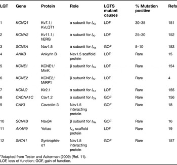
Congenital LQTS is predominantly caused by mutations in 12 known genes (Table 1), which code for, or are directly involved in, the function of ion channel proteins that are important in the heart (Ref. Reference Tester and Ackerman11). Eight genes encode cardiac ion channels and their accessory subunits (Refs Reference Tester and Ackerman11, Reference Romey12, Reference Sanguinetti13, Reference Barhanin14), including KCNE2, which is implicated in LQT6. The remaining genes encode proteins that are involved in trafficking, localisation, anchoring and regulating these channels (Refs Reference Mohler15, Reference Medeiros-Domingo16, Reference Mohler17, Reference Vatta18, Reference Chen19). This list of genes is expected to increase; for example, a mutation in Kir3.4 (G-protein-activated inward rectifier potassium channel 4, KCNJ5) has been recently linked with LQTS (Ref. Reference Yang20). However, LQT1, LQT2 and LQT3 are by far the most common, comprising ~95% of all genotype-positive LQTS (Ref. Reference Kapplinger9), although, even in patients with a compelling diagnosis of LQTS, no mutations are found in ~25% of cases (Ref. Reference Tester and Ackerman11). This might be due to limited screening in most cases (until recently, largely limited to LQT1–LQT3, LQT5 and LQT6), or could result from mutations in as yet undiscovered genes or large deletions or insertions that are not detected by conventional sequencing methods (Ref. Reference Barc21).
Long QT genes and protein roles in cardiac excitabilitya

a Adapted from Tester and Ackerman (2009) (Ref. Reference Tester and Ackerman11).
LOF, loss of function; GOF, gain of function.
Ionic basis of the cardiac action potential
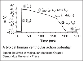
To understand the role of KCNE2 in the heart, one must have some idea of the main ion channels in the heart and their role in the cardiac action potential (AP). A relatively recent, more detailed review of this has been published (Ref. Reference Nerbonne and Kass22), as well as another in the context of inherited cardiac disease (Ref. Reference Amin, Tan and Wilde23), and the reader is referred to these publications for a more thorough discussion of the major ionic currents involved and the ion channels underlying them. During each cardiac cycle of atrial and then ventricular contraction and relaxation, each myocyte in the heart undergoes a cycle of depolarisation and then repolarisation – the AP – which is the result of net fluxes of ions in and out of the cell. In human cardiac cells, depolarisation is carried out by the entry of sodium, largely through Nav1.5 channels (Phase 0; Fig. 1). This is followed by rapid partial repolarisation – a current known as I to (Phase 1, the notch) – as potassium exits the cell, mainly through Kv4 channels. During the plateau of the AP, the net flux of ions is negligible, with calcium entry largely through Cav1.2 channels (I Ca), and some continued sodium entry, balanced by potassium exit as I Kr, I Kur and I Ks currents (Phase 2). Eventually, as I K1 and potassium efflux through these currents increase, and little calcium or sodium enters the cell, the cell repolarises back to the resting potential (Phase 3). The final stage is a resting stage, where again little net ion flux occurs, but where Kir2.1 (the molecular correlate of I K1) remains active to maintain the negative resting membrane potential (Phase 4). There are regional and cell-type differences in the densities of these currents and some cell types lack or have additional currents [e.g. hyperpolarisation-activated current (I f) made up of hyperpolarisation-activated, cyclic-nucleotide-gated (HCN) channels in nodal tissues (Ref. Reference DiFrancesco24) and the relatively atrial-specific I Kur carried by Kv1.5 in humans (Refs Reference Fedida25, Reference Feng26)].
A typical human ventricular action potential. Phases (0-4) and selected contributing currents are shown (redrawn from Ref. Reference Keating and Sanguinetti165).

KCNE proteins
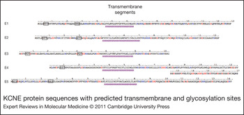
KCNE proteins encode potassium channel β subunits that are incapable of conducting ions when expressed alone, and so require coassembly with a complex of α subunits to have a functional effect. The first to be identified and cloned was KCNE1 (formerly MinK) (Refs Reference Boyle27, Reference Takumi, Ohkubo and Nakanishi28), and since then four more have been identified (Refs Reference Abbott4, Reference Piccini29), all of which are expressed in the heart (Ref. Reference Lundquist30). KCNE2–KCNE4 (formerly MiRP1–MiRP3) were discovered through a search for KCNE1-related sequences, which subsequently identified three genes that encoded single-pass transmembrane proteins of 123, 103 and 167 amino acids (Fig. 2; Ref. Reference Abbott4). The fifth member of this family, KCNE5, was identified based on its homology to KCNE1 as one of the genes deleted in AMME syndrome (Alport syndrome, mental retardation, midface hypoplasia, elliptocytosis; Ref. Reference Boyle27). KCNE5 encodes a 142-amino-acid protein with 56% homology to KCNE1 (95% homology and 34% identity in the transmembrane domain). KCNE2 shows 27% identity and 45% homology to KCNE1 (Ref. Reference Abbott4). KCNE3 and KCNE4 each have 30% and 38% identity with KCNE1, whereas KCNE4 has 30% identity to KCNE2 and KCNE3 (Refs Reference Boucherot, Schreiber and Kunzelmann31, Reference Teng32).
KCNE protein sequences with predicted transmembrane and glycosylation sites. Transmembrane sequence predictions (identified by purple Ms) were made using the PredictProtein website (http://www.predictprotein.org/). Acidic residues are in blue, basic in red. Glycosylation sites are boxed.

The topology of KCNE proteins is such that the N-terminus is extracellular and contains two predicted N-glycosylation sites, except for KCNE4, which has the shortest N-terminus and only one of these predicted N-glycosylation sites (Fig. 2). Removal of glycosylation sites results in protein that does not express at the cell surface (Refs Reference Takumi33, Reference Gage and Kobertz34). The C-terminus of KCNE proteins is intracellular and contains PKC consensus phosphorylation sites (Refs Reference Abbott4, Reference Abbott, Butler and Goldstein35, Reference Teng32) and, in the case of KCNE4, a calmodulin-binding domain (Ref. Reference Ciampa36).
α–β subunit interactions
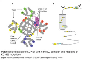
KCNE proteins interact with members of the voltage-gated potassium channel superfamily. These are six transmembrane domain proteins (S1–S6) that contain a voltage sensor domain (VSD; S1–S4) and an ion-conducting pore (S5–P-loop–S6) with a linker between (S4–S5L) that transduces the activated state of the VSD to opening of the channel pore (Fig. 3a; Refs Reference Long, Campbell and MacKinnon37, Reference Long38). Association of KCNE proteins with the α subunits largely takes place in the endoplasmic reticulum (ER) (Ref. Reference Chandrasekhar, Bas and Kobertz39, Reference Vanoye40), but can also occur at the plasma membrane (Ref. Reference Jiang41). Several KCNE proteins can assemble with the channel complex and one KCNE protein might even replace another family member to affect current properties, leaving open the possibility of additional layers of regulation of the channel.
Potential localisation of KCNE1 within the I Ks complex and mapping of KCNE2 mutations. (a) The cartoon structure of the channel is loosely based on the open-state crystal structure of Kv1.2 (Ref. Reference Long, Campbell and MacKinnon166). The view is from the extracellular side with coloured circles placed at the extracellular termination of the six transmembrane domains from each α subunit (1–4, each a different colour). Also in colour are the S4–S5 linkers. (b) KCNE2 mutations mapped onto a representation of the protein. VSD, voltage sensor domain.

Structural analysis and mutation studies, mostly of KCNE1 assembly with KCNQ1, suggest that KCNE proteins interact with the pore domain of voltage-gated potassium (Kv) channels by binding in the cleft between VSDs (Fig. 3a) (Refs Reference Kang42, Reference Melman43). In this position, the protein is positioned to affect activation kinetics by direct interaction with the S4–S5 linker, affecting the coupling of voltage changes to channel opening. This positioning also allows for interactions of KCNE1 with S1 [explaining why gain-of-function mutations in KCNQ1 and KCNE1 can both lead to atrial fibrillation (AF) (Ref. Reference Chen44)], S4 and S6, as suggested by mutational, accessibility and crosslinking experiments (Refs Reference Chung45, Reference Xu46, Reference Shamgar47, Reference Nakajo and Kubo48). The intracellular C-terminus of KCNE1 might also interact with the C-terminus of KCNQ1 subunits (Ref. Reference Haitin49). Large extracellular N-terminal domains in KCNE1, made up of α-helices linked by flexible domains, might interact across the turret region of the channel to sterically inhibit additional KCNE subunits from binding (Ref. Reference Kang42) and explain the experimentally determined 2:4 (KCNE1:KCNQ1) stoichiometry (Refs Reference Chen50, Reference Morin and Kobertz51). However, this stoichiometry has recently been challenged (Ref. Reference Nakajo52).
Review of studies showing the effect of KCNE2 on various potassium channels
Initially, KCNE2 was thought to be part of a complex with hERG channels that recapitulate I Kr (Ref. Reference Abbott4), the rapidly activating delayed rectifier current, which, together with I Ks, is responsible for much of Phase 2 and Phase 3 repolarisation in cardiac myocytes (Fig. 1). Subsequently, KCNE2 has proved to be quite promiscuous, showing an ability to associate with a wide variety of potassium channels in vivo or when coexpressed in heterologous cells. This list includes the I Ks complex (Refs Reference Toyoda53, Reference Jiang54), Kv1.4, Kv2.1 (Ref. Reference McCrossan55), Kv3.1 and Kv3.2 (Ref. Reference Lewis, McCrossan and Abbott56), Kv4.2 (Ref. Reference Zhang, Jiang and Tseng57), Kv4.3 (Refs Reference Zhang, Jiang and Tseng57, Reference Piccini29) and even several members of the HCN family (Refs Reference Yu58, Reference Decher59). The diverse range of associations suggests the potential for a pivotal role in regulating cardiac repolarisation.
I Ks
All five KCNE proteins can assemble with KCNQ1, and much of our understanding of these β subunits has been gained by studying their varied effects on KCNQ1. This channel makes up the slowly activating delayed rectifier current (I Ks) when assembled with KCNE1 in the heart, and is crucial for cardiac AP repolarisation (Refs Reference Sanguinetti13, Reference Barhanin14). Mutations in KCNQ1 or KCNE1, leading to a loss of I Ks function, result in LQTS. In the stomach, KCNQ1 and KCNE2 complexes, in the apical membrane of parietal cells, create a constitutive K+ current required for gastric acid secretion (Refs Reference Dedek and Waldegger60, Reference Grahammer61, Reference Heitzmann62, Reference Lambrecht63, Reference Roepke64). When KCNE2 is coexpressed with KCNQ1 plus KCNE1, the currents are I Ks-like, with perhaps only subtle differences in voltage dependence and kinetics of deactivation (Ref. Reference Toyoda53). In some studies, current amplitude was also found to be reduced by the addition of KCNE2 (Ref. Reference Wu65), suggesting that perhaps KCNE2 was functioning as a negative modulator of I Ks currents. KCNQ1 and KCNE3 coassemble to form a constitutively active K+ current in colonic crypt cells, the K+ flux through which regulates cAMP-stimulated chloride secretion (Refs Reference Dedek and Waldegger60, Reference Bleich and Warth66, Reference Schroeder67). KCNE4 has been shown to assemble with KCNQ1 alone, and in combination with KCNE1 (Ref. Reference Manderfield and George68) in expression systems, resulting in such a shift in the voltage dependence of activation that the channel is silenced in the physiological range of voltages (Ref. Reference Grunnet69). Above +40 mV, this complex results in slowly activating currents that are similar to I Ks (Ref. Reference Bendahhou70). There is no direct evidence of KCNQ1 and KCNE4 assembly in the heart, although KCNE4 mRNA levels are similar or second only to levels of KCNE1 mRNA in various cardiac tissues (Ref. Reference Lundquist30). When KCNE4 and KCNE1 are expressed together with KCNQ1, currents are I Ks-like in kinetics, voltage dependence and amplitude (Ref. Reference Bendahhou70), but if KCNE4 is introduced into an I Ks stable line, the higher levels of KCNE4 expression achieved appear to silence I Ks (Ref. Reference Lundquist30). KCNE5 also shifts the activation voltage of KCNQ1 plus KCNE5 channels, even more so than KCNE4, but mRNA levels for this subunit are significantly lower than those encoding KCNE4 in all regions of the heart (Refs Reference Lundquist30, Reference Bendahhou70). A recent publication has linked a gain-of-function mutation in KCNE5 to AF in a single patient, and postulates that the protein is normally involved in suppressing I Ks in the atrium, where mRNA levels are highest (Ref. Reference Ravn71). Little else is known about the role of KCNE5 in the heart.
Although it is unclear whether KCNE2 coassembles with KCNQ1 in the heart and whether it does so in the presence of other KCNE proteins, one study has looked at the effect of expression of KCNE2 mutants (I57T and Q9E) with KCNQ1 and found that the mutants had quite different effects. Coexpression with I57T resulted in currents similar to I Ks, with slowed down activation and a depolarising shift in the voltage dependence of activation (Ref. Reference Tinel72), wholly unlike the constitutive current observed with coexpression of wild-type (WT) KCNE2 or Q9E KCNE2. Coexpression of Q9E, by contrast, caused a decrease in instantaneous currents compared with WT. Interestingly, KCNE1 and KCNE2 had different effects on a KCNQ1 mutant (Q147R) implicated in AF (Ref. Reference Lundby73). The mutant expressed on its own was not significantly different from WT KCNQ1. However, the presence of KCNE1 resulted in a loss of function, perhaps explaining the patient's 470 ms QTc. In the presence of KCNE2, a gain of function was observed, perhaps explaining the AF – an effect that persisted when KCNE1 and KCNE2 were both coexpressed with the mutant KCNQ1. The two phenotypes are perhaps the result of differential expression of KCNE2 in the various regions of the heart (Ref. Reference Yu58).
hERG
There is still controversy about the molecular correlates that underlie the human rapidly activating delayed rectifier K+ current (I Kr), with studies showing that hERG channels alone do not fully recapitulate native I Kr current (Refs Reference Abbott4, Reference Sanguinetti74, Reference Trudeau75). Specifically, the voltage dependence of activation and rectification of hERG alone is nearly identical to I Kr but the kinetics are slower (Ref. Reference Sanguinetti74). These discrepancies led to a search for a β subunit that might coassemble with hERG to better represent I Kr.
Similar to I Kr, hERG is modulated by extracellular potassium and blocked by lanthanum and cobalt (Ref. Reference Sanguinetti74). Whereas initial experiments showed that, unlike I Kr, hERG was not blocked by methanesulfonanilide antiarrhythmic drugs (e.g. dofetilide, E-4031, MK-99; Refs Reference Abbott4, Reference Sanguinetti74), more recent experiments suggested that this failure of block by methanesulfonanilides was a result of the experimental protocol utilised and the Xenopus oocyte expression system (Refs Reference Trudeau75, Reference Spector76).
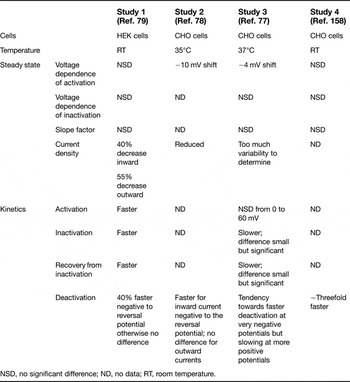
The first study to examine the effects of KCNE2 on hERG found that coexpression in Xenopus oocytes resulted in an ~10 mV depolarising shift in the voltage dependence of activation, a slowing down of activation, a 40% decrease in current amplitude as a result of lower unitary conductance and some voltage-dependent changes in deactivation (Ref. Reference Abbott4). When expressed in mammalian cells, the effects were limited to reduced unitary conductance and faster deactivation, both reportedly similar to those of I Kr (Ref. Reference Abbott4). In addition, this study found that α–β heteromultimers were less sensitive to external potassium changes and showed a biphasic block by the methanesulfonanilide, E-4031, similar to native I Kr, which was not seen when hERG was expressed alone. This has led to the widely accepted notion that KCNE2 is the missing β subunit of I Kr. However, there are discrepancies in reports from different laboratories on the exact effects that coassembly has on hERG currents in mammalian cells (Table 2). From their experience and the literature, Lu and colleagues concluded that the effect of KCNE2 on hERG was to slow down deactivation in the physiological range but speed up deactivation below −90 mV and with modest effects on activation and inactivation (Ref. Reference Lu77). They noted that it was difficult to make comparisons between WT and mutants because of the variability in transfection efficiency, a problem that would be expected to have an impact on all studies using transient transfection, especially those involving multiple cDNAs.
Comparison of the effects of coexpression of KCNE2 on HERG currents in mammalian cells

NSD, no significant difference; ND, no data; RT, room temperature.
Regardless of how exactly KCNE2 modulates hERG in heterologous cells, controversy remains because coexpression failed to produce currents that behave like I Kr recorded from cardiac myocytes in a direct comparison, using the same recording conditions (Ref. Reference Weerapura78). Specifically, hERG and KCNE2 deactivate more slowly than I Kr, and activate at more negative potentials, results that are supported qualitatively by others (Ref. Reference Mazhari79). Pharmacological evaluation did suggest that I Kr, hERG and hERG–KCNE2 complexes were identical in their kinetics and sensitivity to block by quinidine, E-4031 and dofetilide (Ref. Reference Weerapura78). This difference between I Kr and expressed channels may be due to coassembly of splice variants of hERG, because coexpression of hERG1a with hERG1b shows faster kinetics, similar to that of I Kr (Refs Reference Sale80, Reference Larsen81). But again there is some controversy as to whether the 1b form is expressed in the adult human heart (Ref. Reference Pond82). It is uncertain whether the properties of the 1a–1b heteromers have been studied in the presence of KCNE2.
Another issue that calls into question the role of KCNE2 in recapitulation of I Kr is the issue of tissue-specific expression of the β subunit. A few studies have looked at mRNA expression in the heart and found that KCNE2 is the least (Ref. Reference Bendahhou70), or second least (Ref. Reference Lundquist30), expressed KCNE in the heart by a wide margin, with the lowest levels found in the ventricle. In studies of rabbit tissue, the highest expression was found in the SA node, with amounts at 40% and 4% of this level in atrial and ventricular tissues, respectively (Ref. Reference Yu58). Another study found that levels of KCNE2 expression in canine tissue were similar between atrial cells and Purkinje fibres, but levels in the ventricle were 10% that of the other cell types (Ref. Reference Pourrier83). In terms of actual protein, this study showed very little KCNE2 in the ventricle, but this could have been an antibody issue because another group detected a good deal more of the protein in their canine ventricular tissue (Ref. Reference Jiang84). Interestingly, in this latter study, very little KCNE2 appeared to be associated with ERG in coimmunoprecipitation studies, suggesting that the protein has other partners.
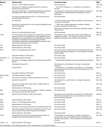
Certainly, studies have shown that KCNE2 mutants identified from patients with acquired or congenital LQTS can affect hERG currents when coexpressed in oocytes or heterologous cells (Table 3). The T8A mutation in KCNE2 caused a 7–10 mV negative shift in the activation voltage (Refs Reference Abbott4, Reference Lu77). Q9E caused a positive shift in activation voltage (Ref. Reference Abbott4), or was not significantly different from WT (Ref. Reference Lu77) and had greater sensitivity to block by clarithromycin (Ref. Reference Abbott4). T10M caused a 27% reduction in current amplitude and slower recovery from inactivation (Ref. Reference Gordon85), and M54T caused faster deactivation and no shift in voltage-dependent activation (Ref. Reference Abbott4), or slower deactivation and a right shift in voltage-dependent activation coupled with a negative shift in the V 1/2 of steady-state inactivation (Ref. Reference Lu77). The V56M mutation was found to accelerate inactivation of coassembled channels (Ref. Reference Isbrandt86). I57T and A116V decrease single-channel conductance and the half-life of the protein at the cell surface (Ref. Reference Abbott and Goldstein87). Only the T10M mutant was coexpressed with WT KCNE2 to mimic the heterozygous state of the patient, and this resulted in characteristics that were not significantly different from WT KCNE2 plus hERG (Ref. Reference Gordon85).
Reported KCNE2 mutations, SNPs and functional affects upon coexpression with hERG versus wild-type KCNE2

AF, atrial fibrillation; TdP, Torsades de pointes; TWA, T-wave alternans; VF, ventricular fibrillation.
Can one make sense of these results in terms of pro-arrhythmia? If mutant KCNE2 is to lead to LQTS through its effects on hERG, it would need to induce a reduction in the repolarisation capability through either a reduction in current density (through reduced channel number or conductance) or less current at the appropriate time during the AP (shifted V 1/2 values of activation/inactivation, slower activation, faster inactivation, slower recovery from inactivation or faster deactivation). Some of these effects are observed for individual mutant KCNE2 proteins expressed with hERG (Table 3), but they are often subtle and are expected to be even more so in the presence of the WT KCNE2 protein, as would be seen in the heterozygous patient. By contrast, mutations in hERG that lead to LQTS often result in at least a 50% reduction in hERG protein at the cell surface due to a failure to fold and traffic (Ref. Reference Sanguinetti88) and thus a significantly greater reduction in I Kr than is observed with the KCNE2 mutants. Perhaps this might explain why many of the symptomatic patients appear to have acquired LQTS, with addition of a hERG blocker unmasking their perhaps already slightly reduced repolarisation reserve (Table 3).
While looking at KCNE2 mutant-dependent mechanisms of arrhythmia, Lu and colleagues found that during closely coupled APs, channels made up from hERG + T8A or Q9E KCNE2 resulted in larger peak currents during a closely coupled second AP, which alone, as they discussed, should protect from premature beats. However, they also suggested it would be difficult to know how this might affect other currents such as I Na to create a situation of pro-arrhythmia (Ref. Reference Lu77). Adding to the confusion is the observation that T8A and Q9E have been found in 1–1.6% of healthy controls (Ref. Reference Ackerman89).
Further complicating the situation is evidence that KCNE1, which is found in abundance throughout the human heart (Refs Reference Szabo90, Reference Szentadrassy91, Reference Melnyk92), can also associate with hERG, and might do so more efficiently as a result of more similar transit times of the protein through the ER to the cell surface (Ref. Reference Um and McDonald93). Coexpression of KCNE1 with hERG results in a doubling of current density by increasing the fraction of functional channels already at the membrane (Ref. Reference McDonald94), and mutations in KCNE1 can also affect hERG currents in heterologous cells (Ref. Reference Bianchi95). At least two laboratories have shown that the rare KCNE1 polymorphism (D85N), in addition to having loss-of-function effects on I Ks, also causes a 30–80% reduction in hERG current density in heterologous cells. In addition, antisense knockdown of KCNE1 in neonatal mouse ventricular cells (Ref. Reference Ohyama96) and AT-1 cells (Ref. Reference Yang, Kupershmidt and Roden97) leads to a reduction in I Kr. Direct interactions between KCNE1 and hERG have been detected in heterologous cells (Ref. Reference McDonald94) and in horse heart (Ref. Reference Finley98); however, because a specific antibody is lacking, an association between EAG1 and KCNE2 could not be explored at the same time.
These results leave open the true nature of I Kr complexes in the heart and thus the impact that KCNE2 mutations might have on this current.
I to
The α subunit of I to, a rapidly activating and inactivating K+ current, is mainly Kv4.3 in the human heart (Ref. Reference Dixon99). There is some region-dependent contribution from Kv1.4 (Ref. Reference Szentadrassy100), which is distinguished from Kv4-based currents (I to,fast) by slower kinetics of recovery from inactivation (and thus designated as I to,slow) and a propensity to accumulate in the inactivated state during repetitive stimulation (Ref. Reference Patel and Campbell101). The role of this current is to set the plateau voltage of the AP, and thus it influences the activation of voltage-gated Ca2+ channels and the balance of inward and outward currents during the plateau (Ref. Reference Niwa and Nerbonne102). It is of particular importance in maintaining a shorter AP duration in the epicardium versus the endocardium to ensure unidirectional propagation of excitation in the heart (Ref. Reference Greenstein103), and is achieved through a gradient of expression of I to,fast from epicardial to endocardial cells (Refs Reference Nabauer104, Reference Wettwer105).
When expressed alone, Kv4 channels inactivate more slowly and recover from inactivation much more slowly than native I to,fast channels (Ref. Reference Niwa and Nerbonne102); therefore, similar to I Ks and I Kr, this current is expected to be made up of a macromolecular complex of α and β subunits. Strong evidence exists for assembly of Kv4.3 with the accessory protein, K+-channel interacting protein-2 (KChIP2), which enhances trafficking of the complex, slows inactivation and accelerates recovery from inactivation (Ref. Reference An106). Kv4.3 and KChIP have been coimmunoprecipitated from mouse cardiac tissue (Ref. Reference Guo107) and, as in the case of I to, a gradient of KCHIP2 mRNA expression exists across the human left ventricular wall (Ref. Reference Rosati108). However, this did not seem to translate into a gradient for the protein, and coexpression does not mimic native I to,fast (Ref. Reference Deschenes109). That is, Kv4.3 and KChIP2 inactivate at more positive voltages and recover more slowly than native I to currents (Ref. Reference Radicke110), suggesting that additional accessory subunits are needed.
In addition to K+ channel accessory proteins (KchAPs) (Ref. Reference Kuryshev111), Kvβs (Refs Reference Yang112, Reference Deschênes and Tomaselli113) and diaminopeptidyl transferase-like protein 6 (DPP6, also known as DPPX) (Ref. Reference Nadal114), KCNE proteins can modulate Kv4.3 expression and kinetics. KCNE1 increases current density fivefold but slows activation, inactivation and recovery from inactivation, whereas KCNE2 enhances Kv4.3 current density threefold and slows inactivation modestly (Ref. Reference Deschênes and Tomaselli113) or decreases current density and slows activation (time to peak) and inactivation significantly (Ref. Reference Wu115). KCNE3 is an inhibitor of Kv4.3; it decreases current density and slows down time to peak, inactivation and recovery from inactivation, and does so even when KChIP is present (Ref. Reference Lundby and Olesen116). KCNE3 can be coimmunoprecipitated with Kv4.3 from human atrial tissue, indicating that the proteins do associate in vivo (Ref. Reference Delpon117), and identification of mutations in KCNE3 and Kv4.3 linked to Brugada syndrome (BrS) has provided further evidence of an association between these two proteins (Refs Reference Delpon117, Reference Giudicessi118).
When KCNE2 and KChIP are coexpressed with Kv4.3, inactivation is still more positive than native I to, but the difference is less than with KchIP and Kv4.3, and an overshoot from recovery of inactivation similar to the native current is present (Refs Reference Radicke110, Reference Radicke119). However, recovery from inactivation remains significantly slower.
Accessory subunits that do seem to accelerate the kinetics of Kv4.3 activation and inactivation, and shift the voltage dependence of activation and inactivation to more negative values, are the DPPs (Refs Reference Nadal114, Reference Ren120, Reference Li121). Coexpression of Kv4.3, KChIP and DPP6 results in channels that have voltage-dependent activation and recovery from inactivation kinetics (Refs Reference Radicke122, Reference Radicke119) similar to that reported for I to in the human ventricle (Ref. Reference Radicke110). DPP6 has been immunoprecipitated with Kv4.2 from the brain (Ref. Reference Nadal114), but so far it has been found alone only in the human heart (Ref. Reference Radicke122). Interestingly, a noncoding mutation in DPP6 has recently been linked to idiopathic ventricular fibrillation (VF) and sudden cardiac death (SCD), with cardiac biopsies indicating that the mutation leads to higher expression of DPP6 mRNA (Ref. Reference Postema123). Addition of KCNE2 to the combination of Kv4.3, KChIP and DPP6 did not affect the negative shifts in voltage dependence, but slightly increased the rate of recovery from inactivation (Refs Reference Radicke124, Reference Radicke110, Reference Radicke122, Reference Radicke119). These results have led to the conclusion that KCNE2 is a component of the I to macromolecular complex (Ref. Reference Radicke110).
Knockout of the KCNE2 gene leads to a decrease in two cardiac repolarising potassium currents in mice, including a 25% reduction in I to (Ref. Reference Roepke125). However, in this study, KCNE2 protein was shown to associate with Kv4.2 and not Kv4.3. In another study, KCNE2 was not identified in mass spectrometry analysis of immunoprecipitates of Kv4.2 from mouse heart, whereas the authors did identify DPP6 in coimmunoprecipitates from mouse brain (Ref. Reference Marionneau, Townsend and Nerbonne126). Although it is possible that KCNE2 was of lower abundance and the hydrophobicity of this membrane protein might have affected the efficiency of detection, the mouse heart is distinct from that of the human, and it is therefore difficult to extrapolate from these data the importance of KCNE2 in the formation of I to in the human heart.
One study did report the results of coexpression of two KCNE2 mutants (I57T and M54T) with Kv4.3 and KChIP2b in CHO cells, which resulted in higher peak current densities, slower inactivation and faster recovery from inactivation than when WT KCNE2 was used (Ref. Reference Wu115). The authors suggested that the gain of function of I to produced by these two mutations creates an arrhythmic substrate in BrS, promoting Phase 2 reentry and fatal ventricular arrhythmias, similar to that seen with a BrS-related KCNE3 mutant (Ref. Reference Delpon117). As pointed out by Wu and co-workers, these two KCNE2 mutants were originally identified in women with borderline prolonged (I57T; 470 ms) or inconsistently prolonged (M54T; 390–500 ms) QTc values, the latter determined after an incidence of VF (Ref. Reference Abbott4), leaving room for the potential of a diagnosis of BrS and not LQTS for carriers of these KCNE2 mutations.
I f
The pacemaker current or ‘funny current’ is responsible for the slow depolarisation of sinoatrial node cells during diastole (Ref. Reference Monfredi127). This leads to activation of the calcium current, which is responsible for the upstroke of the AP in these cells, and the initiation of a cardiac cycle. The pacemaker current is made up of HCN channels, HCN1, HCN2 and particularly HCN4 in humans, probably as heteromultimers (Refs Reference Ludwig128, Reference Chandler129, Reference Ye and Nerbonne130). The expression of HCN channels among cardiac tissues is similar to that of KCNE2, with the highest expression found in the SA node, less in conduction tissue and the least in the ventricle (Ref. Reference Biel131). Coexpression with KCNE2 enhances the expression of HCN2 (Ref. Reference Yu58), and perhaps HCN4 (Refs Reference Decher59, Reference Altomare132) and HCN1 (Refs Reference Yu58, Reference Brandt133). In addition, KCNE2 speeds up the kinetics of activation of HCN1 and HCN2 and has been shown to coassemble with HCN1 in Xenopus oocytes (Ref. Reference Yu58) and with HCN2 in rat ventricular myocytes (Ref. Reference Qu134). KCNE2 has been shown to slow down the activation rate of HCN4 and shift the half activation voltage to more negative values (Ref. Reference Decher59), or to have no effect (Ref. Reference Altomare132). The increases in current density are the result of a KCNE2-mediated increase in single-channel conductance and amplitude for all three HCN isoforms (Ref. Reference Brandt133), with a decrease in the availability of HCN1 accounting for the failure of KCNE2 to enhance currents of this isoform. Human I f activates more quickly than currents recorded from cells expressing HCN4 alone, and thus it is expected that HCN4 must coassemble with a faster activating HCN2 or HCN1. One study found that coexpression of KCNE2 with tandem constructs of HCN1 and HCN4 had no effect on the currents generated, which themselves were not equivalent to native I f (Ref. Reference Altomare132); however, others showed that KCNE2 did speed up the kinetics of activation and deactivation of HCN2. This suggests that recapitulation of I f requires several different subunits and might well be dependent on cellular environment (e.g. ATP levels).
Kv2.1
Although the exact contribution of Kv2.1 to cardiac repolarisation is unclear, Kv2.1 protein has been detected in human atria (Ref. Reference Van Wagoner135), and its mRNA has been detected in the human ventricle (Ref. Reference Ordog136). Studies of rat heart have indicated that Kv2.1 can be immunoprecipitated along with KCNE1 or KCNE2, and coexpression of Kv2.1 with KCNE2 causes a slowing down of activation and deactivation processes (Ref. Reference McCrossan55). The KCNE2 mutants I57T and M54T caused additional slowing down of activation, and M54T also shifted the voltage dependence of activation by +9 mV and reduced inactivation.
KCNE2 and LQTS
A review of the literature has suggested that several potential partners of KCNE2 exist, but in the process we seem to have moved further from being able to define a clear role for KCNE2 and thus how it might specifically contribute to LQTS. Although it remains to be established whether human KCNE2 associates in vivo with any or all of the aforementioned channels in the heart, it is also possible that mutant KCNE2 proteins gain the ability to associate with additional channels and so become even more promiscuous than the WT protein as another mechanism for pro-arrhythmia.
The number of clinical cases also makes it very difficult to draw conclusions. Coding mutations in KCNE2 have been found in less than 1% of mutation-positive LQTS cases (Ref. Reference Kapplinger9). Interestingly, this is less than the 3–5% incidence of copy number variants (large deletions or duplications) found in KCNQ1 and KCNH2 in recent screens of LQTS patients lacking coding or splice site mutations in KCNQ1, KCNH2 and SCN5A (Refs Reference Barc21, Reference Tester137). In addition, it is becoming evident that up to 50% of disease (Ref. Reference Ward and Cooper138) and some LQTS can be the result of RNA splicing errors, as a result of intronic mutations (Refs Reference Crotti139, Reference Bhuiyan140, Reference Zhang141) and synonymous mutations (Ref. Reference Kapplinger142), the contribution of which is only now being recognised. Unfortunately, as our understanding of disease mechanisms expands, it becomes more difficult to interpret the data from LQTS genetic screens in the absence of a clear functional role for KCNE2 and clear consistent functional data on mutation effects. Not only are the clinical presentations variable and possibly highly context specific, but it has also been noted that patients with LQT6 (or LQT5) mutations have a less severe clinical course and lower levels of disease penetrance (Refs Reference Abbott4, Reference Splawski143). For example, in one study of patients having a clinical diagnosis of LQTS and their families, the only three other carriers identified with a specific KCNE2 mutation among family members of one proband were asymptomatic and had a mean QTc of 418 ms (Ref. Reference Napolitano144). This lower level of disease penetrance might indicate that LQT6 is more susceptible to the effects of genetic modifiers or pathophysiological state, or perhaps other unidentified mutations or mechanisms are actually responsible for LQTS in these patients. In addition, mutations in KCNE2 might also lead to cardiac abnormalities as a result of hypo- or hyperthyroidism, a result of association with KCNQ1, in the thyroid (Ref. Reference Roepke145), which might also predispose carriers to arrhythmia (Ref. Reference Purtell, Roepke and Abbott146).
As far as a clear functional defect is concerned, of the 19 mutations reported in the literature (Table 3), 11 have yet to be functionally studied. Moreover, three of them may well be polymorphisms, because they have been found in healthy controls. Although it is possible that these polymorphisms predispose carriers to acquired LQTS, a recurring theme among probands in Table 3 is the occurrence of drug-induced QT prolongation and Torsades de pointes (TdP). There do not appear to be any obvious mutation hot spots, with mutations spread evenly throughout the protein (Fig. 3b). In addition, as a result of the early interest in KCNE2 as a partner for hERG, most functional studies have only looked at the effects on hERG, where mutational effects are inconsistent and subtle.
Conclusions
It seems clear that more study is required and, in particular, native in vivo associations with KCNE2 need to be identified. This will then allow for systematic in vitro studies to clarify the function of KCNE2. However, this too is fraught with difficulty, because the cellular environment of a myocyte is quite different from a heterologous cell, and factors such as phospholipid composition of the membrane and expression of accessory and regulatory subunits can all affect channel behaviour and lead to confusing discrepancies between in vitro and in vivo studies. Until the function of KCNE2 can be ascertained, we will not fully understand the problems associated with its dysfunction.
It is also clear that KCNE2 mutations appear to have an association with acquired arrhythmias (Table 3). Given the number of carriers with drug- or hypokalaemia-related arrhythmias, it would seem prudent that once a KCNE2 mutation is identified, care should be taken to avoid QT-prolonging drugs and to maintain normal electrolyte balance, particularly in women and in those over the age of 65 (Refs Reference Viskin147, Reference Ayad148). Additional risk factors requiring more careful management include bradycardia, presence of organic heart disease and a family history of sudden death. For many cases of drug-induced torsades, removal of the offending drug and correction of electrolyte imbalance appear to be enough to prevent further arrhythmia; however, cases should be assessed on an individual basis and all treatment options (Refs Reference Wehrens149, Reference Schwartz150) should be considered as appropriate.
Acknowledgements and funding
We thank the referees for their kind and careful review and helpful suggestions for improvement. This work was supported by funding from the Heart and Stroke Foundation of British Columbia and the Yukon.