INTRODUCTION
Scuticociliatosis is a parasitic disease caused by around 20 species of ciliates included in the subclass Scuticociliatia Small, Reference Small1967. The ciliates are common free-living members of limnetic and marine ecosystems and can transform into histophagous parasites that cause serious infections in some aquatic animals (Kim et al. Reference Kim, Cho, Kim, Nam and Kim2004; Harikrishnan et al. Reference Harikrishnan, Balasundaram and Heo2010; Fan et al. Reference Fan, Hu, Al-Farraj, Clamp and Song2011; Pan et al. Reference Pan, Zhu, Ma, Al-Rasheid and Hu2013). Scuticociliates can infect a wide variety of teleost fish species: the European seabass Dicentrarchus labrax (Linnaeus, 1758) (Dragesco et al. Reference Dragesco, Dragesco, Coste, Gasc, Romestand, Raymond and Bouix1995; Ramos et al. Reference Ramos, Costa, Barandela, Saraiva and Rodrigues2007); the Southern bluefin tuna Thunnus maccoyii (Castelnau, 1872) (Munday et al. Reference Munday, O’Donoghue, Watts, Rough and Hawkesford1997); the silver pomfret Pampus argenteus (Euphrasen, 1788) (Azad et al. Reference Azad, Al-Marzouk, James, Almatar and Al-Gharabally2007); the black rockfish Sebastes schlegelii Hilgendorf, 1880 (Whang et al. Reference Whang, Kang and Lee2013); hatchery-reared juveniles of the Hapuku wreckfish Polyprion oxygeneios (Schneider and Forster, 1801) and adult kingfish Seriola lalandi Valenciennes, 1833 (Smith et al. Reference Smith, McVeagh, Hulston, Anderson and Gublin2009); the sea dragons Phylloopteryx taeniolatus (Lacepède, 1804) and Phycodurus eques (Günther, 1865) (Umehara et al. Reference Umehara, Kosuga and Hirose2003; Rossteuscher et al. Reference Rossteuscher, Wenker, Jermann, Wahli, Oldenberg and Schmidt-Posthaus2008; Bonar et al. Reference Bonar, Garner, Weber, Keller, Murray, Adams and Frasca2013); the seahorses Hippocampus erectus Perry, 1810, Hippocampus kuda Bleeker, 1852, Hippocampus abdominalis Lesson, 1872, and Hippocampus hippocampus (Linnaeus, 1758) (Thompson and Moewus, Reference Thompson and Moewus1964; Shin et al. Reference Shin, Han, Gómez, Kim, Choresca, Jun and Park2011; di Cicco et al. Reference di Cicco, Paradis, Stephen, Turba and Rossi2013; Declercq et al. Reference Declercq, Chiers, Van den Broeck, Rekecki, Teerlinck, Adriaens, Haesebrouck and Decostere2014; Ofelio et al. Reference Ofelio, Blanco, Roura, Pintado, Pascual and Planas2014); and elasmobranch fish such as the zebra shark Stegostoma fasciatum Hermann, 1783, the shark of Port Jackson Heterodontus portusjacksoni Meyer, 1793, and the Japanese bullhead shark Heterodontus japonicus Micklouho-MaClay and MacLeay, 1884 (Stidworthy et al. Reference Stidworthy, Garner, Bradway, Westfall, Joseph, Repetto, Guglielmi, Schmidt-Posthaus and Thornton2014). Scuticociliates also can colonize crustaceans and echinoderms, acting either as parasites or commensals (Lynn and Strüder-Kypke, Reference Lynn and Strüder-Kypke2005; Small et al. Reference Small, Neil, Taylor, Bateman and Coombs2005). One of the major problems in diagnosing fish scuticociliatosis is the difficulty in identifying the pathogenic species causing the disease. Although morphological and morphogenetic characters of the ciliary pattern are routinely identified by using various silver staining methods, the systematic positions of certain taxa remain ambiguous and their characteristics must be reviewed so that species can be correctly identified (Jung et al. Reference Jung, Kitamura, Song, Joung and Oh2005; Miao et al. Reference Miao, Warren, Song, Wang, Shang and Chen2008; Gao et al. Reference Gao, Katz and Song2013). Biochemical, molecular and immunological techniques must be accompanied by conventional morphological studies based on light microscopic analyses of live and silver-stained material for the correct identification of scuticociliate species, as well as for the reconstruction of the phylogenetic relationships and the analysis of the instraspecific variation between strains (Budiño et al. Reference Budiño, Lamas, Pata, Arranz, Sanmartín and Leiro2011a , Reference Budiño, Leiro, Cabaleiro and Lamas2012; Pan et al. Reference Pan, Zhu, Ma, Al-Rasheid and Hu2013).
Many scuticociliates, especially species of the genera Pseudocohnilembus Evans and Thompson, 1964, Uronema Dujardin, 1841, Miamiensis Thompson and Moewus, Reference Thompson and Moewus1964 and Philasterides Kahl, 1931 (Aescht, Reference Aescht2001), have been associated with infections in flatfish. Such infections seriously affect culture of the turbot Scophthalmus maximus (Linnaeus, 1758) and the olive flounder Paralichthys olivaceus (Temminck and Schlegel, 1846), causing high mortalities and economic losses on fish farms (Iglesias et al. Reference Iglesias, Paramá, Álvarez, Leiro, Fernández and Sanmartín2001; Kim et al. Reference Kim, Cho, Kim, Nam and Kim2004). Philasterides dicentrarchi Dragesco et al., Reference Dragesco, Dragesco, Coste, Gasc, Romestand, Raymond and Bouix1995 has been identified as the main aetiological agent of scuticociliatosis in farmed turbot and olive flounder on the basis of morphological and molecular criteria (Iglesias et al. Reference Iglesias, Paramá, Álvarez, Leiro, Fernández and Sanmartín2001; Kim et al. Reference Kim, Cho, Kim, Nam and Kim2004; Paramá et al. Reference Paramá, Arranz, Álvarez, Sanmartín and Leiro2006). Recently, Jung et al. (Reference Jung, Kitamura, Song, Joung and Oh2005) identified several specimens isolated from the olive flounder as Miamiensis avidus Thompson and Moewus, Reference Thompson and Moewus1964, on the basis of morphological criteria. Synonymy between these ciliate species was suggested after comparison of the morphological characteristics and the SSUrRNA gene sequences of M. avidus and P. dicentrarchi isolates (Jung et al. Reference Jung, Kitamura, Song and Oh2007). Hence, P. dicentrarchi is considered a junior synonym of M. avidus (Song and Wilbert, Reference Song and Wilbert2000; Jung et al. Reference Jung, Kitamura, Song and Oh2007; Song et al. Reference Song, Kitamura, Oh, Kang, Lee, Tanaka and Jung2009a ; Gao et al. Reference Gao, Fan, Yi, Strüder-Kypke and Song2010; Budiño et al. Reference Budiño, Lamas, Pata, Arranz, Sanmartín and Leiro2011a ).
To date, identification and characterization of the species responsible for most outbreaks of scuticociliatosis in turbot and olive flounder have been based on the original morphological descriptions of P. dicentrarchi (Dragesco et al. Reference Dragesco, Dragesco, Coste, Gasc, Romestand, Raymond and Bouix1995) and M. avidus (Thompson and Moewus, Reference Thompson and Moewus1964), or on available data on nucleotide sequences of ribosomal genes obtained by different authors in various ciliate strains isolated from turbot (Paramá et al. Reference Paramá, Arranz, Álvarez, Sanmartín and Leiro2006) and from olive flounder (Jung et al. Reference Jung, Im, Struder-Kypke, Kitamura and Woo2011). However, identification has never been made by comparing the nucleotide sequences of the species M. avidus currently deposited in ATCC (M. avidus ATCC® 50180™). This restricts the accurate identification of this species at the molecular level.
In this study, we (i) describe an outbreak of scuticociliatosis in fine flounder Paralichthys adspersus (Steindachner, 1867) cultured in Peru and (ii) compare the morphological and biometrical characteristics, SSUrRNA, α- and β-tubulin gene sequences, antigenic relationships, tomitogenesis and prey-induced transformation in ciliates isolated from the fine flounder, in the ciliate P. dicentrarchi isolated from turbot, and in the ciliate species M. avidus ATCC® 50180™ (strain Ma/2). The results provide new data that should be considered in the identification of the aetiological agents of scuticociliatosis in flatfish.
MATERIAL AND METHODS
Animals and ethical approval
Twenty specimens of turbot Scophpthalmus maximus of approximately 50 g body weight were obtained from a local fish farm (Galicia, NW Spain). The fish were maintained in 50-l closed-circuit aerated tanks at 17–18 °C. Ten ICR (Swiss) CD-1 mice (between 8 and 10 weeks old) initially supplied by the Charles River Laboratories (USA) were bred and maintained in the Central Animal Facility of the University of Santiago de Compostela (Spain). The mice were held according to the criteria of protection, control, care and welfare of animals and the legislative requirements relating to the use of animals for experimentation (EU Directive 86/609/EEC), the Declaration of Helsinki, and/or the Guide for the Care and Use of Laboratory Animals as adopted and promulgated by the US National Institutes of Health (NIH Publication No. 85-23, revised 1996). The Institutional Animal Care and Use Committee of the University of Santiago de Compostela approved all experimental protocols.
Isolation, ciliate culture and experimental infections
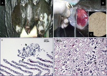
In 2014, an outbreak of scuticociliatosis affecting the fine flounder P. adspersus was detected on a fish farm in Peru (Ancash, Huarmey Province). The farm was equipped with two water flow systems: one with closed and the other with open recirculation. Mortality was very high (between 80 and 100%) in both systems, affecting fish of different sizes. Outbreaks of scuticociliatosis in fine flounder coincided with a series of anomalies in the seawater temperature registered in the eastern Pacific region affected by El Niño resulting in an average temperature that was 3.1 °C higher than the annual average (Comunicado Oficial ENFEN – Estudio Nacional del Fenómeno El Niño-No. 09-2014, Instituto del Mar del Perú – IMARPE). Ciliates were isolated from fish ranging from 16 to 27 cm in size (Fig. 1A). Two isolates of the scuticociliates, denominated Pe5 and Pe7, were obtained from ascitic fluid (with a concentration of approximately 5 × 10⁶ trophozoites per mL) of naturally infected specimens of the fine flounder P. adspersus.
Paralichthys adspersus: clinical signs of infection. (A) Visible changes are observed in the pigmentation of infected fish, and (B) the presence of haemorrhagic lesions on the gills. (C) Ciliates obtained from ascites liquid. Histopathological findings of the infection by scuticociliates: (D) histological section of gills showing the presence of ciliates (arrows), and (E) microphotograph showing a massive infection of ciliates in the myocardium (arrows) causing intense cardiac muscle histophagy. Staining H&E, scale bar = 50 µ m.

Virulent strain I1 of P. dicentrarchi was originally isolated from ascites fluid turbot from infected fish on a farm in Galicia (NW Spain) (Iglesias et al. Reference Iglesias, Paramá, Álvarez, Leiro, Fernández and Sanmartín2001).
Strain Ma/2 of M. avidus deposited by A.T. Soldo and E. B. Small (Veterans Administration Medical Center, Miami, FL) with the name Miamiensis avidus Thompson and Moewus (ATCC® 50180™) was acquired from the American Type Culture Collection (ATCC, USA). The strain Ma/2 of M. avidus belonged to the collection of Dr G.G. Holz and it was originally isolated by Dr L Moewus from infected seahorses and cultivated axenically since 1963 (Moewus, Reference Moewus and Oppenheimer1963; Kaneshiro et al. Reference Kaneshiro, Dunham and Holz1969).
Isolates Pe5, Pe7 and P. dicentrarchi strain I1 were maintained in the laboratory under the culture conditions described by Iglesias et al. (Reference Iglesias, Paramá, Álvarez, Leiro, Aja and Sanmartín2003a ). Strain Ma/2 of M. avidus was cultivated axenically in ATCC® medium 1651 MA (LGC Standards, Spain) at 25 °C and subcultured every 3–5 days.
For experimental infections, turbot was injected intraperitoneally with 0.1 mL of 106 ciliates per mL (M. avidus, strain Ma/2) in phosphate-buffered saline (PBS) containing 10 mm Na2HPO4, 2 mm KH2PO4, 2.7 mm KCl and 137 mm NaCl, as previously described (Paramá et al. Reference Paramá, Iglesias, Álvarez, Leiro, Aja and Sanmartín2003), and the fish were observed daily for signs of infection and mortality. Infection was confirmed post mortem by the presence of ciliates in organs and tissues.
Morphological and histological analyses
Ciliates obtained at the exponential phase of culture (days 2–3) were concentrated by centrifugation at 700 g for 5 min and stained by a modification of the amoniacal silver carbonate method originally described by Fernández-Galiano (Reference Fernández-Galiano1994) and described in detail by Budiño et al. (Reference Budiño, Lamas, Pata, Arranz, Sanmartín and Leiro2011a ).
The nuclear apparatus was visualized by fluorescence, after staining ciliates with an aqueous solution of 0.4 µg mL−1 of 4′−6-diamidine-2-phenylindone (DAPI, Sigma-Aldrich), in a Zeiss Axioplan microscope (Jena, Germany) equipped with a DAPI filter set (BP 365/12; FT 395; LP 397).
Somatic and caudal cilia were measured in parasites fixed in 10% buffered formaldehyde under phase contrast optics.
For histological study, tissues were fixed in 10% buffered formaldehyde solution, dehydrated through an ethanol series, embedded in Paraplast Plus (Sigma-Aldrich), sectioned at 2–5 µm with a Leica RM 2135 rotary microtome (Leica Biosystems, Germany), and stained with haematoxylin and eosin (H&E) for examination by light microscopy (Iglesias et al. Reference Iglesias, Paramá, Álvarez, Leiro, Fernández and Sanmartín2001).
Tomitogenesis and prey induction experiments
For tomite transformation, cultures of 2 × 10³ cells per mL of M. avidus strain Ma/2, P. dicentrarchi strain I1 and isolate Pe5 in stationary phase were centrifuged at 700 g for 5 min, resuspended in non-nutrient synthetic seawater (NSS, 8‰ salinity) and incubated at 25 °C (Gómez-Saladín and Small, Reference Gómez-Saladín and Small1993a ). In prey induction experiments, ciliates of M. avidus strain Ma/2 and P. dicentrachi strain I1 were incubated at a ratio of 1 : 1 in 24-well microplates for 5 days in NSS at 25 °C. The ciliates were observed daily under an inverted optical microscope. When predation phenomena were observed, ciliates were removed and centrifuged at 700 g for 5 min. Some specimens (106 ciliates) were fixed in buffered 10% formaldehyde in phosphate buffer saline for later examination by phase contrast microscopy, while other specimens (106 ciliates) were stained, using a modification of the ammoniacal silver carbonate method, as described previously.
PCR, cloning and phylogenetic analyses
Cultured ciliates (5 × 10⁶ cells per mL) were harvested by centrifugation at 700 g for 5 min. The ciliates were washed twice with PBS and total DNA was purified with DNAeasy Blood and Tissue Kit (Qiagen) according to the manufacturer's instructions. DNA was analysed to estimate its quality, purity and concentration by an A260 measurement in a NanoDrop ND-1000 Spectrophotometer (NanoDrop Technologies, USA.). The DNA was stored at −20 °C until use.
PCR amplification was performed as previously described (Leiro et al. Reference Leiro, Siso, Paramá, Ubeira and Sanmartín2000; Budiño et al. Reference Budiño, Lamas, Pata, Arranz, Sanmartín and Leiro2011a ), with minor modifications. A complete small-subunit ribosomal RNA (SSUrRNA; 1759 bp), a region of 388 bp of the gene coding the β-tubulin and a region of 197 bp of the gene coding the α-tubulin were amplified. The primer sets were designed and optimized by use of the Primer 3Plus program (http://www.bioinformatics.nl/cgi-bin/primer3plus/primer3plus.cgi), with default parameters. The PCR mixtures (25 µL) contained PCR reaction buffer (10 mm Tris-HCl, 50 mm KCl, 1.5 mm MgCl2, pH 9.0), 0.2 mm of each deoxynucleoside triphosphate (dNTPs, Roche), 0.4 µ m of each primer [forward 5′-AATCTGGTTGATCCTGCCAGT-3′/reverse 5′-GATCCTTCCGCAGGTTCA-3′ (SSUrRNA); forward 5′-CCTACCACGGAGACTCTGATT-3′/reverse 5′-CCATAATTCTGTCGGGGTATT-3′ (β-tubulin); forward 5′-ATGCCCTCTGATAAAACCATC-3′/reverse 5′-GAGTCCGGTACAGTTGTCAG-3′ (α-tubulin)]; 0.5 units of high fidelity Taq polymerase (Nzyproof DNA polymerase; Nzytech, Portugal) and 50 ng of genomic DNA. The reactions were run in an automatic thermocycler (iCycler, BioRad, USA) as follows: initial denaturing at 94 °C for 5 min, followed by 35 cycles at 94 °C for 30 s, annealing at 55, 57 and 64 °C (for SSUrRNA, α-tubulin and β-tubulin, respectively) for 45 s, and 72 °C for 1 min; and finally, a 7-min extension phase at 72 °C. The PCR products were analysed on a 4% agarose gel in Tris acetate ethylenediaminetetraacetic acid (TAE) buffer (40 mm Tris-acetate, pH 8.0, 2 mm EDTA) containing Sybr Green at 1× concentration (Intron, Korea), to verify the presence of bands of the correct size, and were photographed with a digital camera. The PCR products were cloned in the pSpark® II DNA cloning vector kit (Canvax Biotech, Spain) according to the manufacturer's instructions. After ligation of the PCR fragment, the Escherichia coli DH5α cells were transformed and then selected by culture on LB agar plates containing 100 µg mL−1 ampicillin, with 50 µL of a stock solution of 20 mg mL−1 of 5-bromo-4-chloro-3-indolyl-β-galactoside (X-Gal) and 20 µL of a 0.5 m solution of isopropylthio-β-D-galactoside (IPTG). Ten E. coli white colonies per ligation sample were amplified in LB medium and plasmid DNA was purified with the QiAaprep® Spin Miniprep kit (Qiagen, Germany) according to the manufacturer's instructions. To confirm the presence of the cloned fragment of the correct size, the fragment was amplified by PCR, with the previously indicated primers and conditions. The PCR-amplified products were then visualized by agarose gel electrophoresis and sequenced in complementary directions (Sistemas Genómicos, Spain).
We used the BLAST interface and the blastn program optimized for very similar sequences (megablast), available at http://blast.ncbi.nlm.nih.gov, to calculate the degree of identity between the nucleotide sequences. Sequence alignment can be performed directly online.
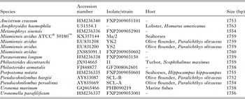
The complete nucleotide sequences of the SSUrRNA gene from isolates Pe5 and Pe7 from fine flounder, strain I1 of P. dicentrarchi isolated from turbot and strain Ma/2 of M. avidus (ATCC® 50180™) were compared with equivalent sequences from strains YK2 and YS2 of M. avidus isolated from olive flounder (accession numbers EU831208 and EU831200; Jung et al. Reference Jung, Im, Struder-Kypke, Kitamura and Woo2011) and with other species of the order Philasterida (Table 1). The sequences were aligned with Clustal Omega (McWilliam et al. Reference McWilliam, Li, Uludag, Squizzato, Park, Buso, Cowley and López2013) and phylogenetic trees were inferred by the neighbour-joining (NJ) method (Saitou and Nei, Reference Saitou and Nei1987). This method was applied to the Kimura two-parameter correction model (Kimura, Reference Kimura1980) by bootstrapping with 1000 replicates (Felsenstein, Reference Felsenstein1985) from multiple alignments and consensus of the study sequences with Clustal Omega software (Sievers et al. Reference Sievers, Wilm, Dineen, Gibson, Karplus, Li, López, McWilliam, Remmert, Söding, Thompson and Higgins2011). The tree is drawn to scale, with branch lengths in the same units as those of the evolutionary distances used to infer the phylogenetic tree. All positions containing gaps and missing data were analysed by use of pairwise deletion method and evolutionary analyses were conducted in MEGA7 (Kumar et al. Reference Kumar, Stecher and Tamura2016).
List of philasterid species included in the phylogenetic analysis, showing the GenBank accession numbers for their SSUrRNA gene sequences, the isolates/strains, host specificity and sequence length in base pairs (bp)

Production of recombinant alpha-tubulin (r-αTub) of P. dicentrarchi in yeast cells
RNA isolated from Philasterides dicentrarchi was purified with a NucleoSpin RNA kit (Macherey-Nagel, Düren, Germany) according to the manufacturer's instructions. After purification of the RNA, the quality, purity and concentration were measured in a NanoDrop ND-1000 Spectrophotometer (NanoDrop Technologies, USA). The reaction mixture used for cDNA synthesis (25 µL/reaction mixture) contained 1.25 µ m random hexamer primers (Promega), 250 µ m of each deoxynucleoside triphosphate (dNTP), 10 mm dithiothreitol (DTT), 20 U of RNase inhibitor, 2.5 mm MgCl2, 200 U of MMLV (Moloney murine leukemia virus reverse transcriptase; Promega) in 30 mm Tris and 20 mm KCl (pH 8.3), and 2 µg of sample RNA. The PCR was carried out with gene-specific primers designed from a partial sequence of the α-tubulin of P. dicentrarchi (forward/reverse primer pair 5′-AAAGAAGAAGGGGTACCTTTGGATAAAAGAatgccctctgataaaaccatc-3′/5′-TGGGACGCTCGACGGATCAGCGGCCGCTTAGTGGTGGTGGTGGTGGTGgagtccggtacagttgtcag-3′). These primers were designed and optimized, using the Saccharomyces Genome Database (http://www.yeastgenome.org/). A hybridization region with the yeast YEpFLAG-1 (Eastman Kodak Company) plasmid and a poly His region (lower case letters correspond with the gene annealing zone) were included. The PCR reaction was initially developed at 95 °C for 5 min and then for 30 cycles of 94 °C for 1 min, 53 °C for 1.5 min and 72 °C for 2 min. At the end of the 30 cycles, a 7-min extension phase was carried out at 72 °C. The PCR products were purified by using the Gene Jet PCR Purification Kit (Fermentas, Life Sciences) according to the manufacturer's instructions.
Purified PCR products were cloned in YEpFLAG-1 (Eastman Kodak Company) yeast expression vector, a plasmid carrying a TRP1 gene that completes the auxotrophy for the tryptophan for the host yeast (López-López et al. Reference López-López, Fuciños, Pastrana, Rúa, Cerdán and González-Siso2010).
Linearized plasmid YEpFLAG-1 was digested with EcoRI and SalI (Takara) and used to transform Saccharomyces cerevisiae cells (strain BJ 3505) by the lithium acetate procedure (Ito et al. Reference Ito, Fukuda, Murata and Kimura1983). The procedure involves co-transformation of yeast cells with the linearized empty plasmid and the PCR-generated DNA fragment, so that a recombination process occurs within the cell to yield a plasmid bearing the desired insert. Positive colonies were selected on complete medium containing glucose (20 g L−1), but without tryptophan (CM-Trp), Yeast Nitrogen Base medium without amino acids (Sigma-Aldrich), except for the amino acids arginine, methionine and threonine at 10 mg L−1; adenine, histidine, leucine, lysine and tyrosine at 40 mg L−1; and isoleucine and phenylalanine at 60 mg L−1.
Plasmid DNA was then extracted with Easy Yeast Plasmid Isolation Kit (Clonetech) according to the manufacturer's instructions. The purified and cloned DNA fragment was subjected to sequencing analysis (Sistemas Genómicos, Spain).
Inoculation and polyclonal mouse antisera
CD-1 mice were inoculated i.p. with 200 µL of a 1 : 1 emulsion composed of 100 µL of a solution of 100 µg of r-αTub of the strain I1 of P. dicentrarchi in PBS and 100 µL of Freund complete adjuvant (Sigma-Aldrich). The same dose of r-αTub protein was prepared in Freund's incomplete adjuvant and injected i.p. in mice 15 and 30 days after the first immunization. The mice were bled via retrobulbar venous plexus 7 days after the second inoculation. The blood was left to coagulate overnight at 4 °C before the serum was separated by centrifugation (2000 g for 10 min), mixed 1 : 1 with glycerol, and stored at −20 °C until use (Piazzon et al. Reference Piazzon, Lamas and Leiro2011).
Immunoassays
For Western-blot assay, integral ciliate membrane-associate proteins (CMP) of scuticociliates were prepared as previously described (Iglesias et al. Reference Iglesias, Paramá, Álvarez, Leiro, Ubeira and Sanmartín2003b ), with minor modifications (Mallo et al. Reference Mallo, Lamas and Leiro2013). Briefly, scuticociliate trophozoites were deciliated by the method described by Dickerson et al. (Reference Dickerson, Clark and Findly1989). The integral proteins were extracted by phase separation in Triton X-114 solution according to the method described by Bordier (Reference Bordier1981). Samples from CMP were separated under non-reducing conditions on linear SDS-PAGE 12.5% gels (Piazzon et al. Reference Piazzon, Lamas, Castro, Budiño, Cabaleiro, Sanmartín and Leiro2008). On completion of the electrophoresis, the gels were stained with Thermo Scientific GelCode Blue Safe Protein Stain (Thermo Fisher, USA) for qualitative determination of the protein bands. At the same time, a gel was immunoblotted at 15 V for 35 min to Immobilon-P transfer membranes (0.45 µm; Millipore, USA) in a trans-blot SD transfer cell (Bio-Rad, USA) with transference buffer (48 mm Tris, 29 mm glycine, 0.037% SDS and 20% methanol, pH 9.2). The membrane was washed with Tris buffer saline (TBS; 50 mm Tris, 0.15 m NaCl, pH 7.4) and immediately stained with Ponceau S to verify transfer. After destaining the membrane with bidistilled water, a blocking solution consisting of TBS containing 0.2% Tween 20 and 3% BSA was added. The membrane was incubated for 1.5 h at room temperature and then washed in TBS and incubated overnight with the mouse polyclonal antisera anti-r-αTub (1 : 500 dilution) at 4 °C. The membrane was washed again with TBS and incubated with rabbit anti-mouse IgG (Dakopatts; dilution 1 : 6000) for 1 h at room temperature. The membrane was then washed five times for 5 min with TBS and incubated for 1 min with enhanced luminol-based chemiluminiscent substrate (Pierce ECL Western Blotting Substrate, Thermo Scientific, USA), before finally being visualized and photographed with a FlourChem® FC2 imaging system (Alpha Innotech, USA).
The enzyme-linked immunosorbent assay (ELISA) was performed as previously described (Iglesias et al. Reference Iglesias, Paramá, Álvarez, Leiro, Ubeira and Sanmartín2003b ), with minor modifications. One μg of CMP in 100 µL of carbonate-bicarbonate buffer (pH 9.6) was added to 96-well ELISA plates (high binding, Greiner Bio-One, Germany) and incubated overnight at 4 °C. The wells were then washed three times with TBS and blocked for 1 h with TBS containing 0.2% Tween 20 (TBS-T1) and 5% non-fat dry milk. The plates were incubated for 30 min at room temperature in a microplate shaker for ELISA (Stuart, UK) at 750 rpm with a 1 : 100 dilution (in TBS-T1 containing 1% non-fat dry milk) of anti-r-αTub, and washed five times with TBS containing 0.05% Tween 20. Bound mouse antibodies were detected with a peroxidase-conjugated anti-mouse Ig polyclonal rabit serum (DAKO) diluted 1 : 1000 in TBS-T1, and incubated for 30 min with shaking. The plates were washed five times in TBS, and 100 µL of o-phenylenediamine dihydrochloride (OPD, Sigma-Aldrich) and 0.003% H2O2 were added to each well. After incubation of the plates for 20 min at room temperature in darkness, the enzymatic reaction was stopped by adding 25 µL of H2SO4 at 3 N. Finally, the absorbance was read at 492 nm in a spectrophotometer microplate reader (Bio-Tek Instruments, USA).
Statistical analysis
Results are expressed as means ± s.e. of means. Data were tested by one-way analysis of variance (ANOVA) followed by a Tukey–Kramer test for multiple comparisons. Differences were considered significant at P < 0.05.
RESULTS
Description of an outbreak of scuticociliatosis in the fine flounder P. adspersus
The outbreak of scuticociliatosis coincided with water temperatures higher than 21–22 °C. The main external symptoms of the affected fish were alterations in skin pigmentation and emaciation (Fig. 1A), gills congested with mucus and abundant aneurysms (Fig. 1B and D), exophthalmia and abdominal distension with ascitic fluid, from which ciliates were isolated (Fig. 1C). At the histopathological level, the main pathology was associated with systemic necrosis affecting several organs including the heart, with severe necrosis of myocardium (Fig. 1D).
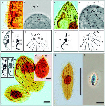
Description of the Peruvian population (isolates Pe5 and Pe7) infecting the fine flounder P. adspersus (Fig. 2A–I; Table 2)
The main characteristics of the cilates isolated from the fine flounder are as follows: cells elongated and spindle-shaped, with a pointed anterior and rounded posterior end, a contractile vacuole and a prominent caudal cilium (Figs 2B, E, G, I and 3C). The buccal apparatus always contained two paroral membranes (P1 and P2) and three oral membranelles or polykinetids (M1, M2 and M3) (Fig. 2A, D, F and H), showing identical morphology to that of Philasterides dicentrarchi (Fig. 2C). P1 extends from the start of M1 to the start of M3, while P2 extends from the middle of M3 to the end of the oral cavity (Fig. 2A, C, D, F and H). M1 is elliptical, M2 is trapezoidal and M3 is smaller than M1 and M2, and irregularly triangular (Fig. 2A, C, D, F and H). The somatic ciliature of the Pe5 and Pe7 isolates consists of 10–13 kineties, and the pore of the subterminal contractile vacuole is at the posterior end of the second kinety (Fig. 2B, E, G and I).
Morphological characteristics of isolates Pe5 and Pe7 from farmed flounder Paralichthys adspersus in comparison with those of Miamiensis avidus strain Ma/2 (ATCC® 50180™). Silver carbonate-stained trophozoites: isolate Pe5 (A, B); isolate Pe7 (F, G); and Miamiensis avidus strain Ma/2 (ATCC® 50180™) (J, M, N). (A, F) Light photomicrographs showing the oral ciliature and the posterior cell portions (B, G) of isolates Pe5 and Pe7, respectively. (C, D, H) Schematic drawings of the buccal apparatus in Philasterides dicentrarchi from sea bass (C), Pe5 (D), and Pe5 (H), showing three oral polykinetids, (M1, M2 and M3) and two paraoral membranes (P1 and P2) (E, I) Drawings of posterior ends of isolates Pe5 and Pe7 showing 12 kineties (1–12), the pore of the subterminal CVP, and a prominent caudal cilium (C, circle). (J) Light microphotograph of silver carbonate-stained specimens of M. avidus strain Ma/2 showing the buccal apparatus. (K, L) Drawings of oral ciliature in M. avidus from original description (K) and strain Ma/2 (L) showing the oral polykinetids M1, M2 and M3, and the paraoral membrane (P). (M) Photomicrograph of a trophozoite of M. avidus strain Ma/2 showing the spine-shaped apical cell portion (arrow). (N, O) Tomites of M. avidus strain Ma/2 stained with silver-carbonate (N), and in vivo under phase contrast optics (O). Scale bars = 10 µm. CVP, contractile vacuole pore.

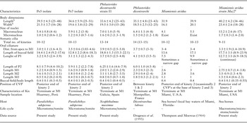
Biometric data for silver-stained ciliates isolates (Pe5 and Pe7) from Paralichthys adspersus, Philasterides dicentrarchi strain I1 isolated from turbot, P. dicentarchi Dragesco et al. Reference Dragesco, Dragesco, Coste, Gasc, Romestand, Raymond and Bouix1995 from seabass, strains T5 and T16 of Miamiensis avidus Thompson and Moewus, Reference Thompson and Moewus1964, and strain Ma/2 of M. avidus (ATCC® 50180™)

a Biometric values obtained from specimens of M. avidus supplied by ATCC and cultured in our laboratory.
b Data obtained from microstomes. The values shown are expressed (in μm) as mean ± s.e., with the minimum-maximum ranges in parentheses. M1–M3: oral polykinetids; P: paroral membrane; P1, P2: paroral membranes 1 and 2; CVP: contractile vacuole pore; Kn: kinety. The measurements were made in 50 specimens. (–) No data available.
The morphometric data of the Pe5 and Pe7 isolates are summarized in Table 2, in which they are compared with the morphometric data available for P. dicentrarchi isolated from turbot (strain I1; present study) and from seabass (Dragesco et al. Reference Dragesco, Dragesco, Coste, Gasc, Romestand, Raymond and Bouix1995) and for M. avidus isolated from seahorses (Thompson and Moewus, Reference Thompson and Moewus1964) as well as with our data from strain Ma/2 of Miamiensis avidus Thompson & Moewus (ATCC® 50180™) stained with ammoniacal silver carbonate and P. dicentrachi strain I1. The ranges of the morphological data on isolates Pe5 and Pe7 generally overlap those of P. dicentrarchi strain I1, of the original species description of M. avidus and of M. avidus held by the ATCC. At the morphological level, isolates Pe5 and Pe7 and P. dicentrarchi strain I1 differ in several morphological features from M. avidus (ATCC), such as the lack of a beak at the frontal part of trophozoites (Fig. 2M), the lack of a continuous paroral membrane (P; Fig. 2J, K and L) and the morphology of the tomites, which are elongated and fusiform in the latter species (Fig. 2N and O). In contrast to M. avidus (ATCC), the P. adspersus isolates Pe5 and Pe7 as well as P. dicentrarchi have a prominent caudal cilium that is longer than the somatic cilia (Fig. 3C). In P. dicentrarchi, the mean length of the caudal cilium was 11.4 ± 2.0 µm (6–14 µm) and of the somatic cilia was 5.6 ± 1.0 µm (5–8 µm). In M. avidus strain Ma/2, the mean length of the caudal cilium was 9.3 ± 2.1 µm (7–11 µm) and that of the somatic cilia, 6.6 ± 1.1 µm (5–9 µm, n = 20).
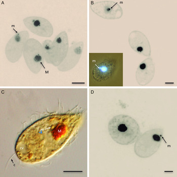
Structure of nuclear apparatus. Epifluorescence photomicrographs (A, B, D) of trophozoites stained with DAPI, and light micrographs of cells stained with silver carbonate (C), showing the position of the macronucleus (M) and micronucleus (m): (A) strain I1 of Philasterides dicentarchi, (B) isolate Pe5, (C) isolate Pe7 and (D) strain Ma/2 of Miamiensis avidus. Figure C also shows the caudal cilium (c) and the posterior contractile vacuole (cv). Scale bars = 10 µm. DAPI, diamidino-2-phenylindole.

The nuclear apparatus of isolates Pe5 and Pe7 and strain I1 of P. dicentrarchi consists of a spherical macronucleus located mainly in the middle or anterior third of the trophozoite, slightly lateralized and between 4 and 8 µm in diameter (Table 2; Fig. 3A–C). The micronucleus, 1–2 µm in diameter, has a variable posterior position in isolates Pe5 and Pe7 and strain I1 of P. dicentrarchi (Fig. 3A–C), (Table 2). In M. avidus strain Ma/2, the macronucleus is irregularly spherical and usually located in the anterior cell half. The micronucleus anterior or laterally of the macronucleus (Fig. 3D).
Tomitogenesis and predatory transformation
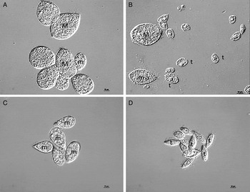
Most polymorphic hymenostomes and scuticociliates have morphologically distinct feeding stages including a bacterivorous microstome, a predatory macrostome, and sometimes a non-feeding, fast swimming tomite (Fig. 4). In culture media containing nutrients, M. avidus strain Ma/2 produced macrostome and microstome forms (Fig. 4A), while all strains of P. dicentrarchi only produced microstome forms (Fig. 4C). Under conditions of nutrient deprivation, M. avidus Ma/2 produced some macrostome and microstome forms and numerous tomites (Fig. 4B) P. dicentrarchi appeared almost exclusively as tomites (Fig. 4D). In M. avidus strain Ma/2, the macrostomes measured 50.1 ± 9.4 × 35.4 ± 5.2 µm (37–65 × 28–44 µm, n = 10) and the tomites 22.4 ± 4.5 × 10.5 ± 2.7 µm (23–28 × 7–13 µm, n = 10). In P. dicentrarchi, the length/width of the tomite was 20.6 ± 2.7 × 13.1 ± 2.1 (17–24 × 11–16 µm, n = 10).
Induction of tomitogenesis in M. avidus and P. dicentrarchi under conditions of starvation. Cultures of M. avidus (A, B) and P. dicentrarchi (C, D), in media with nutrients (A, C) and without nutrients in non-nutrient artificial seawater (B, D). M, macrostomes; m, microstomes; t, tomites. Scale bar = 10 µm.

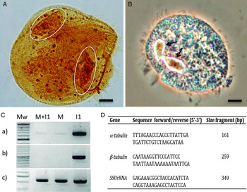
To verify which of the species considered in this study developed predatory macrostome phases, we co-cultured trophozoites of M. avidus strain Ma/2 with trophozoites of P. dicentrarchi strain I1 and trophozoites of strain I1 with isolate Pe7, in media without nutrients. On the second day, the co-cultures of M. avidus strain Ma/2 and P. dicentrarchi strain I1 showed predatory macrostome forms (Fig. 5A and B); however, this phenomenon was not detected in the co-cultures of Pe7 and I1 isolates. We found that M. avidus is a predator of P. dicentrarchi strain I1. To reach this conclusion, we obtained genomic DNA from M. avidus and P. dicentrarchi from 5 day co-cultures at day 5 and amplified it by PCR (Fig. 5C), using the primers designed for P. dicentrarchi DNA genes and showing low (α-tubulin), very low (β-tubulin) and high (SSUrRNA) nucleotide identity with the same genes in M. avidus (Fig. 5D). In all cases, we observed amplification of the SSUrRNA gene, slight amplification of the α-tubulin gene and non-amplification of the β-tubulin gene in DNA samples obtained from cultures of the strain Ma/2 of M. avidus and in DNA samples from co-cultures of M. avidus and P. dicentrarchi (Fig. 5C and a–c). We then cloned and sequenced the amplified fragments of α-tubulin and SSUrRNA genes from co-cultures of M. avidus strain Ma/2 and P. dicentrarchi strain I1, observing that the nucleotide sequence obtained coincided with those of M. avidus Ma/2 (Fig. 5C, a and c).
(A, B) Prey-induced transformation of M. avidus ATCC® 50180™ (strain Ma/2) co-cultivated with the I1 strain from Philasterides dicentrarchi in non-nutrient artificial seawater (NSS). Macrostome formation at day 2 of co-cultures showing ciliates ingested in its interior (circles and arrows): (A) silver staining, (B) phase contrast. (C) PCR from genomic DNA of M. avidus (M), P. dicentrarchi strain I1 (I1) and M. avidus and P. dicentrarchi (M + I1) co-cultivated for five days. (D) Primers designed from the nucleotide sequences of P. dicentrarchi corresponding to (a) the α-tubulin, (b) β-tubulin and (c) the small rRNA subunit (SSUrRNA) gene used for PCR. Mw: molecular size markers. PCR, polymerase chain reaction.

Infectivity of M. avidus strain Ma/2
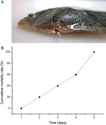
The M. avidus strain ATCC© 50180™ was originally isolated from infected seahorses; however, we did not know whether the species could infect flatfish. In this experiment, we inoculated turbot by intraperitoneal injection with M. avidus, using the same dose as used for P. dicentrarchi strain I1 (105 ciliates per fish). We observed that M. avidus strain Ma/2 is highly pathogenic to turbot, generating abdominal distension with a significant presence of ascitic liquid, from which it was possible to isolate ciliates (Fig. 6A) and causing 100% mortality in infected fish on day 5 (Fig. 6B).
Virulence of the Ma/2 strain of M. avidus ATCC® 50180™ in experimentally infected turbot. (A) Abdominal distension due to accumulation of ascitic fluid in the body cavity. The arrow indicates anal inflammation. (B) Kinetics of cumulative mortality induced after intraperitoneal injection with M. avidus five after infection.

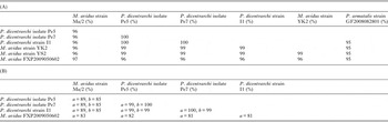
Molecular analysis of the scuticociliates
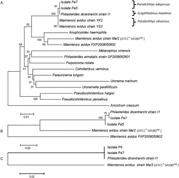
The full length SSUrRNA sequences of Pe5, Pe7 and P. dicentrarchi strain I1 (Accession no. JX914665.1) showed 100% identity with each other and 96% identity with M. avidus strain Ma/2 (Accession KX357144; Table 3(A)). We compared the above- mentioned 18S sequences with those obtained from the GenBank for M. avidus isolate YK2 (Accession no. EU831208.1; Jung et al. Reference Jung, Im, Struder-Kypke, Kitamura and Woo2011), M. avidus isolate YS2 (Accession EU831200.1; Jung et al. Reference Jung, Im, Struder-Kypke, Kitamura and Woo2011), M. avidus isolate FXP2009050602 (Accession JN885091.1; Gao et al. Reference Gao, Katz and Song2012), and strain GF2008082801 of Philasterides armatalis Song, Reference Song2000 (Accession FJ848877; Gao et al. Reference Gao, Fan, Yi, Strüder-Kypke and Song2010). Miamiensis avidus strains YK2 and YS2 showed 99% identity with P. dicentrachi strain I1, Pe5 and Pe7 isolates, 96% identity with M. avidus strain Ma/2 or M. avidus isolate FXP2009050602 and 95% identity with P. armatalis (Table 3(A)). The phylogenetic tree constructed, using the NJ model grouped Pe5, Pe7, P. dicentrachi strain I1 in the same node and closely related to strains YK2, YS2 (Fig. 7A), while M. avidus strain Ma/2 from ATCC and the M. avidus isolate FXP2009050602 grouped in a different node together with the Anophryoides haemophila (Fig. 7A). Analysis of the similarity between the nucleotide sequences corresponding to fragments of the α- (strain I1, accession KX357145; strain Ma/2, accession KX357143) and β-tubulin genes (strain I1, accession CQ342956.1; strain Ma/2 accession KX357147) revealed 99–100% identity between isolates Pe5, Pe7 and strain I1, but only 89% identity for α-tubulin and 85% identity for β-tubulin with M. avidus strain Ma/2 or 81–83% of identity for α-tubulin with M. avidus isolate FXP2009050602 (Table 3(B)). Phylogenetic trees generated after alignments of the nucleotide sequences of the α- and β-tubulin genes for the Pe5 and Pe7 isolates and I1 and the Ma/2 strains, using the model NJ, grouped Pe5, Pe7 and I1 in one node and the M. avidus Ma/2 strain or isolate FXP2009050602 in a different node (Fig 7B and C).
NJ unrooted trees inferred from: SSUrRNA (A), as well as, the α- (B), and β-tubulin (C) nucleotide sequences showing the phylogenetic relationships between isolates Pe5 and Pe7 from the fine flounder, strain I1 of Philasterides dicentrarchi isolated from turbot and strain Ma/2 of Miamiensis avidus ATCC® 50180™. The phylogeny was also inferred by analysis of SSUrRNA gene sequences between the isolates from the fine flounder, strains of Philsterides isolated from turbot, strains of Miamiensis isolated from olive flounder (strains YK2 and YS2), isolate FXP2009050602 of M. avidus, and several species of philasterids (see Table 1) including strain GF200806601 of Philasterides armatalis (A). Nodes represent bootstrap values of 1000 resampled values in the NJ analysis with the Kimura two-parameter correction model, and the scale bar indicates the genetic distance. NJ, neighbour-joining.

Nucleotide identity percentage analyzed by the BLAST alignment program between regions of DNA gene of the small subunit of (A) rRNA (SSUrRNA) and (B) α- and β-subunits of tubulin of isolates Pe5 and Pe7 from the fine flounder Paralichthys adspersus, strain I1 of Philasterides dicentrarchi isolated from turbot and strain Ma/2 of Miamiensis avidus held in ATCC® 50180™

Additionally, the BLAST analysis of the SSUrRNA sequences included the strains YK2 and YS2 of Miamiensis avidus isolated from Paralichthys olivaceus, and the GF2008082801 strain of Philasterides armatalis.
Antigenic relationships between ciliates isolated from flatfish and M. avidus strain Ma/2
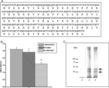
We generated a recombinant protein from a partial sequence of the gene encoding this protein (r-αTub) in P. dicentrarchi strain I1, to compare the antigenic homology between the α-tubulin of the scuticociliates Pe5, Pe7, I1, and M. avidus strain Ma/2 (ATCC® 50180™) (Fig. 8A). The antibodies generated in mice following inoculation with the recombinant protein were used to perform three immunoassays to determine the level of cross-reactivity with this protein in the isolates/strains studied.
Analysis of the antigenic relationships related to the alpha subunit of tubulin, using a gene fragment (A) between isolates from the fine flounder P. adspersus, Philasterides dicentarchi strain I1 and Miamiensis avidus strain Ma/2 (ATCC® 50180™), by various immunoassays using antibodies generated against a fragment of recombinant α-tubulin. . (B) ELISA of the degree of cross-reactivity between α-tubulin from P. dicentrarchi strain I1, isolate Pe7 from the fine flounder P. adspersus and M. avidus strain Ma/2. The results are expressed as mean values of the absorbance at 492 nm ± s.e. (n = 5), and asterisks indicate the statistical significance (P < 0.01). (C) Western blot analysis of ciliary proteins of M. avidus strain Ma/2 (lane 1), P. dicentrachi strain I1 (lane 2) and the Pe7 isolate from P. adspersus showing two bands of recognition corresponding to two polypeptide fragments of α-tubulin (arrows). Mw: molecular weight markers expressed in kD. ELISA, enzyme-linked immunosorbent assay.

Quantitative immunoassays as ELISA revealed that the levels of recognition of native ciliary proteins containing α-tubulin in the Pe7 isolate were similar to those obtained with strain I1; however, these levels were significantly lower when antigens of strain Ma/2 of M. avidus were used (Fig. 8B).
Western blot carried out under reducing conditions revealed two bands of about 50 and 60 kD in ciliary isolated fractions of isolate Pe7 and strain I1. However, no bands were detected when antigens from M. avidus strain Ma/2 were included (Fig. 8C).
DISCUSSION
Here we describe a scuticociliate infection in the fine flounder P. adspersus, cultivated in Peru. The infection occurred on a fish farm with a water recirculation system, which appears to increase the risk of scuticociliatosis (Budiño et al. Reference Budiño, Lamas, González, Pata, Devesa, Arranz and Leiro2011b ) and at high temperatures, another risk factor for development of this disease (Iglesias et al. Reference Iglesias, Paramá, Álvarez, Leiro, Fernández and Sanmartín2001; Moustafa et al. Reference Moustafa, Naota, Morita, Tange and Shimada2010).
We did not conduct a complete histopathological study of the scuticociliatosis in P. adspersus and only observed that the clinical signs and pathology generated by the ciliates are very similar to those produced in Scophthalmus maximus Linnaeus, 1758, and the olive flounder Paralichthys olivaceus Temminck and Schlegel, 1846 (Iglesias et al. Reference Iglesias, Paramá, Álvarez, Leiro, Fernández and Sanmartín2001; Jung et al. Reference Jung, Kitamura, Song and Oh2007; Moustafa et al. Reference Moustafa, Naota, Morita, Tange and Shimada2010; Harikrishnan et al. Reference Harikrishnan, Jin, Kim, Balasundaram and Heo2012). Our research primarily focused on the identification of the causative agent of the disease in P. adspersus. In all samples, the buccal apparatus of the ciliate isolates Pe5 and Pe7 from P. adspersus contained two paroral membranes (P1 and P2) and three oral polykinetids (M1, M2 and M3), with identical arrangement and morphology to that described for Philasterides dicentrarchi (Dragesco et al. Reference Dragesco, Dragesco, Coste, Gasc, Romestand, Raymond and Bouix1995; Iglesias et al. Reference Iglesias, Paramá, Álvarez, Leiro, Fernández and Sanmartín2001; Budiño et al. Reference Budiño, Lamas, Pata, Arranz, Sanmartín and Leiro2011a ). According to the redescription of the genus Philasterides Kahl, 1931 by Grolière (Reference Grolière1980), the type species Philasterides armata has a split paroral membrane and three equidistant adoral poykinetids (M1, M2 and M3). However, the structure of the two paroral membranes is debated as it is uncertain whether it respresents a generic character for Philasterides or may show an intraspecific variability and is thus not suitable for species identification (Jung et al. Reference Jung, Kitamura, Song and Oh2007).
Song and Wilbert (Reference Song and Wilbert2000) redescribed M. avidus, using the dimorphic paroral membrane with its monokinetidal anterior and its dikinetidal posterior part slightly separated as its main morphological characteristic, thereby synonymizing M. avidus and P. dicentrarchi. However, the morphological data of the present study clearly shows that both isolates obtained from P. adspersus (isolates Pe5 and Pe7) have two clearly separated paroral membranes, while M. avidus strain Ma/2 has invariably a single continuous paroral membrane. The original description of M. avidus mentions that a narrow gap ‘sometimes’ appears between the anterior and posterior paroral portions; however, this does not seem to be common in this species, as indicated by the fact that the authors described the paroral as a unit, measuring its length from its anterior to its posterior end (Thompson and Moewus, Reference Thompson and Moewus1964). In contrast to M. avidus, the presence of a bipartite paroral membrane (P1 and P2), seems thus to be a constant morphological feature in P. dicentrarchi (Dragesco et al. Reference Dragesco, Dragesco, Coste, Gasc, Romestand, Raymond and Bouix1995; Iglesias et al. Reference Iglesias, Paramá, Álvarez, Leiro, Fernández and Sanmartín2001; Budiño et al. Reference Budiño, Lamas, Pata, Arranz, Sanmartín and Leiro2011a ).
Jung et al. (Reference Jung, Kitamura, Song and Oh2007) found that a scuticociliate isolate (YS1 strain) from olive flounder, identified as M. avidus, has one or two paroral membranes and two or three oral polykinetids, the authors concluded that the morphology of buccal structures ‘cannot be used as a consistent key for identification of the species’. However, variability reported by Jung et al. (Reference Jung, Kitamura, Song and Oh2007), might reflect different stages in the development of these buccal structures during stomatogenesis, which is very common in several species of scuticociliates (Miao et al. Reference Miao, Wang, Song, Clamp and Al-Rasheid2010). Some studies clearly show that M. avidus has a continuous paroral membrane; only in the first phases of stomatogenesis (during the transformation of microstomes into to macrostomes), is the paroral membrane divided, with a small segment at the posterior end (Gómez-Saladín and Small, Reference Gómez-Saladín and Small1993a ).
The morphology of oral polykinetids M1, M2 and M3 in isolates Pe5 and Pe7 and Philasterides dicentrarchi strain I1 is very similar to that observed in M. avidus; but differs from the oral polykinetids of congeners. The M1 of isolates Pe5 and Pe7 and strain I1 of P. dicentrarchi is elongated like the M1 of M. avidus (Thompson and Moewus, Reference Thompson and Moewus1964), while the M1 of P. armata and P. armatalis is usually triangular (Grolière, Reference Grolière1980; Song, Reference Song2000); the M3 in P. dicentrarchi strain I1 and in the isolates Pe5 and Pe7 is irregularly triangular and similar to M. avidus (Thompson and Moewus, Reference Thompson and Moewus1964), but different from the rectangular M3 of P. armata (Grolière, Reference Grolière1980).
During early and late phases of binary fission in most ciliates, dividers share certain features, such as common position of macronucleus and micronucleus, synchronization of macronuclear amitosis and fission furrow and a specific and well defined dividing size (Long and Zufall, Reference Long and Zufall2010). The position of the micronucleus relative to the macronucleus is not specified in the original descriptions of P. dicentrarchi and M. avidus, which only state that the micronucleus is closely associated with the macronucleus but completely separate from it in P. dicentrarchi (Dragesco et al. Reference Dragesco, Dragesco, Coste, Gasc, Romestand, Raymond and Bouix1995; Paramá et al. Reference Paramá, Arranz, Álvarez, Sanmartín and Leiro2006); however, some later descriptions proposed the anterior position of the micronucleus relative to the macronucleus as a specific characteristic of M. avidus (Hu et al. Reference Hu, Song, Warren, Song, Warren and Hu2009). In this study, we clearly demonstrate that the position of the micronucleus relative to the macronucleus in Pe5, Pe7 isolates and in strain I1 of P. dicentarchi is very variable.
The formation of tomites, which is a general characteristic of scuticociliates, occurs in response to starvation without cyst production, or in response to exposure to certain chemicals (Fenchel, Reference Fenchel1987; Gómez-Saladín and Small, Reference Gómez-Saladin and Small1993b , Morais et al. Reference Morais, Lamas, Sanmartín, Orallo and Leiro2009). Miamiensis avidus is a known tomite-producing scuticociliate (Thompson and Moewus, Reference Thompson and Moewus1964; Gómez-Saladin and Small, Reference Gómez-Saladín and Small1993a , Reference Gómez-Saladin and Small b ). The life cycles of scuticociliates provide further taxonomically significant features. While M. avidus undergoes microstome to macrostome transformation following a considerable change in cell size and the buccal structures (Small, Reference Small1967; Gómez-Saladin and Small, Reference Gómez-Saladín and Small1993a ), such a transformation was not observed in P. dicentrarchi (Dragesco et al. Reference Dragesco, Dragesco, Coste, Gasc, Romestand, Raymond and Bouix1995). In the present study, we performed tomitogenesis and prey-induced transformation experiments with M. avidus strain Ma/2, P. dicentrarchi strain I1 isolated from P. adspersus and isolate Pe7 in a nutrient-depleted medium. We observed induction of the macrostome forms only in M. avidus strain Ma/2, and in co-cultures of this strain Ma/2 with the P. dicentrarchi strain I1; however, this phenomenon does not occur when I1 trophozoites were incubated with trophozoites of isolates (Pe7) of fine flounder, and neither macrostome forms nor any sign of predation/cannibalism could be detected.
The scuticociliate isolates Pe5 and Pe7 from the fine flounder P. adspersus and P. dicentrarchi strain I1 are obviously virulent in their hosts; however, the infective capacity of the M. avidus strain Ma/2, which has been maintained in culture for a long time (more than 40 years) in the ATCC collection (Soldo and Merlin, Reference Soldo and Merlin1972), is unknown. In the present study, we demonstrated the high virulence of the strain Ma/2, causing a mortality of 100% in turbot after experimental infection.
Identification of scuticociliates only on the basis of morphological features may lead to misidentification (Whang et al. Reference Whang, Kang and Lee2013). To solve this problem, molecular techniques, mainly on the analysis of the small subunit rRNA gene (SSUrRNA) are used. Besides barcoding for identification, molecular methods also enable clarification of the phylogenetic relationships and the taxonomy of scuticociliates (Gao et al. Reference Gao, Katz and Song2012). The phylogenetic analyses reveal a cluster of P. dicentrarchi strain I1 and isolates Pe7 and Pe5 as sister group of strains YK2 and YS2, from olive flounder, that had been identified as M. avidus (syn. P. dicentrarchi) (Song and Wilbert, Reference Song and Wilbert2000; Jung et al. Reference Jung, Kitamura, Song and Oh2007) and whose SSUrRNA gene sequence data have been described in previous studies by Jung et al. (Reference Jung, Kitamura, Song, Joung and Oh2005) and Song et al. (Reference Song, Kitamura, Oh, Kang, Lee, Tanaka and Jung2009a , Reference Song, Sasaki, Okada, Sakashita, Kawahami, Matsuoka, Kan, Nakayama, Jung, Oh and Kitamura b ). Miamiensis avidus strain Ma/2, which is considered to represent the ‘real’ M. avidus and the isolate FXP2009050602, a strain that is very morphologically similar to M. avidus described by Gao et al. (Reference Gao, Katz and Song2012), differ in some morphological characteristics and shows significant differences at the molecular level with the strains from Korea (Jung et al. Reference Jung, Im, Struder-Kypke, Kitamura and Woo2011; Gao et al. Reference Gao, Katz and Song2012). Both strains are related to Anoprhyoides haemophila, a species that has already been included with M. avidus in the family Parauronematidae on the basis of SSUrRNA gene treee topologies (Gao et al. Reference Gao, Katz and Song2012). These results therefore indicate that the Peruvian isolates from P. adspersus are similar to P. dicentrarchi and closely related to strains YK2 and YS2; however, they are phylogenetically most distant from strain Ma/2 and isolate FXP2009050602 of M. avidus. To corroborate this, we compared the α/β tubulin gene sequences in P. dicentrarchi and M. avidus strain Ma/2 held in the ATCC and isolate FXP2009050602 of M. avidus since these markers provide valuable information about the taxonomic position of the species analyzed (Budiño et al. Reference Budiño, Lamas, Pata, Arranz, Sanmartín and Leiro2011a ). α- and β-tubulin genes are suitable phylogenetic markers for discriminating strains and investigating the intraspecific genetic variability in P. dicentrarchi (Budiño et al. Reference Budiño, Lamas, Pata, Arranz, Sanmartín and Leiro2011a ). The nucleotide sequences identity of 99 and 100%, respectively in the α- and β-tubulin genes, indicate a conspecificity (Myburg et al. Reference Myburg, Gryzenhout, Wingfield and Wingfield2003). The differences between the P. dicentrarchi strains and M. avidus strain Ma/2 were also confirmed at serological level. Although serological tests are widely used for identification in other protozoa (de Waal, Reference de Waal2012), they are not usually used to diagnose ciliate infections in fish. Nonetheless, they have proved very useful for characterizing and distinguishing antigens from P. dicentrarchi and M. avidus, expressed during infection in turbot and in olive flounder, and thus to differentiate strains (Piazzon et al. Reference Piazzon, Lamas, Castro, Budiño, Cabaleiro, Sanmartín and Leiro2008; Song et al. Reference Song, Sasaki, Okada, Sakashita, Kawahami, Matsuoka, Kan, Nakayama, Jung, Oh and Kitamura2009b ; Budiño et al. Reference Budiño, Leiro, Cabaleiro and Lamas2012). These findings confirm the above-mentioned genetic findings on comparing the nucleotide sequences of the gene encoding the β-tubulin protein.
Final conclusion. The morphological analysis, tomitogenesis and prey-induced transformation, comparisons of SSUrRNA and α/β tubulin gene sequences, and serological analysis all clearly indicate that isolates Pe5 and Pe7 and P. dicentrarchi strain I1 are not conspecific with M. avidus strain Ma/2 (ATCC). We also found that P. dicentrarchi displayed low similarity in SSUrRNA sequences with P. armatalis. The low identity (based on the nucleotide sequences of this gene) between P. dicentrarchi and P. armatalis was also described by Gao et al. (Reference Gao, Katz and Song2012).
Until the nucleotide sequences of the 18S gene of the type species P. armata becomes available, we cannot definitely confirm that this species belongs to the genus Philasterides, or whether it should be transferred to another genus, or even propose its inclusion in a new genus.
The analysis of SSUrRNA gene sequences indicates that P. dicentrarchi obtained from turbot and fine flounder (I1, Pe5 and Pe7) and isolates YK2 and YS2 of Miamiensis avidus from olive flounder are the same ciliate species, suggesting that the latter have been misidentified. Morphological analysis of all of these isolates is therefore urgently required for their correct identification.
In conclusion, the aetiological agent of scuticociliatosis produced in the fine flounder P. adspersus is the same as that described in the olive flounder P. olivaceus and the turbot S. maximus. Due to the lack of information regarding the nucleotide sequence of the SSUrRNA gene in the type species of Philasterides, we suggest that the name P. dicentrarchi should be maintained for the species that causes scuticociliatosis in turbot and fine flounder.
SUPPLEMENTARY MATERIAL
The supplementary material for this article can be found at https://doi.org/10.1017/S0031182017000749.
ACKNOWLEDGEMENTS
We are grateful for the support provided by Jaime Pauro (Headship of Pacific Aquaculture Deep Frozen S A-Perú-) and by Dr Violeta Flores and Marco Medina of the IMERPA (Peru) for technical assistance with scuticociliate isolation.
FINANCIAL SUPPORT
This study was financially supported by grant AGL2014-57125-R from the Ministerio de Economía y Competitividad (Spain), by grant GPC2014/069 from the Xunta de Galicia (Spain) and by PARAFISHCONTROL project. This project received funding from the European Union's Horizon 2020 research and innovation programme under grant agreement No. 634429. This publication reflects the views only of the authors, and the European Commission cannot be held responsible for any use, which may be made of the information contained therein.