Introduction
Bone defects are common diseases, and the number of patients suffering this condition is increasing. Because of the lack of autografts and allografts, synthetic scaffolds have gotten major focus for the bone regeneration of large and load-bearing bone defects [Reference Rustom, Poellmann and Wagoner Johnson1]. In the last decades, several material classes have been considered for bone scaffolds, including metals, polymers, ceramics, and their combinations. Among these materials, calcium phosphate–based systems have been proven effective for bone graft substitutes because of their compositional similarity to bone mineral, which have shown osteoconductivity, biocompatibility, and bioactivity. Because of the brittle nature of calcium phosphate, metals or hard polymers have been used as a support to avoid mechanical failure, and the multiscale porosity with calcium phosphate appeared to have positive effects on osteointegration because of the coating of scaffold [Reference Braem, Chaudhari, Vivan Cardoso, Schrooten, Duyck and Vleugels2, Reference Xiu, Jia, Lv, Yin, Cai, Song, Leng, Zheng, Liu and Cheng3].
Electrospinning produces polymer fibers with diameters ranging from several micrometers down to a few nanometers. The electrospun fibrous membranes exhibited some special characteristics, such as high porosity, large surface area, and mechanically flexible, which are very similar to those of native extracellular matrix and gained increased attractive for potential applications in the biomedical area. For example, it has been already reported that the electrospun scaffolds have the potential to promote osteoblastic cell function and bone regeneration [Reference Jose, Thomas, Johnson, Dean and Nyairo4, Reference Ko, Yin, An and Chung5, Reference Jin, Yang, Zhou, Ma and Nie6, Reference Zhang, Jia, Han, Zhao, Zhao, Fan and Yuan7]. So far, more than 100 different polymers have been successfully electrospun into fibrous membranes for biological engineering, including the common biocompatible polymers such as polylactic acid [Reference Herrero-Herrero, Gómez-Tejedor and Vallés-Lluch8], poly (D, L-lactide-co-glycolide) [Reference Hiep and Lee9], polyurethane (PU) [Reference Yan, Si, Chen, Yuan, Fan, Yao and Zhang10, Reference Nguyen, Padalhin, Seo and Lee11, Reference Ritfeld, Rauck, Novosat, Park, Patel, Roos, Wang and Oudega12, Reference Angeloni, Contessi, De, Bertoldi, Tanzi and Farè13], gelatin (GE) [Reference Wang, Feng, Zhao, Xiao, Lu, Zhang and Guo14, Reference Xu, Wang, Liu, Wang, Jiang, Li, Zhang and Liang15, Reference Sattary, Khorasani, Rafienia and Rozve16, Reference Nagarajan, Belaid, Pochat-Bohatier, Teyssier, Iatsunskyi, Coy, Balme, Cornu, Miele, Kalkura, Cavaillès and Bechelany17], and so on. Additionally, the mineralization of electrospun fibrous membrane, which gets modified with calcium phosphate, helps in improving the stability and biological properties of the polymer scaffold for bone regeneration [Reference Yao, Jin, Liu, Shao, Zhao, Wang and Tang18, Reference Cai, Feng, Liu and Yang19, Reference Rajzer, Menaszek, Kwiatkowski, Planell and Castano20, Reference Chen, Chu and Hsiao21, Reference Yang, Wolke and Jansen22, Reference Meng, Li, Sun, Zheng and Zheng23].
Tourmaline (TM) is a type of multielement silicate mineral, the general chemical formula can be written as XY3Z6Si6O18(BO3)3W4 (where X is Na+, Ca2+, K+, or vacancies; Y is Mg2+, Fe2+, Mn2+, Al3+, Fe3+, Mn3+, and Li+; Z is Al3+, Fe3+, Cr3+, and Mg2+; and W is OH−, F−, and O2−) and has the characteristics of releasing negative ions and irradiating far infrared. The spontaneous polarization is one of its special properties, which would produce electric dipole at both ends of the particles, especially in a small diameter of several microns or less [Reference Yeh, Hsiung, Wei, Zhu, Chen and Jiang24, Reference Tijing, Ruelo, Amarjargal, Pant, Park, Kim and Kim25, Reference Zhang, Lv, Liang and Meng26, Reference Zhang, Li, Hui and Liang27, Reference Qiu, Ma, Yuan and Xu28, Reference Jiang, Radvane, Nakamura, Palmer, Kobayashi, Zhao and Zhao29, Reference Wei, Ma, Wang, Wang and Zhang30]. Leonard D. Tijing has introduced TM nanoparticles into PU electrospun fibers, and the composite membrane has the improved superhydrophilic surface and good antibacterial properties [Reference Tijing, Ruelo, Amarjargal, Pant, Park, Kim and Kim25]. Our previous experiments found that TM particles have some effect on crystal deposition [Reference Zhang, Lv, Liang and Meng26, Reference Zhang, Li, Hui and Liang27]. Therefore, we designed the polymer fibrous membrane, containing TM via electrospinning, as the coating of bone scaffolds, which may regulate the mineralization speed and morphology of calcium phosphate through TM.
In this paper, the hydrophobic PU and hydrophilic GE fibrous membranes with TM nanoparticles have been prepared by electrospinning. These electrospun fibrous membranes containing different proportions of TM were used as organic templates to deposit the calcium crystals through mineralization. The effect of TM on the crystal growth of different polymer electrospun membranes is discussed, and the mineralization regularity of TM/PU and TM/GE electrospun fibrous membranes is demonstrated, which provides a new option for bone tissue engineering scaffold.
Results and discussion
Microstructural characterization
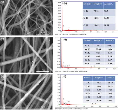
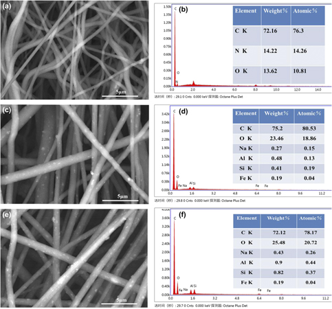
Figure 1 shows the SEM images and EDS spectra of PU membrane; 5% TM/PU membrane and 10% TM/PU membrane, respectively. Though the backscatter probe is used in SEM images, it shows that the TM nanoparticles had uniform distribution in the PU electrospun fibers, with an increase in the content of TM in the composite membrane and that the bright particles (TM) in the fibers increased [Figs. 1(c) and 1(e)]. The EDS spectra of pure PU membrane only contained C, N, O, and H elements [Fig. 1(b)]. While the TM content in the composite electrospun fibrous membranes increased, all elements of TM like Na, Al, Si, and Fe appear in the spectrum and TM elements continue to increase [Figs. 1(d) and 1(f)].
SEM images (a, c, e) and EDS spectra (b, d, f) of PU membrane (a, b); 5% TM/PU membrane (c, d) and 10% TM/PU membrane (e, f), respectively.

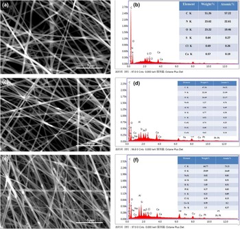
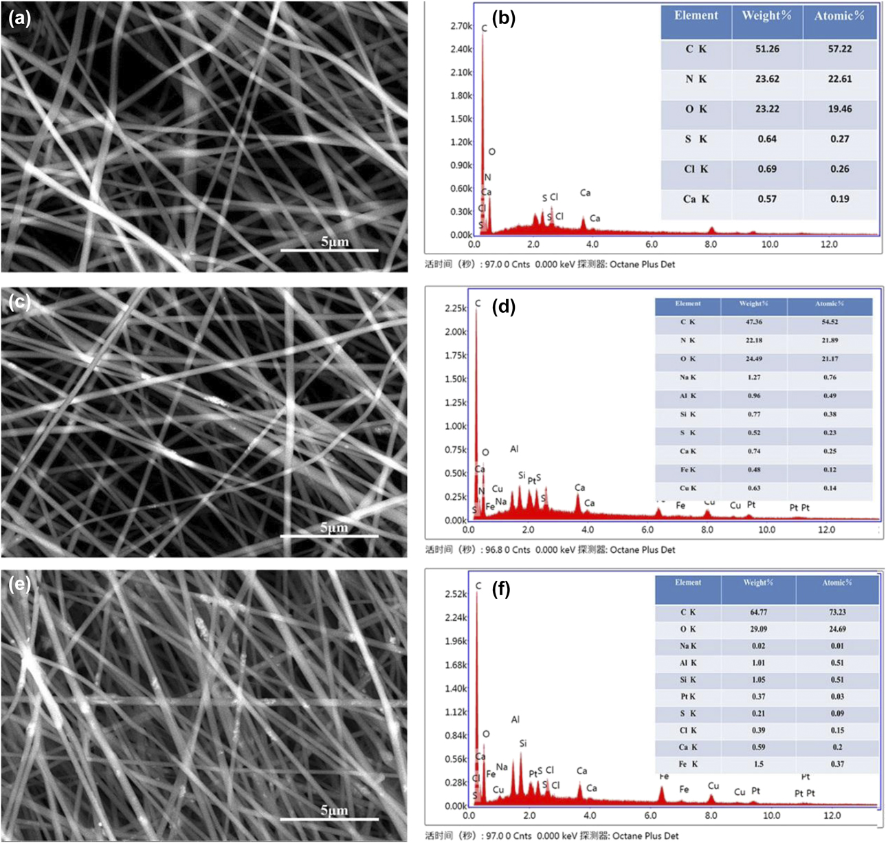
Figure 2 shows the SEM images and EDS spectra of GE membrane; 5% TM/GE membrane and 10% TM/GE membrane, respectively. It shows that with an increase in the TM content of the composite membrane, the bright particles (tourmaline) in the surface of the nanofibers increase [Figs. 2(c) and 2(e)]. Furthermore, the main elements of pure GE electrospun membrane contained C, N, O, S, and Ca [Fig. 2(b)], and the elements such as Na, Al, Si, and Fe from TM appeared in composite membranes [Figs. 2(d) and 2(f)]. It meant that the TM nanoparticles are uniformly dispersed in the GE composite fibers.
SEM images (a, c, e) and EDS spectra (b, d, f) of GE membrane (a, b); 5% TM/GE membrane (c, d) and 10% TM/GE membrane (e, f), respectively.
Mineralization analysis
Figure 3 illustrates the SEM of TM/PU composite membrane (TM content were 0%, 5%, and 10%) at different mineralization periods. After 1 h of mineralization, some flaky crystals were obtained. In addition to flaky crystals on the composite membranes, some small honeycomb-like spheres were also formed on the fibers and flaky crystals [Figs. 3(b) and 3(c)]. Following the mineralization time, the flaky crystals on the pure electrospun nanofibers grew up and the honeycomb-like depositions grew more and more. In addition, the honeycomb-like depositions of 10% TM/PU membrane after 6 h of mineralization [Fig. 3(l)] were larger than that of 5% TM/PU membrane after 6 h of mineralization [Fig. 3(k)]. This shows that with the increase in TM content, the honeycomb spherical depositions on the composite membranes also increase simultaneously.
SEM images of pure PU membranes (a, d, g, j); 5% TM/PU membranes (b, e, h, k) and 10% TM/PU membranes (c, f, i, l) were immersed in 10SBF at 1 h (a, b, c), 2 h (d, e, f), 4 h (g, h, i), and 6 h (j, k, l).

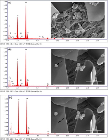
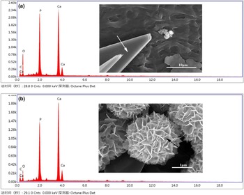
In Fig. 4, EDS analysis of the components of the two different crystals on the membranes is shown. In Fig. 4(a), the Ca/P molar ratio of the honeycomb spherical crystal on the fibers is 1.55:1, and the fibers contained Si, Fe, Al, and Na elements, indicating that the spherical crystals were nucleated on the exposed TM. In Fig. 4(b), the honeycomb spherical crystals in the platelet crystals had a Ca/P molar ratio of 1.31. Therefore, the Ca/P ratio of both honeycomb spherical crystals was relatively close to that of hydroxyapatite (HA) of 1.67:1 [Figs. 4(a) and 4(b)]. However, the Ca/P molar ratio of the flaky crystals was 1.01:1 [Fig. 4(c)], which was the same as that of calcium phosphate (CaHPO4).
EDS spectra of depositions deposited on TM/PU membranes (a): the honeycomb spherical depositions on fiber; (b): the honeycomb spherical depositions on flaky crystals; (c): the flaky crystals on the electrospun fibrous membranes.

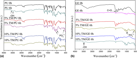
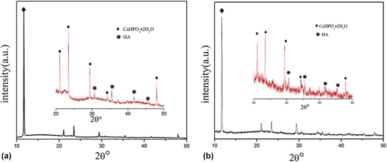
Figure 5(a) shows the FTIR spectra of the TM/PU composite membrane (TM content was 0%, 5%, and 10%) before and after mineralization. On comparing the two cases at 0-h and 6-h incubation in 10SBF solution of each sample, the intensity of PO43− band (572 cm−1, 971 cm−1, and 1053 cm−1) and –OH stretching bands (3488 cm−1 and 3542 cm−1) appeared after 6 h of mineralization, respectively. For further analysis of the mineralization crystals, X-ray diffraction (XRD) measurement was performed [Fig. 6(a)]. The extremely strong characteristic diffraction peaks of hydrated calcium dihydrogen phosphate (CaHPO4·2H2O, DCPD) were found in 2θ = 11.6° (JCPDS no. 9-77), which showed the vertical crystal plane (020) of DCPD, the trend of growth along the b-axis orientation. In the inset of the enlarged XRD patterns (2θ = 20°–50°), the characteristic peaks of DCPD were shown at 2θ = 21.0° and 23.4°. In addition, the diffraction peak of HA was also observed at 2θ = 31.7°, 35.4°, and 45.4° (JCPDS no. 09-0432) [Reference Chen, Chu and Hsiao21, Reference Yang, Wolke and Jansen22, Reference Sekara, Kanchana, Nithyaselvi and Girija31, Reference Zhang, Zhang, Mao, Zhao and Yu32]. The results show that the mineralization products are the mixture of DCPD and HA.
FTIR spectra of different membranes: (a): PU, 5% TM/PU membrane and 10% TM/PU; (b): GE, 5% TM/GE and 10% TM/GE before (0 h) and after (6 h) mineralization.

XRD patterns of depositions on different electrospun fibrous membranes after 4 h of mineralization: (a): 5% TM/PU membrane; (b): 5% TM/GE membrane.

Figure 7 illustrates the SEM of TM/GE composite membrane (TM content was 0%, 5%, and 10%) at different mineralization periods. After 1 h of mineralization, some flaky crystals and honeycomb spherical crystals were obtained. With the increase in the mineralization time, the honeycombed crystals on the pure GE membrane grew up, and the sporadic lamellar crystals also existed. The 10% TM/GE composite membranes at 6 h [Fig. 7(l)] have been wrapped by honeycomb spherical crystals, and the honeycomb-like depositions were larger than that of 5% TM/GE at 6 h [Fig. 7(k)] and 10% TM/GE at 4 h [Fig. 7(i)]. Therefore, with the increase in the TM content and mineralization time, the honeycomb spherical crystals increased.
SEM images of pure GE membranes (a, d, g, j); 5% TM/GE membranes (b, e, h, k) and 10% TM/GE membranes (c, f, i, l) immersed in 10SBF at 1 h (a, b, c), 2 h (d, e, f), 4 h (g, h, i), and 6 h (j, k, l).

Fig. 8 shows that two different crystals were produced, and the components of these two crystals were analyzed by EDS. The Ca/P molar ratio of the flaky crystals was 1.01:1, same as that of calcium phosphate (CaHPO4) [Fig. 8(a)]. The honeycomb spherical crystals on the platelet crystals had a Ca/P molar ratio of 1.30 [Fig. 8(b)], which was similar to that of the honeycomb spherical crystals on the platelet crystals in the TM/PU sample [Fig. 4(b)]. Figure 5(b) shows the FTIR spectra of TM/GE composite membranes (TM content was 0%, 5%, and 10%) before and after 6 h incubation in 10SBF. It can be seen that there was a strong absorption peak of C=O stretching vibration at 1649 cm−1 before mineralization. After mineralization, the following main peaks at 570, 972, and 1053 cm−1 were assigned to PO43−. Furthermore, XRD measurement of depositions was performed [Fig. 6(b)]. On enlarging the pattern in the range of 2θ = 20°–50°, characteristic peaks of DCPD at 2θ = 23.4° can be seen. In addition, the diffraction peak of hydroxyapatite was also observed at 2θ = 31.7°, 35.4°, and 45.3°. The results show that the crystal product is a mixture of DCPD and hydroxyapatite.
EDS spectra of depositions on TM/GE electrospun membranes: (a): the flaky crystals; (b): the honeycomb spherical depositions.

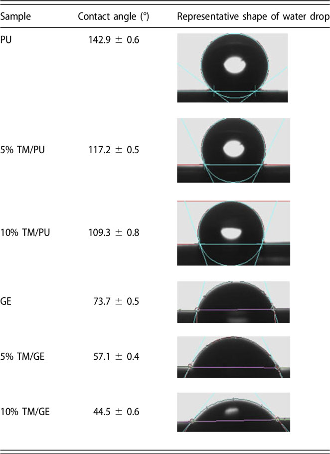
The contact angles of different electrospun samples were detected (Table I). It was known that PU was hydrophobic, and the contact angle of the PU composite membranes with TM nanoparticles became smaller. The hydrophilicity of TM/PU electrospun membranes increased after the addition of TM. GE was hydrophilic, and the contact angle of the GE composite membranes with TM nanoparticles also became smaller. Therefore, TM could improve the hydrophilicity of hydrophobic PU and hydrophobic GE.
Contact angles of different electrospun fibrous membranes.
During mineralization of TM/PU membranes, Ca2+ and HPO42− of 10SBF first reacted to form water-insoluble DCPD platelets (Ca2+ + HPO42−+2H2O = CaHPO4·2H2O). DCPD is a precursor of HA, as rich as Ca2+; DCPD is gradually converted to HA with the increased mineralization time (10CaHPO4·2H2O = Ca10(PO4)6(OH)2 + 4HPO42− + 8H+) [Reference Yang, Wolke and Jansen22]. Therefore, mineralization depositions such as DCPD and HA appeared. Because PU was hydrophobic, there were few nucleation sites for calcium phosphate deposition on its surface. When TM was added, the contact angle of the composite membranes became smaller and the hydrophilicity increased (Table I). TM has a permanent spontaneous polarization, whereby the permanent electrodes appear around TM particles. The Ca2+ would be attracted by TM electrodes at the nucleation sites. Furthermore, as the electric field existed around the TM nanoparticle, water could be decomposed as follows [Reference Qiu, Ma, Yuan and Xu28].
 $${{\rm{H}}_2}{\rm{O}} \Rightarrow {{\rm{H}}^ + } + {\rm{O}}{{\rm{H}}^ - }\quad ,$$
 $$2{{\rm{H}}^ + } + 2{\rm{e}} \Rightarrow {{\rm{H}}_2} \uparrow \quad ,$$
 $${\rm{O}}{{\rm{H}}^ - } + n{{\rm{H}}_{\rm{2}}}{\rm{O}} \Rightarrow {\rm{O}}{{\rm{H}}^ - }{\left( {{{\rm{H}}_{\rm{2}}}{\rm{O}}} \right)_n}\quad .$$
Through the high electrostatic field on the TM surface, H+ and OH− ions were adsorbed to the two poles of TM. The migration rate of H+ was higher than that of OH– ions and redundant H+ was released in the form of hydrogen; thus, the ambience tended to be weakly alkaline and there was an increase in the pH of the solution [Reference Qiu, Ma, Yuan and Xu28], which was beneficial to the formation of hydroxyapatite [Reference Zhao, Zhao, Zhang, Yuan and Yao33]. Therefore, under the same mineralization time, the amount of honeycomb-like depositions on the 10% TM/PU composite membrane was more than that of 5% TM/PU sample. And along the mineralization, the spherical depositions on the composite membrane increased continuously. As the TM content increased, it is easier to form honeycomb spherical depositions during mineralization.
Because GE molecules contain a large number of –NH2 and –COOH groups, they have strong hydrophilicity. When the composite membrane was immersed in 10SBF, –COO− ionized from the –COOH group, which could easily chelate Ca2+ in the solution, and then combined with the phosphate group to form calcium phosphate crystals [Reference Meng, Li, Sun, Zheng and Zheng23]. Because the TM nanoparticles have relatively large surface area and contain –OH group, they can be used as a very good nucleation site. At the same time, TM increases the local supersaturation of ions in nucleation sites in 10SBF and decreases the nucleation free energy of calcium phosphate crystals, resulting in more crystals on the composite membrane. Therefore, at the same mineralization time, the amount of honeycomb-like depositions on the 10% TM/GE composite membrane was more than that of 5% TM/GE sample. And along the mineralization, the spherical depositions in the composite membrane fibers increased continuously. As the TM content increased, it is easier to form honeycomb spherical depositions during mineralization.
Through the mineralization of two kinds of composite membranes, it is shown that TM addition is conducive to the nucleation and growth of calcium and phosphorus depositions. The main mineralized depositions of the PU membrane were hydroxyapatite precursor DCPD, and the HA increased following the TM content, which was in favor of its osteogenesis. Because GE was more hydrophilic than PU and the carboxyl groups of GE could attract and accumulate calcium ions in the solution, the mineralized TM/GE composite membrane is dominated by honeycomb-shaped hydroxyapatite. With the increased TM content in the composite membrane, the mineralized hydroxyapatite also increased.
For investigating the cytotoxicity of TM nanoparticles, the fibroblast L929 cells were cultured on PU and TM/PU membranes for 72 h; the optical density of control, PU, 5% TM/PU, and 10% TM/PU membranes is shown in Fig. S1. It is shown that the proliferation on PU and TM/PU samples were greater than that on the control sample and there was no significant difference between the PU and TM/PU. Therefore, the electrospun membranes containing TM had a good cytocompatibility.
Conclusion
In this paper, hydrophobic PU and hydrophilic GE fibrous membranes with TM nanoparticles were prepared by electrospinning, and the effect of TM nanoparticles on the mineralization was studied. After soaking in 10SBF for 6 h, two types of calcium phosphate crystals, flaky and honeycomb structures, were produced on the composite membrane. EDS analysis showed that the structure of the two types of crystals was different, and the proportion of honeycomb crystals was closer to that of apatite ratio. XRD results showed that the two types of crystals are CaHPO4·2H2O and HA, respectively. With the increase in the added TM in the composite membranes, more calcium phosphate crystals are precipitated. As a promising approach, both TM/GE composite membranes and TM/PU composite membranes had more HA depositions after mineralization and had a good biocompatibility, which shows that the electrospun coating with calcium phosphate crystals regulated by TM can be extended to other biological compounds for bone scaffold.
Experimental
Materials
Polyurethane (PU, Estane® X595A-11, MFI 22 g/10 min, Shore 97A) was purchased from Lubrizol Advanced Materials, Inc., Wickliffe, Ohio. Gelatin (GE, type B, bovine skin, ∼225 Bloom) was purchased from Unite Stars Biotech Co., Ltd., Tianjin, China. The natural mineral material, TM, was received from the Shijiazhuang City, Hebei Province of China, and TM nanoparticles were got by mechanical milling in our lab. All chemicals used for electrospinning, cross-linking, and mineralizing in the experiment were analytical reagents.
Preparation of TM/polymer electrospun membranes
For PU electrospinning solution, 10 wt% PU was dissolved in methyl ethyl ketone (MEK)/N,N-dimethylformamide (DMF) (50/50, wt/wt) for all tests. For TM/PU electrospinning solutions, TM nanoparticles were dispersed in DMF/MEK solvent and slowly dropped in pure PU solution; the mixed solution was under ultrasonication for 2 h. During electrospinning, the applied voltage was 15 kV, the feed rate was at 1 mL/h by a controllable syringe pump (Cole-Parmer, Vernon Hills, Illinois), and the distance of nozzle tip-to-collector was 15 cm. The pure PU membrane and composite membranes containing 5 wt% and 10 wt% TM nanoparticles are marked as PU membrane; 5% TM/PU membrane and 10% TM/PU membrane, respectively. The fibrous membranes were vacuum-dried for standby.
For GE solution, 22 wt% GE was dissolved in acetic acid for all tests. For TM/GE electrospinning solutions, TM nanoparticles were dispersed in acetic acid and slowly dropped in pure GE solution, the mixed solution was under ultrasonication for 2 h. During electrospinning, the applied voltage was 18 kV, the feed rate was at 1 mL/h by a controllable syringe pump (Cole-Parmer), and the distance of nozzle tip-to-collector was 15 cm. The pure GE membrane and composite membranes containing 5 wt% and 10 wt% TM nanoparticles are marked as GE membrane; 5% TM/GE membrane and 10% TM/GE membrane, respectively. The electrospun fibrous membrane was cross-linked at 35 °C for half an hour in this glutaric dialdehyde vapor atmosphere. After cross-linking, the fibrous membranes were vacuum-dried for standby.
Mineralization of TM/polymer membranes
The membrane samples were mineralized in 10× simulated body fluid (10SBF). NaCl (58.433 g), KCl (0.373 g), CaCl2 (2.775 g), MgCl2·6H2O (1.016 g), and KH2PO4 (1.36 g) were dissolved completely, and the clear solution was set to 1 L. The TM/polymer membranes were cut into 2 cm × 2 cm square and separately immersed in 200 mL 10SBF kept at 37 °C for 1, 2, 4, and 6 h; 10SBF was replaced every 2 h. After being removed from 10SBF, the sample was gently washed with ultrapure water, dried, and stored at room temperature.
Characterization
The electrospun fibers modified with the TM nanoparticles were characterized by scanning electron microscopy–energy dispersive spectroscopy (SEM-EDS, Nano SEM450, FEI, Hillsboro, Oregon), and the transmission electron microscopy (TEM) images were taken using an FEI Tecnai F20 transmission electron microscope at an accelerating voltage of 200 kV. For investigating the mineralization of samples, X-ray diffraction (XRD) analysis was on a Bruker D8 Focus diffractometer with Cu Kα radiation (40 kV and 40 mA, λ = 1.5418 Å) at a scanning rate of 2°/min, and Fourier transform infrared spectrum (FTIR, Germany) was taken in the range of 400–4500 cm−1 with a signal resolution of 0.5 cm−1. For cytotoxicity test, 2 × 10⁴ of L929 cells were seeded on the PU and TM/PU membranes and cultured for 72 h in the incubator. 10 μL/well of MTT (5 mg/mL) was added into the cell culture plate, and incubated at 37 °C for 4 h, then the supernatant was removed and 180 μL DMSO was added. The optical density of each sample at a wavelength of 570 nm was measured by enzyme-labeled instrument (Multiskan FC, Thermo Scientific, Waltham, Massachusetts).
Supplementary material
To view supplementary material for this article, please visit https://doi.org/10.1557/jmr.2019.35.
Acknowledgments
The authors are grateful for the financial support from the National Natural Science Foundation of China (Grant No. 51504076) and the Natural Science Foundation of the Hebei Province of China (Grant No. E2016202379).