Introduction
Influenza A is one of three genera of influenza viruses belonging to the family of negative stranded, segmented RNA viruses known as Orthomyxoviridae. In their natural zoonotic reservoirs, the viruses are largely benign, causing asymptomatic enteric infection in the wild aquatic birds of the world. Influenza A viruses are pathogens of major concern in veterinary and public health. They are highly variable, owing to their segmented genome and lack of proofreading during replication. When influenza A viruses spread to domestic poultry, pigs, or people they can evolve into strains that cause mild to catastrophic diseases. While all 16 hemagglutinin (HA) subtypes of influenza A viruses can replicate in domestic ducks and/or quail (Alexander, Reference Alexander2000; Makarova et al., Reference Makarova, Ozaki, Kida, Webster and Perez2003; Iamnikova et al., Reference Iamnikova, Gambarian, Aristova, L'vov, Lomakina, Munster, Lexmond and Foucher2009), only a limited number can infect chickens. In gallinaceous poultry, two of the 16 HA subtypes (H5 and H7) can evolve from low or non-pathogenic strains into highly pathogenic strains that cause systemic infection and death. In pigs and people, influenza is a respiratory disease that varies in pathogenicity from mild to catastrophic.
Here, we consider the structure of the virus and the mechanisms involved in its continuing evolution, including frequent acquisition of mutations and reassortment events. For an influenza virus to successfully evolve from intestinal tropism of a wild aquatic bird with a body temperature of 42°C and an HA binding preference for sialic acid (SA) α2,3 to respiratory tract tropism in humans at a body temperature of 37°C with an HA binding preference for SA α2,6, requires multiple changes in one or more gene segments of the virus. Additionally, pigs have been proposed as the intermediate host (mixing vessel) between avians and humans. The continuing circulation of the HP avian H5N1 influenza virus in sites in Eurasia and the emergence of a novel H1N1 influenza virus to generate the first human pandemic of the 21st century are used to illustrate the continuing evolution of influenza viruses.
Our failure to eradicate HP H5N1 globally and understand its inefficient and sporadic transmission to humans underlines our lack of fundamental knowledge about the pathogenesis and transmissibility of influenza viruses. Here we identify the gaps in our knowledge concerning the molecular determinants of the virus and the hosts that permit efficient transmissibility and pandemic potential. We also identify those areas that currently elude us in control strategies and conclude with a section on future directions that involve the complete resolution of the genomics and proteomics of both the virus and the host. Such knowledge will permit us to engineer influenza viruses with optimal properties for vaccine preparation, to identify novel antiviral targets, and to distinguish the genes in the host that determine resistance, thereby providing the option of generating host animals that are resistant to influenza.
The influenza virus
Types, subtypes, and strains
The influenza virus causes a highly contagious acute respiratory illness affecting birds and mammals. It belongs to a family of negative stranded RNA viruses known as Orthomyxoviridae, which are further divided into five genera: Influenzavirus A, Influenzavirus B, Influenzavirus C, Isavirus, and Thogotovirus. Analysis has shown a high degree of conservation of the nucleotide sequences at the 3′ and 5′ ends of RNA isolated from influenza A, B, and C viruses, suggesting a common ancestry separate from that of other RNA viruses (Desselberger et al., Reference Desselberger, Racaniello, Zazra and Palese1980; Webster et al., Reference Webster, Bean, Gorman, Chambers and Kawaoka1992). Additionally, genome organization and protein homology similarities between influenza A and B but not to influenza C suggest that influenza C viruses diverged from common ancestors before influenza A and B viruses split (Smith and Palese, Reference Smith, Palese and Krug1989; Webster et al., Reference Webster, Bean, Gorman, Chambers and Kawaoka1992).
Despite their common lineage, several differences exist between influenza types A, B, and C, including number, length, and base sequence of RNA segments, degree of overall antigenic variation, pathogenicity, and host specificity. Influenza B and C viruses exhibit less antigenic variation than the A viruses and hence undergo slower evolution (Palese and Young, Reference Palese and Young1982; Webster et al., Reference Webster, Bean, Gorman, Chambers and Kawaoka1992). This difference in genetic diversity is reflected in the fact that influenza A viruses can be divided into 16 HA subtypes and nine neuraminidase (NA) subtypes, and further classified by strains, while influenza B and C (the least common of all three influenza virus types) viruses are classified only by strains (Nobusawa et al., Reference Nobusawa, Aoyama, Kato, Suzuki, Tateno and Nakajima1991; Hay et al., Reference Hay, Gregory, Douglas and Lin2001). HA subtypes are further subdivided into two main groups based on amino acid sequence similarity (group 1 includes H1, H2, H5, H6, H8, H9, H11, H12, H13, H16, and group 2 includes H3, H4, H7, H10, H14, H15) (Nobusawa et al., Reference Nobusawa, Aoyama, Kato, Suzuki, Tateno and Nakajima1991; Russell et al., Reference Russell, Kerry, Stevens, Steinhauer, Martin, Gamblin and Skehel2008). Influenza B and C viruses almost exclusively infect humans, causing mild respiratory disease primarily in children (Webster et al., Reference Webster, Bean, Gorman, Chambers and Kawaoka1992; Hay et al., Reference Hay, Gregory, Douglas and Lin2001; Matsuzaki et al., Reference Matsuzaki, Katsushima, Nagai, Shoji, Itagaki, Sakamoto, Kitaoka, Mizuta and Nishimura2006). The only other known animals susceptible to the B and C viruses are seals (Osterhaus et al., Reference Osterhaus, Rimmelzwaan, Martina, Bestebroer and Fouchier2000), ferrets (Kim et al., Reference Kim, Kim, Cho and Seo2009), pigs (Guo et al., Reference Guo, Jin, Wang, Wang and Zhu1983), and dogs (Manuguerra and Hannoun, Reference Manuguerra and Hannoun1992). In contrast, influenza A viruses, the focus of the remaining discussion, are infectious to a wide variety of avian and mammalian species (Fig. 1).
Overview of known events involving cross-species transmission of influenza A viruses. Influenza viruses containing all 16 hemagglutinin (HA) and nine neuraminidase (NA) subtypes of influenza A viruses have been isolated from wild aquatic birds, which are the natural reservoir of influenza A and the source of virus that crosses the species barrier to infect domestic birds and mammals (green circle). Solid black lines indicate events of cross-species transmission of influenza A viruses and red lines indicate the most likely cycle of transmission through which a new influenza A pandemic strain would arise. Parallel to the lines are subtypes transmitted between species; all avian influenza viruses are low pathogenic (LP), unless otherwise specified as highly pathogenic (HP) (Nielsen et al., Reference Nielsen, Clavijo and Broughen2001; Myers et al., Reference Myers, Olsen and Gray2007; Lee et al., Reference Lee, Pascua, Song, Baek, Kim, Choi, Sung, Webby, Webster, Poo and Choi2009; Peiris, Reference Peiris2009; Reperant et al., Reference Reperant, Rimmelzwaan and Kuiken2009; Harder and Vahlenkamp, Reference Harder and Vahlenkamp2010; Sponseller et al., Reference Sponseller, Strait, Jergens, Trujillo, Harmon, Koster, Jenkins-Moore, Killian, Swenson, Bender, Waller, Miles, Pearce, Yoon and Nara2010). The 16 HA subtypes of influenza A are divided into two main groups based on amino acid sequence similarity. Group 1 HA subtypes are indicated in orange and group 2 HA subtypes are indicated in blue.

Wild aquatic birds are the natural reservoir of all 16 HA and nine NA subtypes of influenza A circulating throughout the world (Webster et al., Reference Webster, Bean, Gorman, Chambers and Kawaoka1992; Fouchier et al., Reference Fouchier, Osterhaus and Brown2003; Van Reeth, Reference Van Reeth2007). In birds, influenza A viruses are not only classified by HA and NA subtypes, but also as having low pathogenicity (LP) or high pathogenicity (HP). Influenza A viruses largely cause asymptomatic or low pathogenic infection in wild aquatic birds; however, influenza strains can evolve very rapidly after they cross the species barrier to the domestic avian and mammalian population, leading to the development of pandemic and/or highly pathogenic strains (Swayne and Suarez, Reference Swayne and Suarez2000).
Structure and function
To fully understand the mechanisms of variation and evolution of influenza A viruses, one must first have a basic knowledge of the overall structure and function of the influenza virus and its genome. The genome of the influenza virus is comprised of eight segments of RNA, which encode for 10 or 11 different polypeptides. In addition to the two surface proteins (HA and NA) and two matrix proteins (M1 and M2), the encoded polypeptides include three viral polymerase proteins (PB1, PB2, and PA), PB1-F2 (Chen et al., Reference Chen, Calvo, Malide, Gibbs, Schubert, Bacik, Basta, O'Neill, Schickli, Palese, Henklein, Bennink and Yewdell2001), a nucleocapsid (NP), a non-structural protein (NS1), and a nuclear export protein (NEP). Each RNA segment is bound by the three polymerase proteins at their termini and NP that encapsidates the viral genome for RNA transcription, replication, and packaging. The resulting structure is termed a ribonucleoprotein (RNP) and is helical in shape.
Of the 11 polypeptides, PB1-F2 and NEP are the most recently discovered. The PB1-F2 protein is thought to play a role as a virulence factor by compromising mitochondria function and eventually leading to cell death (Gibbs et al., Reference Gibbs, Malide, Hornung, Bennink and Yewdell2003). The NEP protein, previously known as NS2 (for non-structural protein 2), has been shown to mediate the nuclear export of viral RNA, hence the change in nomenclature (O'Neill et al., Reference O'Neill, Talon and Palese1998).
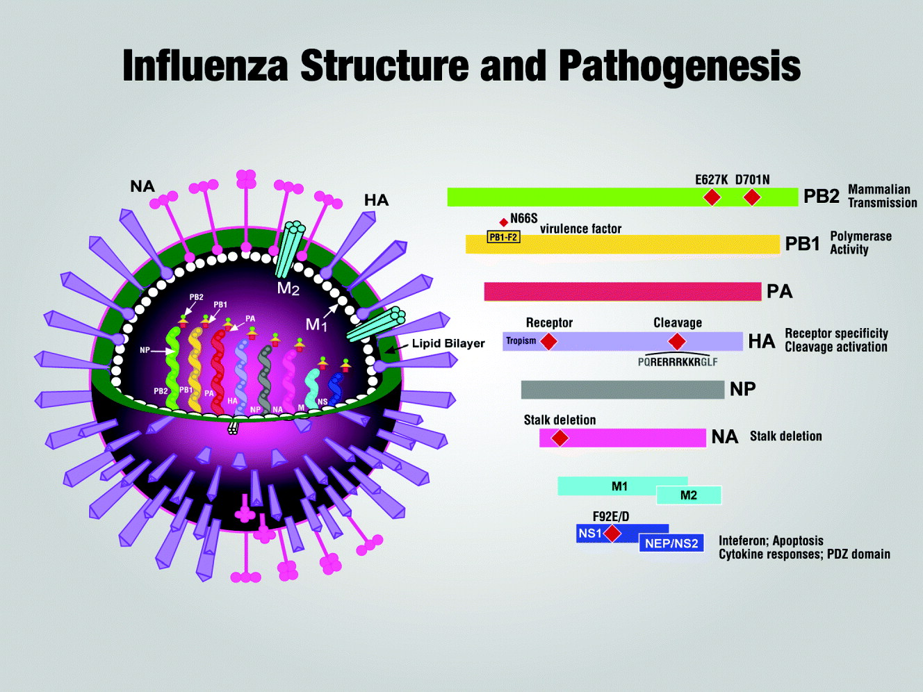
Influenza A viruses are morphologically variable. In their host, they consist of a heterogenous population of small spherical particles (80–120 nm in diameter) and long filamentous particles; however, after multiple passages in eggs and/or tissue culture, the virus population is mainly spherical in shape (Bourmakina and Garcia-Sastre, Reference Bourmakina and Garcia-Sastre2003). The surface of the virus particle is comprised of a lipid bilayer envelope that is predominantly made up of HA and NA, with a small number of copies of M2, a small transmembrane proton channel (Fig. 2). HA protein, the predominant surface antigen of influenza, is responsible for the binding of the virus particle to sialic acid residues on the surface of the host cell and subsequent fusion of the virion envelope and host cell membrane, releasing the viral genome into the target cell. NA protein functions to release the budding virus from the infected cell through cleavage of host cell sialic acid residues. It is in the HA and NA that the continuing evolution of influenza A viruses is most prominent (Scholtissek, Reference Scholtissek1979; Webster et al., Reference Webster, Bean, Gorman, Chambers and Kawaoka1992; Portela and Digard, Reference Portela and Digard2002). Lining the inner layer of the lipid membrane is a shell of matrix protein (M1). M1 is the major structural component of the virion and is thought to bridge the outer lipid envelope and the internal RNPs, thereby playing a critical role in virus budding (Portela and Digard, Reference Portela and Digard2002).
Structure of influenza A virus. The surface of the influenza virion is composed of a lipid bilayer that contains the surface glycoproteins (HA and NA), and a transmembrane ion channel (M2). The HA glycoprotein is a rod-shaped trimer, and each of its three HA molecules is comprised of two subunits: HA1 that forms the globular head where the receptor binding and antigenic sites are located, and HA2 that, together with HA1, forms the stalk connecting to the viral envelope (Lamb, Reference Lamb, Krug, Fraenkel-Conrat and Wagner1989). The NA polypeptide consists of four monomers that anchor the polypeptide to the lipid envelope via a stalk-shaped projection with a box-shaped head on top (Garman and Laver, Reference Garman, Laver and Fischer2005). The matrix protein (M1) lies beneath the lipid bilayer, and is bound by nuclear export protein (NEP). Internal to M1, at the core of the influenza virion, are the eight segments of RNA that encode for the 11 different polypeptides. Individual RNA segments are bound by a polymerase complex, consisting of the three proteins PA, PB1, and PB2, at their termini and encapsidated by the NP into a helical structure (RNP). The eight RNA segments vary in size, with the two segments encoding PB1 and PB2 being the largest (2341 nucleotides each) and the segment encoding NS1/NEP being the smallest (890 nucleotides). The remaining five segments encoding for PA, HA, NP, NA, and M1/M2 are 2233, 1778, 1565, 1413, and 1027 nucleotides in length, respectively (Hoffmann et al., Reference Hoffmann, Stech, Guan, Webster and Perez2001). The major residues associated with pathogenicity are shown diagrammatically on each of the gene products. Pathogenesis is a polygenic trait involving multiple genes in the virus and the host.

Evolutionary strategies
Historical evidence suggests that influenza has afflicted humans as far back as 412 BC, when Hippocrates described a disease epidemic that modern doctors now believe to have been influenza (Ghendon, Reference Ghendon1994). The evolution of influenza viruses is a continuing process, and with the advent of modern science, the expansion of our knowledge on the evolution of this unique pathogen is continual as well. A thorough understanding of the evolutionary biology of influenza is necessary not only from a historical perspective, but most importantly from a human and veterinary public health viewpoint.
Influenza A viruses are capable of a substantial amount of genetic diversity, owing to their lack of ‘proofreading’ mechanisms of their polymerase proteins, and therefore inability to repair any errors occurring during the replication process. Additionally, the segmented genome of an influenza virus allows for exchange of RNA segments between genotypically different influenza viruses resulting in the generation of a novel strain and/or subtype (Reid and Taubenberger, Reference Reid and Taubenberger2003). The three mechanisms through which influenza viruses can undergo evolutionary change include mutation (resulting in antigenic drift), reassortment (resulting in antigenic shift), and, in rare instances, recombination.
Antigenic drift and its impact
Antigenic drift is a continuous process in influenza A viruses whereby point mutations arise in the viral genome during the replication cycle. This process is evident in all gene products of influenza; however, it is most pronounced at the antibody binding sites of the HA and/or NA glycoproteins. Because of the lack of a proofreading mechanism, these mutations are unable to be repaired, and the consequential accumulation of amino acid sequence changes alters antigenic sites to such an extent that they are less recognizable by the host's antibody response. These virus variants, which are no longer neutralized by host antibodies, are naturally selected for because of increased fitness (Treanor, Reference Treanor2004; Boni, Reference Boni2008). The process of antigenic drift is generally most evident in human influenza viruses, less so in swine and equine influenza, and least apparent in avian influenza (Gorman et al., Reference Gorman, Bean and Webster1992; Ghendon, Reference Ghendon1994; de Jong et al., Reference de Jong, van Nieuwstadt, Kimman, Loeffen, Bestebroer, Bijlsma, Verweij, Osterhaus and Class1999; Manuguerra et al., Reference Manuguerra, Zientara, Sailleau, Rousseaux, Gicquel, Rijks and van der Werf2000).
Studies on the HA of H3N2 suggest that antigenic drift of influenza is more punctuated in nature than the underlying process of genetic evolution (Smith et al., Reference Smith, Lapedes, de Jong, Bestebroer, Rimmelzwaan, Osterhaus and Fouchier2004). There are long periods of antigenic stasis that are marked by neutral sequence evolution without substantial antigenic change. These periods are interspersed with shorter intervals of strong selection for antigenic change and the replacement of old lineages with new dominant ones of increased fitness (Wolf et al., Reference Wolf, Viboud, Holmes, Koonin and Lipman2006; McHardy and Adams, Reference McHardy and Adams2009). Additionally, there is a large body of research that suggests new variants of human H3 influenza A viruses are selected for more rapidly than the H1 influenza viruses (Ferguson et al., Reference Ferguson, Galvani and Bush2003). The rapid replacement of old variants of H3 viruses with new antigenic drift variants is emphasized in the phylogenetic tree of human influenza A H3 HA, which is very linear and has evolved as such since the introduction of H3N2 viruses into the human population in 1968 (Bush et al., Reference Bush, Bender, Subbarao, Cox and Fitch1999; Hay et al., Reference Hay, Gregory, Douglas and Lin2001; Treanor, Reference Treanor2004). In contrast, new antigenic drift variants of the human influenza A H1 often co-circulate with existing variants, which is reflected in its phylogenetic tree that displays a recent diversion from the main lineage (Ferguson et al., Reference Ferguson, Galvani and Bush2003; Treanor, Reference Treanor2004).
Antigenic drift has a significant impact on the effectiveness of the human seasonal influenza vaccine. Co-circulation of antigenically distinct variants, as discussed above, can add additional complications to the recommendations for vaccine composition (Hay et al., Reference Hay, Gregory, Douglas and Lin2001). The impact of antigenic drift on vaccine effectiveness was illustrated most recently during the 1997–1998 influenza season in Europe and the United States, which saw particularly severe disease outbreaks due to a poorly matched vaccine and new antigenic drift variant A/Sydney/05/97 (H3N2), and again in 2003–2004 when A/Panama/2007/99 drifted to A/Fujian/411/2002 (Carrat and Flahault, Reference Carrat and Flahault2007). For this reason, annual evolution of the vaccine formulation is necessary to ensure the closest match between the vaccine and the currently circulating virus strain (Carrat and Flahault, Reference Carrat and Flahault2007; WHO, 2010a). To achieve this, the World Health Organization (WHO) set up the Global Influenza Network in 1948, which is composed of influenza collaborating centers from around the globe. The network meets twice a year to make recommendations for the upcoming season's vaccine based upon information that they have gathered on circulating strains. Since 1972, 39 changes have been made to the vaccine formulation, including 23 changes to the influenza A H3 component and eight changes to H1 (Hay et al., Reference Hay, Gregory, Douglas and Lin2001; Carrat and Flahault, Reference Carrat and Flahault2007; WHO, 2010a). The frequency that the influenza vaccine has required reformulation emphasizes the importance of antigenic drift to the evolution of influenza and the need for continuing research on the development of broadly cross-reactive vaccines so that we can better prepare for the drifted, immune evading strains of influenza A viruses.
Antigenic shift and its impact
The process of antigenic shift is associated with the introduction of novel viruses into the human population from other host species either through reassortment between gene segments of an influenza virus present in humans and those found in another host (McHardy and Adams, Reference McHardy and Adams2009) or through direct animal-to-human transmission. Reassortment occurs when genotypically different influenza A viruses infect the same host cell and an exchange of RNA segments occurs. The result is the generation of a novel combination of gene segments derived from different parental strains and to which the population is immunologically naive (Nelson and Holmes, Reference Nelson and Holmes2007). Within the avian reservoir, individual birds are often infected with multiple viruses, resulting in numerous gene reassortment events and continuous generation of a nearly endless array of new viruses that could potentially cross the species barrier (Morens et al., Reference Morens, Taubenberger and Fauci2009).
Two of the three major human influenza A pandemics of the 20th century (1957 and 1968) and the major pandemic of the present century (2009) occurred as a result of reassortment between human influenza A viruses and those of other host species. Genetic evidence indicates that the HA, NA, and PB1 gene segments of the H2N2 1957 pandemic strain and the HA and PB1 segments of the H3N2 1968 pandemic strain were all avian in origin, while the remaining segments were likely directly descended from the 1918 strain (the third major pandemic of the 20th century) (Reid and Taubenberger, Reference Reid and Taubenberger2003). In contrast, the origin of the 1918 H1N1 pandemic strain, the most devastating of the pandemics to date, has yet to be fully established. There is evidence to suggest that it may not have jumped from avians to humans through a simple reassortment event, but instead circulated and adapted first in an intermediary mammalian host, such as swine, prior to the 1918 pandemic (Nelson and Holmes, Reference Nelson and Holmes2007; Smith et al., Reference Smith, Bahl, Vijaykrishna, Zhang, Poon, Chen, Webster, Peiris and Guan2009a). Sequence analysis has revealed that the H1 of the 1918 strain is more closely related to the avian, although much less so than the 1957 and 1968 strains, and with no clear similarity to currently available avian H1 sequences. Additionally, phylogenetic analysis has placed the 1918 strain in the mammalian clade (Reid and Taubenberger, Reference Reid and Taubenberger2003). Because of its great virulence, it is important for us to thoroughly understand the origins of the 1918 pandemic strain, so that our surveillance efforts can be better directed at identifying potential pandemic strains and preventing their spread (Reid and Taubenberger, Reference Reid and Taubenberger2003).
Analysis of genome sequences sampled worldwide between 1918 and 2006 showed that the 1918-derived human H1N1 influenza viruses have not acquired gene segments from non-human sources for 88 years (since 1918) (Zimmer and Burke, Reference Zimmer and Burke2009). They have, however, accumulated mutations over time, resulting in a substantial degree of antigenic drift away from the 1918 virus. The one exception to this was the reintroduction of the H1N1 influenza virus to the human population in 1977 that was closely related to a 1950 reference virus strain. It has been suggested that the 1977 outbreak may be attributable to an accidental laboratory release, since it is unlikely that the H1N1 virus could have been maintained in an animal reservoir since 1950 without undergoing detectable mutation (Kendal et al., Reference Kendal, Noble, Skehel and Dowdle1978). The antigenic drift of 1918-derived human H1N1 influenza viruses is in sharp contrast to the classical swine H1N1 influenza viruses, first isolated in 1930, that had been in relative antigenic stasis until 1998 when a circulating human H3N2 virus and a North American lineage avian influenza virus reassorted with the classical swine virus generating a triple reassortant H3N2 swine influenza A virus (Zhou et al., Reference Zhou, Senne, Landgraf, Swenson, Erickson, Rossow, Liu, Yoon, Krauss and Webster1999; Garten et al., Reference Garten, Davis, Russell, Shu, Lindstrom, Balish, Sessions, Xu, Skepner, Deyde, Okomo-Adhiambo, Gubareva, Barnes, Smith, Emery, Hillman, Rivailler, Smagala, de Graaf, Burke, Fouchier, Pappas, Alpuche-Aranda, López-Gatell, Olivera, López, Myers, Faix, Blair, Yu, Keene, Dotson, Boxrud, Sambol, Abid, St George, Bannerman, Moore, Stringer, Blevins, Demmler-Harrison, Ginsberg, Kriner, Waterman, Smole, Guevara, Belongia, Clark, Beatrice, Donis, Katz, Finelli, Bridges, Shaw, Jernigan, Uyeki, Smith, Klimov and Cox2009). The relative antigenic stability of classical swine H1N1 viruses (between 1930 and 1998) during the same time that human seasonal H1N1 viruses were experiencing antigenic drift has created an antigenic gap between classical swine H1N1 and seasonal human H1N1 viruses. Swine therefore represent a reservoir of H1 viruses with the potential to cause major respiratory outbreaks in humans (Garten et al., Reference Garten, Davis, Russell, Shu, Lindstrom, Balish, Sessions, Xu, Skepner, Deyde, Okomo-Adhiambo, Gubareva, Barnes, Smith, Emery, Hillman, Rivailler, Smagala, de Graaf, Burke, Fouchier, Pappas, Alpuche-Aranda, López-Gatell, Olivera, López, Myers, Faix, Blair, Yu, Keene, Dotson, Boxrud, Sambol, Abid, St George, Bannerman, Moore, Stringer, Blevins, Demmler-Harrison, Ginsberg, Kriner, Waterman, Smole, Guevara, Belongia, Clark, Beatrice, Donis, Katz, Finelli, Bridges, Shaw, Jernigan, Uyeki, Smith, Klimov and Cox2009).
While the H3N2 reassortant and H1N1 classical swine viruses have occasionally been isolated from humans, the viruses have not exhibited sustained transmissibility in the human population. Then, in March and early April 2009, a new swine origin influenza A H1N1 virus emerged in Mexico and the United States, and has now become the first influenza pandemic of the 21st century (Perez-Padilla et al., Reference Perez-Padilla, de la Rosa-Zamboni, Ponce de Leon, Hernandez, Quiñones-Falconi, Bautista, Ramirez-Venegas, Rojas-Serrano, Ormsby, Corrales, Higuera, Mondragon and Cordova-Villalobos2009; Reed et al., Reference Reed, Angulo, Swerdlow, Lipsitch, Meltzer, Jernigan and Finelli2009). The pandemic (H1N1) 2009 strains contain a combination of gene segments that had not been previously reported in swine or humans. The NA and M gene segments are in the Eurasian swine lineage, the HA, NP, and NS in the classical swine lineage, and the PB1, PB2, and PA in the swine triple reassortant lineage (Garten et al., Reference Garten, Davis, Russell, Shu, Lindstrom, Balish, Sessions, Xu, Skepner, Deyde, Okomo-Adhiambo, Gubareva, Barnes, Smith, Emery, Hillman, Rivailler, Smagala, de Graaf, Burke, Fouchier, Pappas, Alpuche-Aranda, López-Gatell, Olivera, López, Myers, Faix, Blair, Yu, Keene, Dotson, Boxrud, Sambol, Abid, St George, Bannerman, Moore, Stringer, Blevins, Demmler-Harrison, Ginsberg, Kriner, Waterman, Smole, Guevara, Belongia, Clark, Beatrice, Donis, Katz, Finelli, Bridges, Shaw, Jernigan, Uyeki, Smith, Klimov and Cox2009). The fact that the closest ancestral gene of all eight gene segments is of swine origin suggests that the pandemic virus may have circulated undetected in swine herds; however, where the reassortment events occurred that led to the pandemic (H1N1) 2009 virus remains unclear (Garten et al., Reference Garten, Davis, Russell, Shu, Lindstrom, Balish, Sessions, Xu, Skepner, Deyde, Okomo-Adhiambo, Gubareva, Barnes, Smith, Emery, Hillman, Rivailler, Smagala, de Graaf, Burke, Fouchier, Pappas, Alpuche-Aranda, López-Gatell, Olivera, López, Myers, Faix, Blair, Yu, Keene, Dotson, Boxrud, Sambol, Abid, St George, Bannerman, Moore, Stringer, Blevins, Demmler-Harrison, Ginsberg, Kriner, Waterman, Smole, Guevara, Belongia, Clark, Beatrice, Donis, Katz, Finelli, Bridges, Shaw, Jernigan, Uyeki, Smith, Klimov and Cox2009). The suggestion that these viruses, with pandemic potential, circulated unnoticed in the swine population indicates how critical surveillance is not only in the human population but also in non-human influenza A hosts.
The highly pathogenic avian influenza (HPAI) H5N1 viruses that crossed the host species barrier from birds to humans in 1997 have also evolved through a considerable number of reassortment events. The Asian lineage HPAI H5N1 first emerged in 1996 in geese in Guangdong, China (A/goose/Guangdong/1/96) and since that initial outbreak, has spread to over 60 countries in Eurasia and Africa (Guan et al., Reference Guan, Smith, Webby and Webster2009). Although outbreaks of HPAI H5N1 have occurred in poultry since 1959 (Alexander, Reference Alexander2007), only the descendants of the A/goose/Guangdong/1/96 virus have spread so far geographically and persisted for such a long period of time (Duan et al., Reference Duan, Bahl, Smith, Wang, Vijaykrishna, Zhang, Zhang, Li, Fan, Cheung, Huang, Poon, Shortridge, Webster, Peiris, Chen and Guan2008). The origin and evolutionary history of the A/goose/Guangdong/1/96 strain remains relatively unknown, although there is evidence to suggest that the original virus was introduced directly into poultry as a non-reassortant low pathogenic avian influenza (LPAI) H5N1 virus from wild migratory birds and developed its pathogenicity after introduction into domestic poultry (Guan et al., Reference Guan, Smith, Webby and Webster2009).
In 1997, a reassortant variant of A/goose/Guangdong/1/96 emerged and infected humans in Hong Kong, killing six of the 18 individuals infected. This virus genotype, which had acquired internal gene segments from an H9N2 virus found in quail and the neuraminidase gene segment from an H6N1 virus in teal (Webster et al., Reference Webster, Peiris, Chen and Guan2006), was successfully eradicated by the culling of all poultry in Hong Kong; however, H5N1 reassortants continued to emerge from apparently healthy geese and domestic duck reservoirs in the coastal provinces of China between 1999 and 2002 (Chen et al., Reference Chen, Deng, Li, Tian, Li, Jiao, Zhang, Liu, Webster and Yu2004).
In late 2002, H5N1 infection resulted in a lethal disease outbreak in waterfowl in two Hong Kong nature parks (Ellis et al., Reference Ellis, Bousfield, Bissett, Dyrting, Luk, Tsim, Sturm-Ramirez, Webster, Guan and Malik Peiris2004), which was closely followed by the re-emergence of lethal H5N1 infection in humans in 2003 (Wuethrich, Reference Wuethrich2003). The H5N1 virus began to expand geographically throughout China in 2002 (Guan et al., Reference Guan, Smith, Webby and Webster2009). This expansion was coupled with the emergence of 11 (out of the recognized 15 in 2002) new H5N1 reassortants (or genotypes) and the beginning of significant amounts of antigenic drift in the HA gene segment of the H5N1 viruses (Webster and Hulse, Reference Webster and Hulse2004; Guan et al., Reference Guan, Smith, Webby and Webster2009). The viruses circulated throughout Southeast Asia beginning in 2003, and it was not until 2005, when the first H5N1 outbreak in migratory wild birds occurred at Qinghai Lake China, that they rapidly spread westward to central Asia, Europe, and Africa (Salzberg et al., Reference Salzberg, Kingsford, Cattoli, Spiro, Janies, Aly, Brown, Couacy-Hymann, De Mia, Dung do, Guercio, Joannis, Maken Ali, Osmani, Padalino, Saad, Savić, Sengamalay, Yingst, Zaborsky, Zorman-Rojs, Ghedin and Capua2007).
Interestingly, because of the swiftness with which H5N1 spread westward, all the isolates analyzed have been genetically and antigenically similar, which was in sharp contrast to the genotypic variation seen in China and Southeast Asia (Guan et al., Reference Guan, Smith, Webby and Webster2009). Since its origin in 1996, the Asian lineage HPAI H5N1 viruses have been associated with 474 human infections, 60% of which have been fatal (WHO, 2010b), and to great devastation in the poultry industry with the loss of hundreds of millions of birds. Although the virus has not exhibited sustained transmission in mammalian species, its wide geographic range and rapid evolution represent a continued pandemic threat (Peiris et al., Reference Peiris, de Jong and Guan2007). There are over 40 HPAI H5N1 reassortants (genotypes) that have been identified, and they are believed to have arisen from two major reassortant events. The first reassortant event was marked by the genesis of the genotype B virus, the second was marked by genotype X virus, and then additional variants, including the two genotypes currently most prevalent, Z and V, were generated by further interaction or reassortant events (Duan et al., Reference Duan, Bahl, Smith, Wang, Vijaykrishna, Zhang, Zhang, Li, Fan, Cheung, Huang, Poon, Shortridge, Webster, Peiris, Chen and Guan2008; Vijaykrishna et al., Reference Vijaykrishna, Bahl, Riley, Duan, Zhang, Chen, Peiris, Smith and Guan2008).
All the gene segments from the Asian lineage H5N1 viruses have continually reassorted with other influenza viruses, with the exception of the HA gene that has remained constant in all isolates characterized since 1996 and instead has continually accumulated mutations that have given rise to antigenic drift (Campitelli et al., Reference Campitelli, Ciccozzi, Salemi, Taglia, Boros, Donatelli and Rezza2006). The emergence of so many new antigenic variants led the WHO to establish an H5N1 nomenclature system, which groups circulating viruses into clades (currently 10) and sublineages based on HA sequence homology and provides a unified way of evaluating the evolution of the H5N1 HA gene (WHO, 2009).
In regions of countries where HPAI H5N1 has become endemic, including China, Vietnam, and Indonesia (Domenech et al., Reference Domenech, Dauphin, Rushton, McGrane, Lubroth, Tripodi, Gilbert and Sims2009), multiple distinct sublineages have become established (Guan et al., Reference Guan, Smith, Webby and Webster2009). It has been proposed that in these endemic regions, avian influenza viruses from the natural gene pool of wild birds are introduced into the domestic duck where they undergo frequent reassortment with the endemic H5N1 viruses. These reassortants are then transmitted between the population of domestic ducks and other species of domestic poultry, resulting in continued reassortment and subsequent antigenic drift. This process likely selects for fit viruses with a broad host range that are able to expand into other geographical regions (Vijaykrishna et al., Reference Vijaykrishna, Bahl, Riley, Duan, Zhang, Chen, Peiris, Smith and Guan2008).
The fact that the outbreak in wild birds at Lake Qinghai was subsequently followed by the rapid spread of HPAI H5N1 outside of Southeast Asia has led to the development of several large-scale surveillance programs to elucidate the role these birds and their interaction with domestic poultry may play in the spread of H5N1 (Munster and Fouchier, Reference Munster and Fouchier2009). The rapid evolution of the HPAI H5N1 viruses raises great concern over whether these viruses will adapt to maintain sustained human to human transmission, and emphasizes the utmost importance of developing strategies that can be employed to attempt to reduce the evolution of these viruses (Nelson and Holmes, Reference Nelson and Holmes2007).
The H9N2 virus is another subtype of influenza A with pandemic potential and one that has been shown to spread to multiple countries (Li et al., Reference Li, Xu, Peiris, Poon, Yu, Yuen, Shortridge, Webster and Guan2003; Wan et al., Reference Wan, Sorrell, Song, Hossain, Ramirez-Nieto, Monne, Stevens, Cattoli, Capua, Chen, Donis, Busch, Paulson, Brockwell, Webby, Blanco, Al-Natour and Perez2008). These viruses have been isolated in turkeys and shorebirds of North America, and ducks in Asia since 1966 (Li et al., Reference Li, Yu, Tian, Yu, Liu, Jing, Ping and Chen2005), but it was not until the mid-1990s that two distinct lineages of H9N2 became established in terrestrial poultry (A/Duck/Hong Kong/Y280/97 and A/Quail/Hong Kong/G1/97) (Choi et al., Reference Choi, Ozaki, Webby, Webster, Peiris, Poon, Butt, Leung and Guan2004). Since its establishment in poultry, viruses of the H9N2 subtype have spread to Korea, Europe, and the Middle East, infecting primarily poultry, but with isolated infections in both humans and pigs in Hong Kong (Xu et al., Reference Xu, Li, Smith, Li, Tai, Zhang, Webster, Peiris, Chen and Guan2007). The H9N2 lineage virus A/Quail/Hong Kong/G1/97 shares six internal genes with the H5N1 human isolates from Hong Kong in 1997, and as a result of continued co-circulation of these two viruses in southern China, multiple reassortant viruses have emerged (Yen and Webster, Reference Yen and Webster2009). Additionally, studies have shown that H9N2 viruses in terrestrial poultry can spill over to domestic ducks and reassort with viruses in the duck generating double and triple reassortants (Xu et al., Reference Xu, Li, Smith, Li, Tai, Zhang, Webster, Peiris, Chen and Guan2007). Because of the propensity for H9N2 virus to reassort and cross the host species barrier to both humans and pigs, concerns have been raised about the possibility of the virus further reassorting with human-like viruses in the ‘mixing vessel’ (Scholtissek, Reference Scholtissek1990) that is the pig (Xu et al., Reference Xu, Li, Smith, Li, Tai, Zhang, Webster, Peiris, Chen and Guan2007). As a result of the pandemic potential of H9N2 viruses, prevention and intensive surveillance for H9N2 in poultry is a critical component in helping to avoid and/or predict the emergence of a pandemic strain.
Recombination
A third but infrequent means through which influenza A viruses can evolve is recombination. Recombination in influenza A viruses can occur through two main mechanisms: non-homologous recombination that occurs between two different RNA segments, and the more controversial homologous recombination that is thought to involve template switching while the polymerase is copying the RNA (Boni et al., Reference Boni, Zhou, Taubenberger and Holmes2008).
Non-homologous recombination between the HA and NP genes of a LPAI virus is thought to be responsible for an outbreak of HPAI H7N3 in chickens that occurred in Chile in 2002, marking the first time avian influenza virus was isolated from birds in South America (Suarez et al., Reference Suarez, Senne, Banks, Brown, Essen, Lee, Manvell, Mathieu-Benson, Moreno, Pedersen, Panigrahy, Rojas, Spackman and Alexander2004). Two years later, a similar scenario unfolded in British Columbia, when a virulence shift of an H7N3 virus in domestic poultry was attributed to non-homologous recombination. In vitro studies, primarily in chicken embryo cells, also provide evidence for various forms of non-homologous recombination in influenza viruses and subsequent increased virulence (Khatchikian et al., Reference Khatchikian, Orlich and Roitt1989; Orlich et al., Reference Orlich, Gottwald and Rott1994). Available evidence from the field and the laboratory therefore suggests that, although rare, non-homologous recombination does take place in influenza viruses, and may play an important role in altering the pathogenicity of circulating strains.
In contrast to the large body of evidence for non-homologous recombination, homologous recombination in influenza viruses has yet to be definitively demonstrated (Nelson and Holmes, Reference Nelson and Holmes2007). Analyses of gene sequences from various influenza A subtypes deposited in GenBank have provided some support for homologous recombination among the PB2, PB1, PA, HA, and/or NP segments of swine (H1N2 and H1N1) (He et al., Reference He, Han, Douglas and Lin2008), human (H1N1 and H3N2) (Boni et al., Reference Boni, Zhou, Taubenberger and Holmes2008; He et al., Reference He, Han, Douglas and Lin2008), and avian (H9N2) influenza A viruses (He et al., Reference He, Xie, Han, Dong, Wang, Liu, Ma, Tang, Liu, Pang and Li2009); however, it should be taken into consideration that some of the recombinants detected in GenBank may in fact have been laboratory-generated artifact mosaics representing cases of mixed infection (Boni et al., Reference Boni, Zhou, Taubenberger and Holmes2008). The most compelling evidence for homologous recombination is provided by three H9N2 isolates obtained from dead chickens in Guangxi Province, China. Complete sequence analysis on plaque purified samples indicated that all three H9N2 isolates had similar mosaic PA genes that were descended from H9N2 and H5N1 (He et al., Reference He, Xie, Han, Dong, Wang, Liu, Ma, Tang, Liu, Pang and Li2009). Full elucidation of the role recombination holds in the evolution of influenza is necessary, as if it does occur, it would facilitate the rapid generation of novel influenza genotypes, potentially including antigenic and drug resistance variants (Boni et al., Reference Boni, Zhou, Taubenberger and Holmes2008).
The continual evolution of influenza and its ongoing pandemic threat are issues of high importance for both public and veterinary health. The most likely cycle of transmission through which a new influenza A pandemic strain would arise includes reassortment between avian influenza and circulating human strains, direct adaptation of an avian influenza virus from birds to humans, reassortment of an avian and a human influenza virus in pigs followed by transmission to humans, or transmission of a new strain of avian influenza to an intermediate mammalian host (most likely swine) where the viruses acquire the ability to transmit and efficiently replicate in humans (Horimoto and Kawaoka, Reference Horimoto and Kawaoka2001). Much remains to be learned about influenza, including its evolutionary processes and mechanisms of interspecies transmission, and these gaps, which require the timely attention of researchers, will be discussed in the following section.
Gaps in knowledge and research
The gaps in our knowledge of influenza viruses are perhaps best illustrated by the emergence of pandemic H1N1 in April 2009 while HPAI H5N1 continued to circulate in poultry in Eurasia. The emergence of an H1N1 influenza virus in humans while a seasonal H1N1 virus is still circulating is a paradigm shift in our thinking. This 2009 pandemic H1N1 virus acquired gene segments from the aquatic bird reservoir, the swine reservoir, and from humans by reassortment (Dawood et al., Reference Dawood, Jain, Finelli, Shaw, Lindstrom, Garten, Gubareva, Xu, Bridges and Uyeki2009). Evidence suggests that the reassortment occurred between Eurasian and American swine viruses (Neumann et al., Reference Neumann, Noda and Kawaoka2009) and circulated undetected for up to 17 years (Smith et al., Reference Smith, Vijaykrishna, Bahl, Lycett, Worobey, Pybus, Ma, Cheung, Raghwani, Bhatt, Peiris, Guan and Rambaut2009b). After transmission to humans in Mexico (Dominguez-Cherit et al., Reference Dominguez-Cherit, Lapinsky, Macias, Pinto, Espinosa-Perez, de la Torre, Poblano-Morales, Baltazar-Torres, Bautista, Martinez, Martinez, Rivero, Valdez, Ruiz-Palacios, Hernández, Stewart and Fowler2009; Perez-Padilla et al., Reference Perez-Padilla, de la Rosa-Zamboni, Ponce de Leon, Hernandez, Quiñones-Falconi, Bautista, Ramirez-Venegas, Rojas-Serrano, Ormsby, Corrales, Higuera, Mondragon and Cordova-Villalobos2009), the H1N1 2009 virus spread rapidly and was declared a pandemic on June 11, 2009. In contrast, the HPAI H5N1 virus, which was first transmitted to humans over a decade ago, has killed hundreds of millions of domestic poultry in Eurasia and infected over 400 persons (in the past 13 years) with up to 60% lethality (Peiris et al., Reference Peiris, de Jong and Guan2007).
The pandemic H1N1 2009 virus is highly transmissible in humans and pigs (Brookes et al., Reference Brookes, Nunez, Choudhury, Matrosovich, Essen, Clifford, Slomka, Kuntz-Simon, Garcon, Nash, Hanna, Heegaard, Quéguiner, Chiapponi, Bublot, Garcia, Gardner, Foni, Loeffen, Larsen, Van Reeth, Banks, Irvine and Brown2010) and has spread to turkeys, ferrets, dogs, and cats in nature. In contrast, the HPAI H5N1 viruses have not acquired consistent transmissibility in mammals (Peiris et al., Reference Peiris, Cheung, Leung and Nicholls2009); however, they have spread to domestic poultry in over 60 countries in Eurasia, become endemic in domestic poultry in Indonesia and Egypt, and continue to circulate in China and Vietnam. The reservoir of the HPAI H5N1 viruses that has permitted perpetuation of the virus in Eurasia but not spread into the Americas is unresolved. Here we will chronicle the gaps in our knowledge of influenza without extensively reviewing existing knowledge, as that will be done in the specific articles that follow.
Molecular determinants of transmissibility and pandemic potential
The molecular determinants involved in the transfer of influenza viruses from the aquatic bird reservoir to domestic poultry and mammals, including humans, are polygenic traits and differ for each type of host transmission. Influenza viruses in their natural reservoir, the aquatic birds of the world, are in equilibrium with the host, cause no apparent disease, replicate predominantly in the intestinal tract, and are shed through the fecal–oral route (Webster et al., Reference Webster, Bean, Gorman, Chambers and Kawaoka1992). For an avian influenza virus to become a successful mammalian influenza virus, alterations in temperature of replication, preferred site of replication (lung versus intestines), receptor specificity, and method of spread must all take place. Many molecular changes would be expected to occur to permit an avian influenza virus that replicates at 42°C in the bird intestinal tract to infect the respiratory tract of a mammal (37°C) (Neumann et al., Reference Neumann, Noda and Kawaoka2009). Because the specificity of the HA of avian influenza viruses is for SA α 2,3 receptors found in the intestinal tract of the bird, whereas predominantly SA α 2,6 receptors are found in the upper respiratory tract of humans, it is perhaps not surprising that multiple changes in gene segments of the avian influenza virus are required to adapt it to a mammalian host. Lastly, for an avian virus to adapt to a mammalian host, selection from a fecal–oral waterborne transmission mode to a respiratory airborne mode of transmission would have to occur. All these molecular changes seem almost too complex to achieve, and it is therefore understandable that transmissibility of the influenza virus is a complex polygenic trait, and avian to mammalian host adaptation is something that is rarely achieved. It is critical that we work to fully understand the polygenic nature of host adaptation to help prevent the introduction of novel influenza viruses into the human population.
Surveillance for influenza viruses
Wild bird/domestic bird interphase
The successful acquisition of all of the requisite changes for conversion from an avian to mammalian host likely involves passage through multiple hosts over an extended time period. Spread of avian influenza viruses into the high-density poultry-raising operations that humans have created, such as the mega chicken and turkey operations of the domestic poultry industry, would represent a large population of potentially susceptible domestic avian hosts. Other agricultural practices also provide alternative mechanisms for spread, including open range duck and goose farming where wild and domestic birds can co-mingle with backyard poultry consisting of mixtures of ducks, quail, turkeys, and chickens. The ultimate manmade mixers are the live poultry markets (wet markets) (Webster, Reference Webster2004). These markets provide optimal conditions for ducks and geese, which have contact with the wild aquatic reservoir, to mix with chicken, quail, or turkey from industrial farms.
Domestic bird/lower mammalian host interphase and the human/lower mammal interphase
Adaptation of avian influenza virus to a new mammalian host involves the spread of virus to hosts that have dual SA receptors and slightly lower average body temperatures than most birds. The quail, pheasant, and turkey are avian hosts that express both SA α2,3 and SA α2,6 receptors (Matrosovich et al., Reference Matrosovich, Stech and Klenk2009). The pig, which is often described as an intermediate host for transmission of influenza viruses from the avian reservoir to other mammalian species, also expresses dual receptors and has a lower core body temperature (39°C).
To successfully monitor the movement of an influenza virus from the wild bird reservoir to mammals, we must be more proactive at conducting surveillance at each of the interphases. While extensive prospective surveillance for influenza viruses is done in wild birds, limited prospective surveillance is done in apparently healthy swine. There is an urgent need for a surveillance system for influenza in swine similar to the Global Influenza Surveillance Network (GISN) conducted for human influenza by the WHO.
Molecular determinants of high pathogenicity in poultry, pigs, and people
In their natural reservoir, influenza viruses, including the H5 and H7 subtypes, are largely benign and cause asymptomatic infections. During adaptation to domestic avian species, however, the H5 and H7 subtypes have the ability to acquire high pathogenicity, and other subtypes, including H6 and H9, can acquire moderate pathogenic potential. Those avian influenza viruses that successfully make the transition to mammals, including H1, H2, and H3, may also vary from highly pathogenic to moderately pathogenic to mildly pathogenic, as exemplified by H1N1 1918 Spanish influenza which was a killer while the H1N1 2009 pandemic influenza virus has been mildly pathogenic (Neumann et al., Reference Neumann, Noda and Kawaoka2009). The viral genes involved in high pathogenicity to birds and to mammals are similar in that they are polygenic traits aimed at binding to novel host cells and replicating and overcoming the defense strategies of the bird or the mammal. In avian species, lethal strains cause systemic infections, and the major change is in acquisition of basic amino acids in the cleavage site of the HA (Fig. 2). In mammals, the molecular changes associated with high pathogenicity are polygenic traits and specific mutations have been identified in PB2 (E627K), PB1-F2 (N66S), HA (basic amino acids inserted), NA (deletion), and NS (F92D) (Fig. 2). The absence of any of these residues in pandemic H1N1 2009 explains in part why the pandemic has been mildly pathogenic and establishes that other viral residues are also involved in pathogenesis. An additional mutation of G590S in PB2 has been associated with mammalian pathogenicity in pandemic H1N1 2009 (Mehle and Doudna, Reference Mehle and Doudna2009). Although the polygenic nature of pathogenicity of avian and mammalian influenza viruses is more resolved than that of transmissibility, further work is still needed to identify specific mutations and RNA segments associated with changes in pathogenicity.
Host susceptibility to influenza
Host susceptibility to infection with influenza virus varies greatly between animal species. Some animal species are completely resistant to influenza infection (e.g. rabbit and sheep), others are susceptible to infection and some strains show no disease signs (e.g. mallard ducks) (Swayne and Pantin-Jackwood, Reference Swayne, Pantin-Jackwood and Swayne2008) while still other species are highly susceptible and, depending on virus strain, can succumb to infection (humans and pigs). This is well illustrated by the pandemic H1N1 2009 influenza virus and the HPAI H5N1 virus. While the H1N1 2009 virus infects 100% of naive humans, the HPAI H5N1 virus infects a yet undetermined small percentage of exposed humans; however, HPAI H5N1 results in up to a 60% mortality rate in the more than 400 humans infected. The available information indicates that family clusters of infection occur with H5N1 in humans (Olsen et al., Reference Olsen, Ungchusak, Sovann, Uyeki, Dowell, Cox, Aldis and Chunsuttiwat2005). These numbers are in comparison with the hundreds of millions of gallinaceous poultry that have been infected with HPAI H5N1 and died. Thus, there exist vast differences in the susceptibility of animals to different influenza viruses, and the genetic basis of this susceptibility remains largely unresolved.
Experimental infections in mice have provided some insight into determinants of host susceptibility to influenza. Although wild mice are naturally resistant to influenza infection, the majority of influenza viruses can be adapted to replicate in mouse strains lacking the MX1 gene (Salomon et al., Reference Salomon, Staeheli, Kochs, Yen, Franks, Rehg, Webster and Hoffmann2007; Tumpey et al., Reference Tumpey, Szretter, Van Hoeven, Katz, Kochs, Haller, García-Sastre and Staeheli2007). Studies in inbred mice show marked differences in susceptibility of C57BL/6J and DBA/2J mice (4.0log10 infectious doses) that are attributable to differences in proinflammatory cytokines CCL2 and tumor necrosis factor alpha (Aldridge et al., Reference Aldridge, Moseley, Boltz, Negovetich, Reynolds, Franks, Brown, Doherty, Webster and Thomas2009; Boon et al., Reference Boon, deBeauchamp, Hollmann, Luke, Kotb, Rowe, Finkelstein, Neale, Lu, Williams and Webby2009) involving multiple genetic components. It is noteworthy that these are the same genes upregulated in macaques infected with 1918 Spanish influenza (Kobasa et al., Reference Kobasa, Jones, Shinya, Kash, Copps, Ebihara, Hatta, Kim, Halfmann, Hatta, Feldmann, Alimonti, Fernando, Li, Katze, Feldmann and Kawaoka2007).
Antigenic variability
Antigenic variation creates a continuing problem regarding vaccine recommendations for humans globally and for the development of H5N1 vaccine stockpiles, which have been put into place in several countries. Once an influenza virus has successfully acquired the changes to permit host range transmission, the battle is on between the host defense mechanisms and the highly variable virus. In the host, innate, cell-mediated, and humoral responses are mounted to control the virus while different viral gene products modulate the host responses. The innate immune response of the host is inhibited by the NS1 gene of the virus (Garcia-Sastre et al., Reference Garcia-Sastre, Egorov, Matassov, Brandt, Levy, Durbin, Palese and Muster1998; Gack et al., Reference Gack, Albrecht, Urano, Inn, Huang, Carnero, Farzan, Inoue, Jung and García-Sastre2009) while the antigenic domains on the HA and NA glycoproteins change under selective pressure to evade the humoral response. The pandemic H1N1 2009 and the highly pathogenic H5N1 provide unusual examples of antigenic variability. The pandemic H1N1 2009 influenza virus, over the course of nearly 1 year, has remained antigenically stable in humans due to the vast number of naive individuals in the world with little or no immunity. Thus there was insufficient antibody pressure to select mutants. This will certainly change as global immunity increases. It is anticipated that variation will be faster in humans than in pigs, as was the case with classical H1N1 swine influenza virus.
In contrast, the HP H5N1 influenza virus has shown marked antigenic variation and has evolved into at least 10 different clades (WHO/OIE/FAO 2008). The majority of the clades originate from Southeast Asia (Chen et al., Reference Chen, Smith, Li, Wang, Fan, Rayner, Vijaykrishna, Zhang, Zhang, Guo, Cheung, Xu, Duan, Huang, Qin, Leung, Wu, Lu, Chen, Xia, Naipospos, Yuen, Hassan, Bahri, Nguyen, Webster, Peiris and Guan2006). Viruses isolated in Europe, Africa, and the Middle East have all belonged to clade 2.2, and the H5N1 viruses that spread to Egypt have further evolved and diverged into subclades 2.2.1. The immune pressure selecting this variation is probably in the domestic avian population and could be the result of continuous use of vaccine. The other contributing factor is the host range of the H5N1 viruses that transfer between different avian and mammalian species and acquire a plethora of different gene constellations (Peiris et al., Reference Peiris, de Jong and Guan2007). Therefore, host range, as well as immune selection, has likely contributed to the variability in highly pathogenic H5N1 influenza viruses.
Why have agricultural vaccines failed to control HP H5N1?
Vaccination is the approved method for prevention and control of seasonal and pandemic influenza in humans, and has been successful for pandemic H1N1 despite initial production delays. Vaccination is also recommended by the World Organisation for Animal Health (OIE) as part of the control strategy for HPAI in domestic poultry (Swayne and Kapczynski, Reference Swayne, Kapczynski and Swayne2008). Vaccine strategies for use in lower animals, including poultry, have the potential to be equally efficacious as in humans. However, it is noteworthy that HPAI H5N1 has not been eradicated in countries in Southeast Asia, such as Vietnam, China, Indonesia, and Egypt, who are using vaccine in an attempt to control outbreaks (Chen, Reference Chen2009). While vaccines against HPAI H5N1 are efficacious and markedly reduced the incidence of HP H5N1 in Vietnam in 2006 both in poultry and in people, the use of vaccine without intensive surveillance, improved biosecurity, culling, and compensation will not lead to eradication of HPAI H5N1.
The philosophy of vaccine use for controlling influenza in humans is quite different from that in lower animals. In humans, vaccine use is aimed at reducing disease impact but not at eradication, while in lower animals and birds the stated goal is as an adjunct to eradication. If vaccine use in poultry is only to reduce disease impact rather than to augment eradication, then antigenic drift and vaccine strain changes are inevitable without achieving eradication. Unfortunately continued vaccination of poultry and control of disease rather than eradication is being practiced for HPAI H5N1 in some areas where it is endemic.
Are wild birds the continuing source of HPAI H5N1?
There is no doubt that both long-distance migratory waterfowl and resistant wild waterfowl have the capacity to spread HPAI H5N1. Passerine birds, like sparrows and starlings (and potentially all avian species), also have the ability to spread HPAI H5N1 viruses. It is probable that wild waterfowl (bar-headed geese and duck species) were involved in the spread of HPAI H5N1 from Qinghai Lake in China westward to India, Europe, and Africa (Yen et al., Reference Yen, Guan, Peiris, Webster, Klenk, Matrosovich and Stech2008). The question that remains unresolved is whether the wild birds are the reservoirs of Asian HPAI H5N1 or whether the reservoirs are in domestic avian species, and wild birds contribute to the spread. Studies in China and Russia have correlated the occurrence of HPAI H5N1 outbreaks in domestic poultry with migratory birds. These studies correctly point out that several species of passerine birds (e.g. pigeons) and duck species (e.g. mallards) can be infected with HPAI H5N1, show no disease signs, and act as spreaders. To the contrary, the absence of the Asian lineage HPAI H5N1 influenza viruses in the Americas in 2010 argues against migratory waterfowl being the reservoir of HPAI H5N1. Additionally, despite intensive surveillance in wild migratory waterfowl in Europe and Japan since the emergence of HPAI H5N1 and HPAI H7N7, highly pathogenic viruses have not been isolated in waterfowl during southern migration from the breeding grounds. The failure to detect HPAI H5N1 during migration from the breeding sites in the Northern Hemisphere is in keeping with the failure to detect HPAI viruses during long-term surveillance in wild birds.
The problems with influenza vaccines for people, pigs, and poultry
The problem with the development of influenza vaccines is that influenza virus is inherently variable with bursts of antigenic drift (Smith et al., Reference Smith, Lapedes, de Jong, Bestebroer, Rimmelzwaan, Osterhaus and Fouchier2004; Rappuoli et al., Reference Rappuoli, Medini, Siena, Budroni, Dormitzer and Del Giudice2010) and frequent reassortment. The WHO network constantly monitors this variation in humans and makes recommendations for antigenically matched vaccines for the Northern Hemisphere (February) and for the Southern Hemisphere (September). However, it currently takes up to 6 months to change a vaccine strain because production is largely in embryonated chicken eggs and the use of modern adjuvants is not universally accepted. Cell-based and adjuvant technologies are currently being tested for safety and immunogenicity in humans, and have been developed and accepted by the European Medical Agency (Rappuoli et al., Reference Rappuoli, Medini, Siena, Budroni, Dormitzer and Del Giudice2010). It is probable that in the future the ancient egg-based influenza vaccine technology will be replaced by the more flexible cell-based vaccines.
Live attenuated vaccines have been shown to be genetically stable, can produce multiple doses per egg, and are safe and efficacious (Chen and Subbarao, Reference Chen and Subbarao2009). Their value was illustrated during the early months of the H1N1 2009 pandemic. Although live attenuated vaccines in humans are currently only approved for use in people aged 2–49, extension to all age groups is being evaluated. The development of the next generation of recombinant influenza vaccines is also in progress, but for the foreseeable future, human influenza vaccines will continue to be produced in chicken eggs.
Agricultural influenza vaccines are even more difficult than human vaccines to change strains, as many countries require complete revalidation of the product. An advantage of agricultural influenza vaccines for poultry is that the use of water in oil emulsion type adjuvants permits vaccines to be formulated with strains antigenically distant from circulating strains and still remain efficacious (Swayne and Kapczynski, Reference Swayne, Kapczynski and Swayne2008). A shortcoming of agricultural influenza vaccines is the lack of an international requirement for standardization of antigen content, a requirement that has been in place for human vaccines for over a decade.
Seasonality
We have no satisfactory explanation for the seasonal periodicity of influenza activity. Patterns of influenza transmission vary, depending on the area of the world. For example, in the temperate climates of the world, there is one peak disease activity in the cooler months. Two peaks of disease activity occur in the subtropics, and in the tropics human influenza occurs year round (Monto, Reference Monto2008). This pattern is often different during the initiation of a new pandemic, as exemplified by the summer wave from April to September 2009 that occurred in temperate countries after the emergence of the H1N1 2009 pandemic.
With HPAI H5N1, outbreaks in domestic poultry peak in the cooler months but continue year round in areas where the virus has become endemic such as Indonesia and Egypt (Peiris et al., Reference Peiris, de Jong and Guan2007). While lower temperatures promote viral stability, and lower humidity and seasonal crowding in winter may explain, in part, the peak activity in cooler climates, a fundamental understanding of the seasonality of influenza in cooler climates remains an enigma (Lipsitch and Viboud, Reference Lipsitch and Viboud2009).
Co-infections
Influenza in humans is frequently complicated by concomitant or subsequent bacterial infections and, as a result, it has been proposed that influenza results in transient immunosuppression. A significant amount of the high lethality of Spanish 1918 H1N1 influenza was associated with bacterial pneumonia (Taubenberger et al., Reference Taubenberger, Hultin and Morins2007), and the pandemic H1N1 2009 influenza has also been complicated with subsequent bacterial infections. Animal models show that infection with bacteria following influenza infection can lead to death while neither agent alone is lethal (McCullers, Reference McCullers2006). In poultry, co-infection of influenza with bacteria and other viruses can convert a low pathogenic influenza virus into severe disease. For example, the H9N2 influenza viruses that alone cause mild disease outbreaks in domestic chickens can become serious disease outbreaks when bacteria are present (Kishida et al., Reference Kishida, Sakoda, Eto, Sunaga and Kida2004).
Anti-influenza drug resistance
Currently there are two families of drugs approved for use to treat influenza infection in humans: the adamantanes (amantadine and rimantidine) that target the M2 ion channel and the antineuraminidase inhibitors (oseltamivir and zanamivir) (Monto, Reference Monto2003). Rapid development of resistance occurs with the adamantanes. The pandemic H1N1 2009 viruses are completely resistant to the adamantanes as are most of the seasonal H3N2 influenza viruses. The emergence of oseltamivir-resistant H1N1 influenza viruses in Norway in 2007 came as a shock to the influenza experts when the resistant virus rapidly spread globally. Fortunately these viruses are still sensitive to zanamivir, another neuraminidase inhibitor that fits more tightly into the NA molecule active site. It is inevitable that resistance to both neuraminidase inhibitors will emerge. While monotherapy continues to be used for influenza, HIV taught us that monotherapy is an unacceptable strategy that eventually leads to selection of resistant mutants (White et al., Reference White, Webster, Govorkova and Uyeki2009). There is an urgent need for combination chemotherapy for influenza, and studies on drugs that target the polymerase complex and the HA are under investigation.
Future directions
The future goal of influenza research is to fully understand the molecular interactions of the virus and the host. The resolution of the genomics that control the interactions of influenza viruses and their hosts will provide new insight into our knowledge of pathogenesis and provide novel strategies to control infection. The sequence of many influenza viruses has already been established; the sequencing of the human genome is complete and the sequencing of pig, chicken, and duck genomes is in progress. As DNA sequencing becomes cheaper, the idea of a complete understanding of the human microbiome will move from a concept to reality. Thus all of the organisms that reside in or impact a host along with the host's genomic sequence will be resolved. The companion fields of proteomics, structural analysis, and chemical biology fit under the general field of ‘-omics’ and will provide insight into novel diagnostic tools, treatments, and disease control strategies. While we currently consider it routine to request complete sequence analysis of influenza genomes, it is still a challenge to sequence the host (human, pig, duck, or chicken). The future holds routine complete sequencing of the host to search for risk factors, perhaps on a daily basis after infection. The challenge will be bioinformatics, data management, and ethical issues of privacy concerning the human genome.
While the above projections may be considered pipe dreams, they are achievable within the next decade. It is worth considering the advances made in our understanding of the 1918 Spanish influenza virus over the past decade. This virus was ‘resurrected from the grave’ based on the patience and brilliant sequences of small pieces of influenza genome from samples isolated from individuals who had died in 1918 and had lung samples preserved in tissue blocks or in permafrost by Jeffrey Taubenberger (Taubenberger et al., Reference Taubenberger, Hultin and Morins2007). Based on historical records, we know that the 1918 Spanish influenza was the ‘mother of all pandemics’ (Taubenberger and Morens, Reference Taubenberger and Morens2006), having killed between 50 and 100 million persons globally. It is noteworthy that the complete sequence told us little more than that the 1918 virus was of avian origins and emphasizes the need to link sequences to function. It was necessary to remake the virus from the sequence information and conduct studies on pathogenesis in animal models in high-level biosecurity facilities to learn how pathogenicity of the virus was mediated.
Studies in mice, ferrets, and macaques have established that the Spanish 1918 influenza virus successfully manipulated the host responses to prevent control of virus replication by the innate immune response (Tumpey et al., Reference Tumpey, Basler, Van Hoeven, Katz, Kochs, Haller, García-Sastre and Staeheli2005; Kobasa et al., Reference Kobasa, Jones, Shinya, Kash, Copps, Ebihara, Hatta, Kim, Halfmann, Hatta, Feldmann, Alimonti, Fernando, Li, Katze, Feldmann and Kawaoka2007). Consequently, the host produced an exacerbated cytokine response, which was responsible for the rapid onset of pathology.
The remarkable advances in the past decade include the technology to sequence the 1918 Spanish influenza virus genome (Taubenberger et al., Reference Taubenberger, Reid, Krafft, Bijwaard and Fanning1997), the ability to synthesize the infectious 1918 influenza virus to create a synthetic virus (Wimmer et al., Reference Wimmer, Mueller, Tumpey and Taubenberger2009), and to determine which host genes are up- or down-regulated and the identification of the gene products (the cytokines) involved (Kash et al., Reference Kash, Tumpey, Proll, Carter, Perwitasari, Thomas, Basler, Palese, Taubenberger, García-Sastre, Swayne and Katze2006). The ability to synthesize and manipulate a viral genome and manipulate the host genome holds enormous potential for constructive as well as destructive use. Manipulation of the viral genome to ‘neuter’ the pathogenic potential yet produce high levels of innocuous virus provide options for improved vaccines; identification of active domains on the polymerase proteins and their interactions with each other and the host provide potential for new antivirals. Modulation of the over-exuberant host cytokine responses holds the potential for management of clinical disease signs (Aldridge et al., Reference Aldridge, Moseley, Boltz, Negovetich, Reynolds, Franks, Brown, Doherty, Webster and Thomas2009).
Each of the gaps in our knowledge identified here is amenable to resolution. The prospect is for the development of viruses that are beneficial from the perspective of disease control and the development of domestic animals that are genetically resistant to influenza viruses. While strategies can be envisioned to eradicate influenza viruses from the aquatic bird reservoir of the world, an immediate option may be to incorporate the host gene(s) from the wild duck that modulate the disease signs of HPAI into the domestic chicken (Barber et al., Reference Barber, Aldridge, Webster and Magor2010).
Acknowledgements
This study was supported by the National Institute of Allergy and Infectious Diseases, National Institutes of Health, Department of Health and Human Services, Contract No. HHSN266200700005C, and by the American Lebanese Syrian Associated Charities (ALSAC). We thank Jerry Aldridge and Jacco Boon for reviewer comments and helpful advice, Elizabeth Stevens and Julie Gholson Groff for preparation of diagrams, and James Knowles for manuscript preparation.