Introduction
Assisted reproduction technologies in domestic animals have achieved great advances over the last decades, the majority of which are due to a better understanding of the physiological processes occurring in oocytes and embryos of different species. This situation is the case for in vitro embryo production (IVP) from cattle, a technology utilized by several commercial companies all over the world and especially in Brazil, where over 70% of bovine embryos are produced by this technique (Viana & Camargo, Reference Viana and Camargo2007). Although widely used in bovine, the IVP still has some limiting factors. It is generally accepted that in vitro embryo production systems are far from optimal. The proportion of embryos that reach blastocyst stage after in vitro fertilization is also strongly influenced by oocyte developmental competence, which in turn has significant influence of follicular origin (Lonergan et al., Reference Lonergan, Monaghan, Rizos, Boland and Gordon1994). Oocyte competence is obtained during oogenesis and folliculogenesis; a period in which the oocyte grows and accumulates transcripts and proteins (Crozet et al., Reference Crozet, Kanka, Motlik and Fulka1986; Hyttel et al., Reference Hyttel, Fair, Callesen and Greve1997). This accumulation supports the nuclear development, but more so cytoplasmic maturation and early embryonic development through maternal to zygote transition (Gandolfi & Gandolfi, Reference Gandolfi and Gandolfi2001).
Morphological criteria have been widely used as the main method to select competent cumulus–oocyte complexes (COCs) because most oocytes are obtained from follicles from different origins and thus may show different developmental competence (Gandolfi, Reference Gandolfi1996; Machatkova et al., Reference Machatkova, Krausova, Jokesova and Tomanek2004). However, morphological selection is a subjective analysis that still requires improvements, especially in identifying competent oocytes. This improvement is particularly important for emerging technologies such as somatic cells nuclear transfer and intracytoplasmic sperm injection.
During follicular growth the oocytes synthesize and accumulate RNA and proteins (Fair et al., Reference Fair, Hyttel and Greve1995; Fair & Hyttel, Reference Fair and Hyttel1999), including glucose-6-phosphate dehydrogenase (G6PDH) (Tsutsumi et al., Reference Tsutsumi, Satoh, Taketani and Kato1992). This enzyme is the first and key regulatory step of the pentose phosphate pathway (PPP) and its main role is to provide ribose phosphate for nucleotide synthesis and reduced nicotinamide adenine dinucleotide phosphate (NADPH), necessary for the synthesis of fatty acids in the cells (Tian et al., Reference Tian, Braunstein, Pang, Stuhlmeier, Xi, Tian and Stanton1998). Moreover, the PPP is also involved in different steps of fertilization (Urner & Sakkas, Reference Urner and Sakkas2005). When oocytes complete their growth and maturation, G6PDH activity reduces (Mangia & Epstein, Reference Mangia and Epstein1975; Tsutsumi et al., Reference Tsutsumi, Satoh, Taketani and Kato1992). This metabolic pattern allows the selection of oocytes with different levels of G6PDH by exposing them to brilliant cresyl blue (BCB), a dye that can be broken down by G6PDH enzyme (Ericsson et al., Reference Ericsson, Boice, Funahashi and Day1993). BCB-treated oocytes with high G6PDH activity, indicating the growth phase, show a colourless cytoplasm, whereas fully grown oocytes show a blue cytoplasm (Pujol et al., Reference Pujol, López-Béjar and Paramio2004; Alm et al., Reference Alm, Torner, Löhrke, Viergutz, Ghoneim and Kanitz2005). As growing oocytes are known to be less competent than fully grown oocytes, this BCB dye could be useful to select oocytes with greater developmental competence. Previous studies have evaluated the potential of BCB as a tool for selection of more competent immature oocytes in several species such as pig (Ericsson et al., Reference Ericsson, Boice, Funahashi and Day1993; Roca et al., Reference Roca, Martinez, Vazquez and Lucas1998; Wongsrikeao et al., Reference Wongsrikeao, Otoi, Yamasaki, Agung, Taniguchi, Naoi, Shimizu and Nagai2006; Ishizaki et al., Reference Ishizaki, Watanabe, Bhuiyan and Fukui2009), cattle (Pujol et al., Reference Pujol, López-Béjar and Paramio2004; Alm et al., Reference Alm, Torner, Löhrke, Viergutz, Ghoneim and Kanitz2005; Opiela et al., Reference Opiela, Kątska-Książkiewicz, Lipiński, Słomski, Bzowska and Ryńska2008), buffalo (Manjunatha et al., Reference Manjunatha, Gupta, Devaraj, Ravindra and Nandi2007) and goat (Rodríguez-González et al., Reference Rodríguez-González, López-Béjar, Velilla and Paramio2002; Urdaneta et al., Reference Urdaneta, Jiménez-Macedo, Izquierdo and Paramio2003). However the results for cattle oocytes have been controversial with regard to the usefulness of the dye in selecting more competent immature oocytes, when compared with oocytes selected morphologically but not exposed to the BCB stain (Alm et al., Reference Alm, Torner, Löhrke, Viergutz, Ghoneim and Kanitz2005; Opiela et al., Reference Opiela, Kątska-Książkiewicz, Lipiński, Słomski, Bzowska and Ryńska2008).
Oocyte developmental competence is associated with its ability to accumulate transcripts and proteins and undergo nuclear and cytoplasmatic maturation, which in turn is associated with molecular and cellular changes (Hyttel et al., Reference Hyttel, Fair, Callesen and Greve1997; Sirard et al., Reference Sirard, Richard, Blondin and Robert2006). Several maternal transcripts and proteins accumulated throughout oogenesis influence the initial divisions during embryonic development (Schultz, Reference Schultz2002). The MATER and ZAR1 genes are known to have a maternal effect and their transcripts accumulate during oogenesis (Dean, Reference Dean2002; Penetier et al., Reference Pennetier, Uzbekova, Perreau, Papillier, Mermillod and Tran2004; Uzbekova et al., Reference Uzbekova, Roy-Sabau, Dalbiès-Tran, Perreau, Papillier, Mompart, Thelie, Pennetier, Cognie, Cadoret, Royere, Monget and Mermillod2006). The products of those genes, initially identified in mice, have been shown to play a crucial role during the first cleavages since embryos from ZAR1 and MATER null mice arrest development at the 1- to 2-cell stage (Tong et al., Reference Tong, Gold, Pfeifer, Dorward, Lee, Bondy, Dean and Nelson2000; Wu et al., Reference Wu, Viveiros, Eppig, Bai, Fitzpatrick and Matzuk2003). In bovines, a decrease in the number of MATER and ZAR1 transcripts is observed during oocyte maturation (Penetier et al., Reference Pennetier, Perreau, Uzbekova, Thélie, Delaleu, Mermillod and Dalbiès-Tran2006; Uzbekova et al., Reference Uzbekova, Roy-Sabau, Dalbiès-Tran, Perreau, Papillier, Mompart, Thelie, Pennetier, Cognie, Cadoret, Royere, Monget and Mermillod2006). Nevertheless, there is no information on whether variations in expression of these genes in immature cattle oocytes may be associated to oocyte developmental competence. As BCB is able to distinguish immature oocytes with different G6PDH activity, which in turn is associated to developmental competence, it was decided to evaluate the expression of MATER and ZAR1 genes in oocytes screened for their competence to generate embryos by BCB.
Thus, the aim of this study was to provide additional information about the use of BCB as a method to select immature bovine oocytes and to evaluate the relative expression of MATER and ZAR1 genes in oocytes with different developmental competence, according to G6PDH activity indicated by their colouration after exposure to BCB.
Materials and Methods
All chemicals used were from Sigma Chemical Co. unless otherwise stated.
Experimental design
This study was constituted of four groups: a control group with COCs not exposed to BCB and in vitro matured immediately after selection (control); a holding group with COCs not exposed to BCB but incubated for 60 min at 38.5 °C in mDPBS plus bovine serum albumin (BSA) (holding control); a BCB group with COCs with cytoplasm coloured with BCB dye (BCB+); and a BCB group with COCs with a colourless cytoplasm (BCB−).
Oocyte recovery and selection
Ovaries were collected from crossbred Bos taurus × Bos indicus cows at a local slaughterhouse and transported to the laboratory in a warm saline solution (supplemented with 0,1g/l streptomycin). COCs were obtained by aspiration of 3 to 8 mm follicles using a 21-gauge needle attached to a 10 ml syringe and manipulated in TALP–HEPES medium (Gordon, Reference Gordon1994) supplemented with 0.4% BSA. The COCs were classified morphologically according to oocyte cytoplasm aspect and morphology of cumulus cell layers, following the criteria described by Viana and collaborators (Reference Viana, Camargo, Ferreira, Sa, Fernandes and Marques Junior2004). Only COCs with a compact cumulus cell layers and oocyte with homogenous (grade I) or slightly heterogeneous (grade II) cytoplasm were used.
BCB staining
The COCs were pooled into fresh manipulation medium and thereafter placed in 26 μM of BCB diluted in mDPBS (Nutricell Nutrientes Celulares, Campinas, Brazil) supplemented by 0.4% BSA, in which they were incubated for 60 min at 38.5 °C in humidified air. After exposure to BCB, the COCs were washed twice in mDPBS with 0.4% BSA and classified into two groups: oocytes with coloured (blue) cytoplasm (BCB+ group) and oocytes with colourless cytoplasm (BCB− group). After incubation, COCs were washed twice in manipulation medium and once in maturation medium before starting in vitro maturation.
In vitro maturation and fertilization
Procedures of in vitro maturation and fertilization were adapted from a previous study (Camargo et al., Reference Camargo, Viana, Ramos, Serapião, Sa, Ferreira, Guimarães and do Vale Filho2007). Briefly, COCs were in vitro matured in TCM199 with 10% inactivated estrous cow serum and 20 μg/ml of FSH (Pluset) for 22 to 24 h, in a humidified atmosphere of 5% CO2 and 38.5 °C in air. For in vitro fertilization (IVF), in vitro matured COCs were separated into groups of 25 to 30, washed and transferred to 100 μl drops of fertilization medium under mineral oil. Motile spermatozoa were obtained after centrifugation in a Percoll discontinuous density gradient (45–90%) and added to the fertilization drop at a final concentration of 1 × 10⁶ ml−1. IVF was performed in Fert–TALP medium (Gordon, Reference Gordon1994), supplemented with penicillamine, hypotaurine, epinephrine and heparin (10 μl/ml), for 22 h at same conditions of maturation.
In vitro embryo culture
After fertilization, oocytes were partially stripped by mechanical pipetting in TALP–HEPES medium until one or two cumulus cells layers were remaining. Groups of 15–20 presumptive zygotes with their respective cumulus cells were then cultured in 50 μl CR2aa medium supplemented with 10% fetal calf serum (Nutricell Nutrientes Celulares, Brazil) and 1 mg/ml BSA (Camargo et al., Reference Camargo, Viana, Ramos, Serapião, Sa, Ferreira, Guimarães and do Vale Filho2007), covered with mineral oil. Embryo culture was performed in 5% CO2 and a humidified atmosphere at 38.5 °C in air. Half of the medium was replaced at 72 h post-insemination (hpi), when cleavage rates were evaluated. The blastocyst rate was assessed at 192 hpi (D8).
RNA extraction, reverse transcription and real-time PCR amplification
COCs were obtained from each group after staining with BCB (BCB+ and BCB−) and control (control and holding control). Cumulus cells were removed by gentle pipetting of the COCs in TALP–HEPES plus 3 mg/ml BSA. Oocytes were washed three times and then pooled randomly (three pools of 12 immature oocytes for each group); they were rapidly frozen in liquid nitrogen, stored at –80 °C and subsequently thawed for RNA extraction. Total RNA extraction was performed using RNeasy Micro kit (Qiagen), treated with DNase and the first strand was synthesized using Superscript III First-strand supermix kit (Invitrogen). Relative quantification was performed in duplicate using real-time PCR (ABI Prism1 7300, Applied Biosystem) and reactions using a mixture of Power SYBR Green PCR Master Mix (Applied Biosystem) with cDNA equivalent to 0.9 oocytes and gene-specific primers. Template cDNA was denatured at 95 °C for 3 min, followed by 40 cycles of 95 °C for 15 s; gene-specific primer annealing temperature for 30 s and elongation at 60 °C for 30 s. After each PCR run, a melting curve analysis was performed for each sample to confirm that a single specific product was generated. Amplicon size was confirmed by ethidium bromide staining and 2% agarose gel electrophoresis. Negative controls, comprised of the PCR reaction mix without nucleic acid, were also run with each group of samples. Expression of the housekeeping gene beta-actin (ACTB) was used as an endogenous reference. Calculations of relative quantification were performed by the comparative Ct method, using the highest value found in control group oocytes as a calibrator and values were shown as n-fold difference relative to the calibrator.
The primers quantifying MATER (GenBank accession number NM_001007814) were forward 5′-AATGACGACGCTGTGTTCTG-3′ and reverse 5′-GCGGTTCTCAGGTTCTTCAG-3′ with an annealing temperature of 53 °C to amplify a 206-bp fragment. The ZAR1 (GenBank accession number NM_001076203) primers were forward 5′-TGCCGAACATGCCAGAAG-3′ and reverse 5′-TCACAGGATAGGCGTTTGC-3′ with an annealing temperature of 53 °C to amplify a 188-bp fragment. The ACTB (GenBank accession number NM_173979) primers were forward 5′-GACATCCGCAAGGACCTCTA-3′ and reverse 5′-ACATCTGCTGGAAGGTGGAC-3′ with an annealing temperature of 53 °C to amplify a 205-bp fragment. Primers efficiency was calculated using the program LinRegPCR (Ramakers et al., Reference Ramakers, Ruijter, Deprez and Moorman2003) for each reaction. The average efficiency of primers for each gene was calculated taking into account all groups, being 1.86 ± 0.07 for ACTB (reference), 1.86 ± 0.05 for MATER and 1.91 ± 0.09 for ZAR1.
Statistical analysis
The cleavage and blastocyst rates after eight replicates were assessed by chi-squared (×2) test. Differences in MATER and ZAR1 transcripts level were assessed using Student's t-test. Differences with p < 0.05 were considered significant.
Results
In vitro development of COCs exposed to BCB
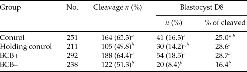
This experiment evaluated the developmental competence of bovine oocytes exposed to BCB. From 530 oocytes exposed to BCB, 292 (60.4%) were classified as BCB+ and 238 (39.6%) as BCB−. Their development after in vitro fertilization was compared with oocytes not exposed to BCB (control and holding control group). The cleavage and blastocyst rates are shown in Table 1. There was significant difference (p < 0.05) for cleavage among groups. Cleavage for control (65.3%) and BCB+ (64.4%) groups were similar to each other, but greater than holding control (49.8%) and BCB− (51.3%) groups. No difference (p > 0.05) was found for blastocyst rate between BCB+ (18.5%) and control (16.3%) groups, although these rates were greater (p < 0.05) than BCB− (8.4%) group. The blastocyst rate in the holding control group (14.2%) was not different from control and BCB+ groups but there was a tendency (p = 0.051) to be higher than BCB− group. The proportion of cleaved embryos reaching blastocyst stage was also higher (p < 0.05) in BCB+ (28.7%) than in BCB− (16.4%) group, however no statistical difference was found (p > 0.05) between control (25.0%) and BCB− (16.45%) groups.
Cleavage (72 hpi) and blastocyst (196 hpi) rate of embryos in vitro produced from immature oocytes selected by brilliant cresyl blue (BCB).
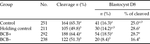
a, bDifferent letters in the same column differ statistically (p < 0.05) by chi-squared test.
Control: oocytes not exposed to BCB; holding control: oocytes not exposed to BCB and incubated for 60 min at 38.5 °C in mDPBS plus BSA; BCB+: oocytes with blue cytoplasm; BCB−: oocytes without blue cytoplasm.
Relative quantification of MATER and ZAR1 transcripts
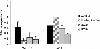
In order to identify differences on expression of MATER and ZAR1 genes among oocytes with different developmental competence, BCB+ and BCB− immature oocytes were evaluated using reverse transcription and real-time PCR. Oocytes from control and holding group were also included in this evaluation. Despite relative expression of MATER in the holding control, BCB+ and BCB− were downregulated in comparison with the control group, no statistical differences (p > 0.05) were found among them. There was also no difference (p > 0.05) in the relative expression of ZAR1 transcripts among groups (Fig. 1).
Relative quantification of MATER and ZAR1 transcripts (mean ± SE) in immature bovine oocytes after selection by brilliant cresyl blue (BCB). No significant difference (p > 0.05) was verified in relative expression for each gene among groups. BCB+, oocytes with blue cytoplasm; BCB−, oocytes without blue cytoplasm.

Discussion
The developmental competence and expression of two maternal genes, MATER and ZAR1, in bovine oocytes exposed to BCB dye was evaluated and it was found that oocytes with coloured cytoplasm have greater competence to develop after in vitro maturation and fertilization and that such competence seems not to be associated with differences in expression of both genes. Other studies reported higher cleavage and blastocysts rates with BCB-coloured cytoplasm oocytes compared with the colourless cytoplasm oocytes (Pujol et al., Reference Pujol, López-Béjar and Paramio2004; Alm et al., Reference Alm, Torner, Löhrke, Viergutz, Ghoneim and Kanitz2005; Opiela et al., Reference Opiela, Kątska-Książkiewicz, Lipiński, Słomski, Bzowska and Ryńska2008). This dye can stain oocytes with low activity of G6PDH enzyme, a feature of fully grown oocytes (Pujol et al., Reference Pujol, López-Béjar and Paramio2004; Alm et al., Reference Alm, Torner, Löhrke, Viergutz, Ghoneim and Kanitz2005). Besides, BCB-stained oocytes also showed greater diameter (Pujol et al., Reference Pujol, López-Béjar and Paramio2004), increased volume, high number of DNA mitochondrial copies (El Shourbagy et al., Reference El Shourbagy, Spikings, Freitas and St John2006) and gluthathione synthase concentration (Wu et al., Reference Wu, Liu, Zhou, Lan, Han, Miao and Tan2007) – features associated positively with developmental competence. Thus, BCB seems to be useful in identifying oocytes with different developmental competence.
Early studies suggested that this dye could be used to select bovine oocytes for in vitro fertilization and to increase efficiency of in vitro embryo production, as BCB+ oocytes had higher blastocyst rates than the control group, i.e., oocytes not exposed to BCB dye (Pujol et al., Reference Pujol, López-Béjar and Paramio2004, Alm et al., Reference Alm, Torner, Löhrke, Viergutz, Ghoneim and Kanitz2005). Similar findings were also reported for buffalo oocytes (Manjunatha et al., Reference Manjunatha, Gupta, Devaraj, Ravindra and Nandi2007). Nevertheless, those findings are different from the ones of the present study. Despite higher blastocyst production with BCB+ oocytes than with BCB− oocytes, no difference was found between BCB+ and control group, which is in agreement with recent studies with bovine (Opiela et al., Reference Opiela, Kątska-Książkiewicz, Lipiński, Słomski, Bzowska and Ryńska2008), ovine (Kątska-Książkiewicz et al., Reference Kątska-Książkiewicz, Opiela and Ryńska2007) and porcine (Wongsrikeao et al., Reference Wongsrikeao, Otoi, Yamasaki, Agung, Taniguchi, Naoi, Shimizu and Nagai2006) oocytes. The reason for such discrepancy among studies is not clear. It may be associated with different morphological criteria used to select oocytes before exposing to BCB or control group. A very exigent criterion of selection of only high-grade oocytes may reduce the difference between these groups as most oocytes in both groups would have greater developmental competence. Another possible reason could be that the low G6PDH activity in BCB+ oocytes would not be the only factor associated with developmental competence. A recent study in mice oocytes showed that the competence of BCB+ oocytes can vary according to their diameter (Wu et al., Reference Wu, Liu, Zhou, Lan, Han, Miao and Tan2007), suggesting that diameter may be critical for further development than G6PDH activity.
To verify the influence of the storage in mDPBS itself, the COCs were incubated in mDPBS without BCB for 60 min as a holding control group. Unlike, Alm et al. (Reference Alm, Torner, Löhrke, Viergutz, Ghoneim and Kanitz2005) we found lower cleavage rate than control and BCB+ groups, but similar to that of the BCB− group. However, when embryos at day D8 were evaluated the blastocyst rate was similar among other groups. The reason for this finding is not clear but it is possible that the incubation effect in cleavage rate was not severe enough to compromise the blastocyst rate. On the other hand, the competence of BCB+ oocytes may overcome some possible incubation effects whereas BCB− oocytes may be more sensitive to incubation.
Our data, together with that from other studies, suggest that, although BCB dye can distinguish two populations of oocytes with different developmental competence, this selection is not enough to predict increased blastocysts production efficiency when compared with exclusively morphological criteria. Even for improving embryo quality, BCB dye might not be indicated, as observed by studies without difference in cell number between blastocysts generated from BCB+ and control oocytes (Rodríguez-González et al., Reference Rodríguez-González, López-Béjar, Velilla and Paramio2002; Alm et al., Reference Alm, Torner, Löhrke, Viergutz, Ghoneim and Kanitz2005). Probably for an in vitro embryo production system this dye may not be as useful as previously thought. Instead, it may be an additional step in the whole process without an increase in the number and quality of embryos. However, Bhojwani et al. (Reference Bhojwani, Alm, Torner, Kanitz and Poehland2007) showed that the BCB could be used to select developmentally competent oocytes for nuclear transfer and this finding supports its application in techniques that require more COCs manipulation.
The present study also evaluated the relative expression of MATER and ZAR1 genes in oocyte exposed to BCB. This dye had already been used as a model to screen oocytes for their developmental competence in order to detect differential gene expression between bovine oocytes (Ghanem et al., Reference Ghanem, Hölker, Rings, Jennen, Tholen, Sirard, Torner, Kanitz, Schellander and Tesfaye2007). No difference in expression was found between BCB+ and BCB− oocytes, despite the greater cleavage and blastocyst rate found in the BCB+ group. This result indicates that lower developmental competence of BCB− oocyte is not associated with alteration in the relative expression of these two genes in the immature oocyte, although MATER and ZAR1 are recognized as maternal-effect genes required for proper embryo development during the early stages of post-fertilization period (Tong et al., Reference Tong, Gold, Pfeifer, Dorward, Lee, Bondy, Dean and Nelson2000; Wu et al., Reference Wu, Viveiros, Eppig, Bai, Fitzpatrick and Matzuk2003).
It is known that the presence of MATER and ZAR1 in oocytes is needed for further embryo development, since null mutant mice embryos arrest cleavage at the 1- or 2-cell stage (Tong et al., Reference Tong, Gold, Pfeifer, Dorward, Lee, Bondy, Dean and Nelson2000; Wu et al., Reference Wu, Viveiros, Eppig, Bai, Fitzpatrick and Matzuk2003). However, it is not clear whether the embryo development is associated with different levels of MATER and ZAR1 transcripts in immature oocytes. Wood et al. (Reference Wood, Dumesic, Abbott and Strauss2007) showed that human oocytes with low developmental competence, collected from women with polycystic ovary syndrome, have express MATER and another two maternal-effect genes were up-regulated, suggesting a negative effect of over-expression of these genes on embryo development. On the other hand Thélie et al. (Reference Thélie, Papillier, Pennetier, Perreau, Traverso, Uzbekova, Mermillod, Joly, Humblot and Dalbiès-Tran2007) found no variation in MATER and ZAR1 expression between in vitro and in vivo matured bovine oocytes, despite differences in developmental competence between them, which suggests that variation in MATER and ZAR1 transcripts expression may not be associated with the capacity of a matured oocyte to develop into an embryo after fertilization. Nevertheless, the present result suggests that there is no association between the capability of an immature oocyte to reach blastocyst and variations in mRNA expression of MATER and ZAR1; therefore expression of these genes may not be a good marker for developmental competence unless subtle differences on their expression are enough to disrupt embryo development. However, it does not mean that expression of other genes are not involved in differences between BCB+ and BCB− oocytes competence as other studies reported differential gene expression between BCB+ and BCB− oocytes (Ghanem et al., Reference Ghanem, Hölker, Rings, Jennen, Tholen, Sirard, Torner, Kanitz, Schellander and Tesfaye2007; Torner et al., Reference Torner, Ghanem, Ambros, Hölker, Tomek, Phatsara, Alm, Sirard, Kanitz, Schellander and Tesfaye2008). Moreover, there may be other mechanisms impairing embryo development between BCB+ and BCB− oocytes than differences on expression of MATER. The activation of embryonic genome is preceded by a transitory transcription and translation of polypeptides which form the Transcription Related Complex (TRC; Minami et al., Reference Minami, Suzuki and Tsukamoto2007). Some of these polypeptides are sensitive to alpha-amanitin (Conover et al., Reference Conover, Temeles, Zimmermann, Burke and Schultz1991), which in turn is acknowledged to block embryo development around maternal zygotic transition (Seshagiri et al., Reference Seshagiri, McKenzie, Bavister, Williamson and Aiken1992; Liu & Foote, Reference Liu and Foote1997). It was shown that 2-cell arrested embryos from MATER null mice were able to synthesize TRC (Tong et al., Reference Tong, Gold, Pfeifer, Dorward, Lee, Bondy, Dean and Nelson2000), indicating that absence of MATER protein does not interfere in the synthesis of polypeptides belong to TRC and thus predicting the existence of different mechanisms of embryo development inhibition.
In conclusion, even though BCB dye detects immature oocyte populations with different developmental competence, its usefulness to improve in vitro embryo production is questionable. Developmental competence of immature oocytes exposed to BCB does not seem to be associated with variations in expression of MATER and ZAR1 transcripts.
Acknowledgements
The authors thank EMBRAPA Dairy Cattle-CNPGL and CAPES for providing a scholarship for the first author.