Cardiac magnetic resonance imaging can provide high-resolution morphological data, as well as physiological assessment of ventricular function, valvular function, and great artery flow.Reference Puranik, Muthurangu, Celermajer and Taylor1 This, combined with echocardiography, can obviate the need for diagnostic cardiac catheterisation in young patients with congenital cardiac disease. In our own centre, catheterisation is largely reserved for those patients with a high probability of requiring intervention. However, there are few clinical data directly comparing cardiac magnetic resonance and cardiac catheterisation findings. Moreover, there is anecdotal suggestion that cardiac magnetic resonance imaging can exaggerate great artery stenosis.
In this study, we sought to retrospectively examine our own data, by comparing the cardiac catheterisation findings of all children in whom prior cardiac magnetic resonance had found important great artery stenosis.
Methods
Design
We performed a retrospective analysis of all patients, between January 2006 and August 2008, in whom important great arterial stenosis had been diagnosed during cardiac magnetic resonance assessment, and who had subsequently undergone cardiac catheterisation within 6 months. The aim of the cardiac catheterisation was to confirm the diagnosis and perform catheter intervention if indicated. The decision to plan catheter intervention was made during a formal, clinical, multidisciplinary meeting, in which all clinical and imaging data concerning the patient are presented. The decision was made on accepted criteria for intervention, based on clinical, echocardiographic, and cardiac magnetic resonance findings. The cardiac magnetic resonance findings helped to characterise the location and type of narrowing in the artery, as well as the associated ventricular and valvular parameters. Informed, signed consent for imaging and subsequent research data analysis were obtained from each patient's parents at the time of admission to hospital for the cardiac magnetic resonance procedure. All data were collected from inpatient medical records, departmental computer databases, and archived cardiac magnetic resonance and angiography images.
Cardiac magnetic resonance imaging studies
Scanning was performed using a 1.5-Tesla magnetic resonance scanner (Avanto; Siemens Medical Systems, Erlangen, Germany) with post-processing on a dedicated magnetic resonance workstation (Leonardo, Siemens Medical Solutions, Erlangen, Germany). Patients less than 7 years of age were imaged under general anaesthesia. Older patients were imaged without sedation. The spatial and temporal resolution of the imaging was optimised according to each patient's heart rate and body size; imaging parameters are similar to those published previously.Reference Muthurangu, Taylor and Hegde2 Anatomy was initially mapped using an axial stack of Half-Fourier Acquisition Single-Shot Turbo Spin-Echo images. A set of steady-state free-precession cine images was then acquired during serial breath holding. Slice positions for these two-dimensional cine images included the cardiac long axis, an anatomical short-axis stack, approximately 10 slices, with no gap, and orthogonal planes through the ascending, transverse, descending aortic arch and branch pulmonary arteries, respectively.
For a three-dimensional morphologic assessment of the great vessels, two sequences were used. The three-dimensional gadolinium-enhanced cardiac magnetic resonance angiography was performed using a coronal three-dimensional fast-field echo sequence. In addition, for each patient, three-dimensional “white-blood” imaging was performed with a respiratory-navigated and electrocardiogram-gated, single-slab three-dimensional steady-state free-precession sequence. For all three-dimensional data, the sequence parameters were optimised to achieve isotropic voxels, allowing multiplanar reformatting and measurement of structures in any plane.
At the time of the scan, the vessels in which significant narrowing was noted were additionally imaged in long- and short-axis planes with turbo spin-echo black-blood images. These were acquired during systole, in orthogonal long-axis planes. This sequence is less susceptible to artefact from magnetic field inhomogeneity or blood flow turbulence.
Through-plane magnetic resonance phase-contrast flow quantification data were acquired at the ascending aorta, the main pulmonary artery, and in the branch pulmonary arteries. For vessels with stenosis, an image plane was selected distal to the stenosis and proximal to any branching. If flow acceleration or turbulence was thought to potentially confound the measurement, then flow volumes in the pulmonary veins and or caval veins were recorded in order to optimise the internal accuracy of the measured flow volumes.
Arterial morphological measurements were made from the isotropic angiographic data, using multi-planar reformatting. For each measurement position on each vessel, two perpendicular, truly cross-sectional diameters were measured.
Structured cardiac magnetic resonance reporting included all great vessel dimensions, flow mapping of the net proportional forward flow in each branch pulmonary artery, flow velocity mapping through observed arterial stenoses, and a qualitative assessment – mild, moderate, and severe – of any stenoses observed. Obstruction was categorised as severe – obstruction more than 40% of the normal distal vessel – moderate – obstruction 20–39% – and mild – obstruction less than 20% – on morphological images. A redistribution of the relative flow proportions in the pulmonary arteries supported these categorisations.
Cardiac catheterisation
Standard catheterisation procedures using general anaesthesia were performed, including pressure measurements and angiography. All patients underwent angiography with standard projection to evaluate the morphology of arterial stenosis and to compare with cardiac magnetic resonance findings. Pressure measurements were recorded across the stenosed arterial segment by the standard pull-back method from the distal to proximal stenosed area. Peak systolic pressure difference more than 30 millimetres of mercury and mean pressure difference more than 15 millimetres of mercury were considered significant for catheter intervention in patients with pulmonary valve stenosis; however, for patients with arch obstruction, peak systolic pressure difference more than 20 millimetres of mercury and mean pressure difference more than 10 millimetres of mercury were considered significant for catheter intervention, if associated with clinical or echocardiographic findings.
Patient selection
Of 134 patients who had undergone cardiac catheterisation within 6 months of cardiac magnetic resonance, there were 45 patients with congenital cardiac disease in whom cardiac catheterisation was performed with the specific intention to confirm and intervene on great artery stenosis. The clinical cardiac magnetic resonance report was assessed for each of these 45 patients. The key diagnoses involving haemodynamically significant changes of the great arteries were recorded for each patient. For some patients, there was a single key finding, whereas for others there were three to four key findings, involving different vessels. For each patient, these key cardiac magnetic resonance findings were then compared with the specific findings related to that artery at cardiac catheterisation.
Concordant observations. were those in which moderate-to-severe stenoses were confirmed at cardiac catheterisation.
Discordant observations. were those in which moderate-to-severe stenosis was NOT confirmed at cardiac catheterisation. That is, at cardiac catheterisation, there was a small pressure gradient or mild narrowing on angiography, and thus intervention was not performed.
Non-parametric statistical methods were used. A p-value less than 0.05 was considered significant.
Result
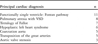
Of the 45 patients studied, cardiac magnetic resonance found 60 key lesions involving haemodynamically significant great artery stenoses. The 45 patients had a wide range of cardiac diagnoses (Table 1). Cardiac catheterisation was performed at a median of 84 days, with an interquartile range of 4–149 days, after cardiac magnetic resonance.
Principal cardiac diagnosis.

VSD = ventricular septal defect
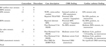
On comparison of the specific cardiac catheterisation findings for the 60 cardiac magnetic resonance observations, 53 (88%) findings were concordant and seven (12%) were discordant.
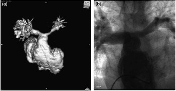
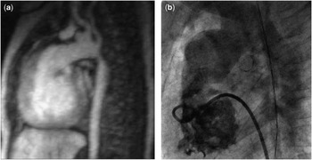
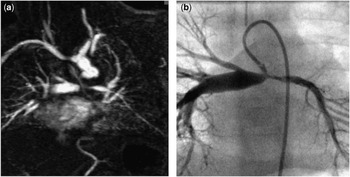
Of the discordant observations, the predominant reason for discordance – that is, in six of seven (86%) cases – was that cardiac magnetic resonance defined moderate–severe great vessel stenosis, which was not confirmed by cardiac catheterisation. In these patients, cardiac catheterisation revealed mild stenosis and haemodynamic gradients that were insufficient to indicate intervention (Figs 1 and 2). These six cases involved three branch pulmonary arteries and three aortic arch lesions (Table 2).
(a and b) This is a three-dimensonal volume-rendered image from the cardiac magnetic resonance angiogram (a) and a frame from the pulmonary artery angiogram during subsequent cardiac catheterisation (b). These images illustrate apparent discordant findings from a 16-year-old patient following arterial switch repair of transposition of the great arteries. The cardiac magnetic resonance found a long-segment stenosis of the proximal left pulmonary artery, with distal hypoplasia of the vessel, and only 35% of the total pulmonary flow going to the left side. The catheterisation found a 16-millimetre gradient across the proximal left pulmonary artery and no significant vessel stenosis. No intervention was performed.

(a and b) This is a single-plane maximal intensity projection from the cardiac magnetic resonance angiogram (a) and a frame from the aortic angiogram during subsequent cardiac catheterisation (b). These images illustrate apparent discordant findings from a 3-month-old child with hypoplastic left heart syndrome, following Norwood Stage 1 repair. The cardiac magnetic resonance found severe, discrete coarctation of the aorta. At subsequent cardiac catheterisation, there was a 5-millimetre of mercury gradient across this narrowing; no intervention was performed.

Key discordant findings.

Ao = aorta; CMR = cardiac magnetic resonance; CoA = coarctation of aorta; DORV = double outlet right ventricle; DKS = Damus Kaye stansel; HLHS = hypoplastic left heart syndrome; LPA = left Pulmonary artery; MPA = main pulmonary artery; RPA = right Pulmonary artery; RV = right ventricle; TCPC = total cavo-pulmonary connection; TGA = transposition of the great arteries; VSD = ventricular septal defect
In a single case, cardiac catheterisation found proximal right pulmonary artery narrowing, which had not been mentioned in the cardiac magnetic resonance report (Fig 3).
(a and b) This is a single-plane maximal intensity projection from the cardiac magnetic resonance angiogram (a) and a frame from the pulmonary angiogram, with injection from the Blalock–Taussing shunt, during subsequent cardiac catheterisation (b). The cardiac magnetic resonance and catheterisation were concordant in their finding of left pulmonary artery stenosis, but at catheterisation there was an additional finding of mild stenosis of the right pulmonary artery just distal to the Blalock–Taussing shunt insertion. This narrowing was not specifically commented upon in the cardiac magnetic resonance report.

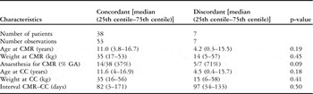
There was no difference between discordant and concordant groups on the basis of patient age, weight, time interval between cardiac magnetic resonance and cardiac catheterisation, or type of lesion (Table 3).
Patient characteristics of concordant and discordant findings.

CC = cardiac catheterisation, CMR = cardiac magnetic resonance, GA = general anaesthesia
Discussion
The use of cardiac magnetic resonance imaging for the evaluation of congenital cardiac disease has increased significantly over the last decade. Despite the fact that cardiac magnetic resonance remains an adjunct to echocardiographic evaluation of the heart, in many situations cardiac magnetic resonance can replace diagnostic cardiac catheterisation. Cardiac catheterisation remains essential predominantly when the ascertainment of pressure is mandatory and for interventional procedures. Cardiac magnetic resonance is an important investigative tool throughout the stages of surgery for hypoplastic left heart syndrome. Previous studies discussed the use of cardiac magnetic resonance replacing cardiac catheterisation before bidirectional Glenn anastomosis in children with a functionally univentricular heart.Reference Brown, Gauvreau and Powell3–Reference Muthurangu, Taylor and Hegde6 In previous studies of patients with coarctation of the aorta, it has been shown that anatomic and flow data obtained by cardiac magnetic resonance have a high sensitivity and specificity for predicting a catheterisation gradient greater than 20 millimetres of mercury.Reference Nielsen, Powell, Gauvreau, Marcus, Prakash and Geva7, Reference Oshinski, Parks and Markou8. Cardiac magnetic resonance is also useful for long-term follow-up of arterial lesions.Reference Tsai-Goodman, Geva, Odegard, Sena and Powell9
In terms of using cardiac magnetic resonance as a diagnostic screening tool to guide the decision for interventional cardiac catheterisation, our study shows that the majority of findings in cardiac magnetic resonance are concordant with the subsequent invasive findings.
However, there was a minority of discordant observations, in which cardiac magnetic resonance apparently “over diagnosed” the importance of arterial narrowing. It is conceivable that cardiac catheterisation can “under diagnose” important arterial narrowing, especially if the measured pressure drop is the main variable assessed, as may sometimes be the case with aortic arch obstruction. Particularly in the context of impaired ventricular function, anaesthetic effects that decrease cardiac output, and therefore decrease any gradient across an arterial narrowing, may confound interpretation of the cardiac catheterisation gradient data.
It is equally important not to uncritically convert the peak flow velocity measured distal to a great artery stenosis to the estimated pressure drop, using the simplified Bernoulli equation. Measurement of pressure drop at a great artery stenosis must take account of flow volume diversion. Cardiac catheterisation does not fully account for flow volume diversion. For example, in unilateral pulmonary artery stenosis, the larger proportion of forward flow will enter the non-stenosed pulmonary artery. A lower volume of flow into the stenosed artery – diagnosed on cardiac magnetic resonance – results in a lower pressure gradient across the narrowed segment.
Of the discordant diagnoses in this study, two were proximal branch pulmonary artery narrowing in which there was hypoplasia of the distal vessel; thus, little gradient was measured at cardiac catheterisation, which contributed to the decision not to dilate the vessel.
Limitation
This is a retrospective cohort analysis, and cardiac catheterisation was not performed in those patients in whom cardiac magnetic resonance found no significant narrowing. Therefore, no ascertainment was possible of the sensitivity and specificity of cardiac magnetic resonance diagnosis for great vessel stenosis.
No systematic outcome data are available – for example, surgical confirmation or follow-up studies – with which to assess the discordant observations, and with which to give a final assessment of diagnostic accuracy for each modality.
Conclusion
In this consecutive clinical cohort, there was a high level of concordance between cardiac magnetic resonance-diagnosed and cardiac catheterisation-measured great artery stenosis. The discordant findings were from a variety of patient types and vessels, with no particular trend. The predominant reason for discordance – that is, in 86% of discordant findings – was a lower catheterisation gradient than predicted by morphologic and functional cardiac magnetic resonance assessment. The level of concordance in the assessment of clinically important lesions confirms the value of cardiac magnetic resonance in the selection of patients for invasive catheter intervention for great artery stenoses. Importantly, no clinically significant great artery stenoses were detected at cardiac catheterisation, which had been overlooked at cardiac magnetic resonance.
Prospective assessment and refinement of comparative cardiac magnetic resonance and cardiac catheterisation data may further improve the validity of non-invasive imaging thresholds for intervention.