Atrial septoplasty is a life-saving procedure performed in children with transposition of great arteries when immediate surgery is not feasible. It is usually performed from the femoral or umbilical vein. In exceptional circumstances, atrial septoplasty may have to be performed via internal jugular venous access. We report the case of a 3-month-old infant, wherein we accomplished successful atrial septoplasty through internal jugular venous access. We highlight the technical challenges faced and plausible solutions for the successful performance of this procedure.
Case
A 3-month-old infant, noted to have cyanosis since birth, was referred to our hospital with worsening central cyanosis (oxygen saturation of 65%), tachypnoea, and poor feeding for 1 week. Cardiac auscultation revealed a single, loud second heart sound and grade 3/6 ejection systolic murmur at the left upper sternal edge.
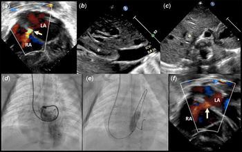
Transthoracic echocardiography confirmed the diagnosis of transposition of great arteries. A restrictive foramen ovale was shunting from left-to-right atrium (Fig 1a), while there were two small apical ventricular septal defects shunting from right-to-left ventricle. Abdominal long-axis view showed a near-complete occlusion of inferior caval vein due to a homogenous echogenic mass, likely a thrombus (Fig 1b and c). The thrombus measuring 27 × 8 mm was present 2.5 cm below the inferior caval vein–right atrial junction. In view of severe hypoxaemia and poor general condition, the child was put on mechanical ventilation and an urgent balloon atrial septostomy was planned to stabilise the child.
(a) Echocardiogram in subcostal view with colour Doppler study showing restrictive interatrial septum (yellow arrow) with left-to-right flow. (b) Abdominal long-axis view showing near-total occlusion of intrahepatic inferior caval vein due to thrombus (asterisk). (c) Abdominal short-axis view showing the thrombus (asterisk) in inferior caval vein. (d) Angiogram in shallow left anterior oblique view with cranial tilt showing contrast injection through Cobra catheter opacifying the left atrium and left ventricle. (e) Fluoroscopic view in shallow left anterior oblique view with cranial tilt showing partially inflated Tyshak balloon with a waist in the middle due to restrictive interatrial communication. (f) 2D echocardiogram with colour Doppler study in subcostal view showing enlarged atrial septal defect with low-velocity left-to-right flow. ICV-RA jn = inferior caval vein–right atrial junction; LA = left atrium; RA = right atrium.

In the catheterisation laboratory, a right internal jugular venous access was obtained using a 5-Fr introducer sheath. Attempts at negotiating a 4-Fr Fogarty balloon catheter (Edwards Lifesciences, Irvine, CA) through the foramen ovale, by pre-shaping the stylet to 90° angulation were unsuccessful. Subsequent attempts at crossing the foramen ovale using a straight tip 0.035” hydrophilic wire (Terumo Co., Tokyo, Japan) introduced through various curved catheters (Multipurpose, Judkin’s 2.5 right coronary, and Picard catheters) also failed. Eventually, foramen ovale was crossed with a 5-Fr Cobra C3 curve catheter (Cook Medical LLC., Bloomington, IN, United States of America) over a 0.025” hydrophilic wire. Catheter position in the left atrium was confirmed on angiography (Fig 1d). The catheter was exchanged for a 9 × 20 mm Tyshak II balloon catheter (Numed Inc., Hopkinton, NY, United States of America) over 0.018” exchange length wire and the foramen ovale was dilated twice (Fig 1e). The saturation improved to 80% and interatrial defect showed non-restrictive flow on echocardiography (Fig 1f). There were no complications during the procedure. After the procedure, Heparin infusion was continued due to the large thrombus in inferior caval vein.
The child had a seizure the following day. Neuroimaging revealed thrombosis of the superior sagittal sinus with accompanying venous infarct and haemorrhagic transformation, possibly due to an underlying hypercoagulable state. In view of the high risk of poor neurological outcomes after open-heart surgery, parents opted to provide only palliative care and transferred their child to a healthcare facility near their residence.
Discussion
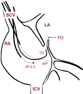
Creation of a good interatrial communication by balloon atrial septostomy is a life-saving intervention performed in the transposition of great arteries. It improves the mixing between systemic and pulmonary circulations, both of which function as parallel circuits. Reference Feltes, Bacha and Beekman1 In older infants, the interatrial septum is thick and balloon atrial septostomy may not be effective in creating a reasonably sized defect. In such circumstances, balloon atrial septoplasty is preferred. Femoral and umbilical veins are the most preferred vascular access for this procedure. Very rarely trans-hepatic venous access may be used. Negotiation of foramen ovale from the inferior caval vein is deemed extremely easy due to the favourable orientation of foramen ovale with respect to inferior caval vein ostium. Additionally, the valve of inferior caval vein helps by preferentially directing the blood towards the foramen ovale. In the index case, a large thrombus in inferior caval vein precluded a trans-femoral approach. A trans-hepatic venous approach has all the advantages of trans-femoral approach, but we did not use it due to the high chance of engaging the inferior caval vein thrombus and the risk of subsequent systemic embolisation. Therefore, we decided to perform atrial septoplasty through the internal jugular venous approach. The jugular venous approach is usually not preferred since the anatomical orientation of interatrial septum and the position of foramen ovale in relation to the orifice of superior caval vein make it quite difficult to enter the left atrium. In order to overcome this anatomical disadvantage, we used a catheter with a larger secondary curve, the Cobra C3 curve, wherein the tip of the catheter is curved more than 90° back towards the direction of the shaft. This provided a favourable position to cross the foramen ovale (Fig 2). Furthermore, crossing into the left atrium is challenging due to the small size of the cardiac structures in early infancy. This could be tackled by the use of smaller curve catheters as it is the usual practice while performing cardiac interventions in small infants. Though we used a straight tip hydrophilic guidewire for crossing the defect, a J-tipped or a deflectable guidewire may be safer for use in neonates and young children, and should be preferred if available in the laboratory. We used a balloon catheter with lower rated burst pressure due to its low profile and as the aim of our procedure was only to bail out the child and perform early arterial switch operation.
The orientation of various curved catheters in relation to the foramen ovale when entering the right atrium from the superior vena cava. C3 = Cobra C3 curve catheter; FO = foramen ovale; ICV = inferior caval vein; JR 2.5 = Judkins 2.5 curve right coronary catheter; LA = left atrium; MP = multipurpose catheter; RA = right atrium; SCV = superior caval vein.

There are only two previously described reports of successful balloon atrial septostomy through the jugular approach. Reference Padhi, Bakshi and Londhe2,Reference Mackie, Aiyagari and Zampi3 Padhi et al Reference Padhi, Bakshi and Londhe2 performed successful balloon atrial septostomy in a 45-day-old infant with transposition of great arteries and occluded inferior caval vein while Mackie et al Reference Mackie, Aiyagari and Zampi3 chose to use this approach in a newborn with hypoplastic left heart syndrome and a superiorly located, restrictive atrial septal defect. Both authors reported an anticipated risk of injuring the sinoatrial node when forcefully “pulling” the catheter towards superior caval vein during balloon atrial septostomy. Reference Padhi, Bakshi and Londhe2,Reference Mackie, Aiyagari and Zampi3 However, there is unlikely to be any such risk when performing balloon dilation of interatrial septum. To the best of our knowledge, ours is the first case reporting the use of jugular venous access for performing an atrial septoplasty.
Conclusion
Atrial septoplasty is an emergency procedure performed in children with transposition of great arteries when immediate surgery is not feasible. Femoral and umbilical veins are the most preferred vascular access, however, in exceptional circumstances, atrial septoplasty may have to be performed via internal jugular vein. Crossing the foramen ovale through the jugular venous approach is technically challenging, but can be accomplished with the use of appropriate hardware as demonstrated by our case.
Acknowledgements
None.
Financial support
This research received no specific grant from any funding agency, commercial, or not-for-profit sectors.
Conflicts of interest
None.
Ethical standards
The authors assert that all procedures contributing to this work comply with the ethical standards of the relevant Indian guidelines on human experimentation and with the Helsinki Declaration of 1975, as revised in 2013, and a need for formal approval was waived off by the Ethics Committee of All India Institute of Medical Sciences, New Delhi, India.