The typical stem cells, usually referred to as embryonic stem cells (ESCs), derive from the inner cell mass of the blastocyst and can be propagated indefinitely in an undifferentiated state. ESCs can also be differentiated into all cell lineages both in vivo and in vitro. However, the use of these cells for research and/or therapy is impeded by ethical and legal considerations. On the other hand, postnatal (adult) stem cells retain a broad differentiation potential, even though their multipotency is more restricted than in ESCs. Until recently, bone marrow has been the major source of human adult stem cells. The use of bone marrow stem cells (BMSCs), which include haematopoietic stem cells and mesenchymal stem cells (MSCs), is impractical because of technical issues and a limited yield. In contrast, the adipose tissue has been recently established as an alternative, abundant and accessible reservoir of adult stem cells. Adipose tissue is derived from the embryonic mesoderm and contains a cell-enriched supportive stroma, which can be easily isolated in large quantities. The existence of adipose-derived stem cells (ASCs) capable of differentiating into mature adipocytes is highly relevant to the understanding of the biology of adipose tissue and the development of its specific regional depots. In this regard, it should be recognised that the adipose tissue located in the abdominal region displays distinct metabolic features as compared with fat from the subcutaneous depot (Refs Reference Giorgino, Laviola and Eriksson1, Reference Laviola2). It has been suggested that the different metabolic properties of visceral and subcutaneous adipocytes may also be due to inherent characteristics of the cells that are resident in each fat depot. Relevant to this concept, human adipocytes differentiated in vitro from ASCs obtained from visceral or subcutaneous fat biopsies showed depot-specific differences for genetic, biochemical and metabolic endpoints (Refs Reference Tchkonia3, Reference Katz4, Reference Perrini5). Thus, the biochemical and metabolic specificity of various adipose tissue depots may reside in different biological characteristics of resident cells, including the undifferentiated ASCs. This is of importance, since the distribution of body fat appears to be more important than the total amount of fat for the development of metabolic diseases, and visceral adiposity is strongly linked to obesity-related comorbidities such as insulin resistance, type-2 diabetes, hypertension, dyslipidaemia and cancer.
To add complexity to this scenario, it has been recently proposed that ASCs consist of heterogeneous cell populations. When human subcutaneous and visceral fat biopsies are processed to obtain undifferentiated precursors, at least three distinct subsets of ASCs can be isolated and maintained in culture, all exhibiting the potential of unlimited proliferation and of differentiation along the mesenchymal lineage to produce adipocytes, osteoblasts and chondrocytes (Refs Reference Miyazaki6, Reference Matsumoto7, Reference Perrini8). In comparison with BMSCs, ASCs are equally capable of differentiating towards mesodermal lineage. Thus, fat depots are an alternative source of stem cells for different applications that will be highlighted in this review. In addition, the definition of ASCs biological properties may shed light on the origin of obesity and its associated pathologies.
Definition of ASCs
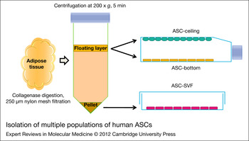
Adult stem cells can be identified in most tissues, including haematopoietic, neural, gastrointestinal, epidermal and adipose tissues. Adipose tissue, such as bone marrow, is derived from the mesenchyme and contains a supportive stroma that is easily isolated in relatively large quantities. Stromal cells that have preadipocyte characteristics can be isolated from adipose tissue of adult subjects in the stromal vascular fraction (SVF), spread in vitro and induced to differentiate into adipocytes (Refs Reference Gimble and Guilak9, Reference Zuk10, Reference Zuk11, Reference Rodriguez12). Initially, these stromal-derived cells were defined as ‘preadipocytes’, even though they were shown to be capable of forming not only fat, but also bone, cartilage and muscle (Refs Reference Gimble and Guilak9, Reference Zuk10, Reference Zuk11, Reference Rodriguez12, Reference Gimble and Guilak13, Reference Lee14, Reference Gronthos15). Then, a variety of definitions have been used, including ‘ASCs’, ‘adipose-derived adult stem cells’, ‘adipose-derived adult stromal cells’, ‘adipose-derived stromal cells’, ‘adipose mesenchymal stem cells’ and ‘processed lipoaspirate cells’, among others. In 2004, the International Fat Applied Technology Society adopted the term ‘adipose-derived stem cells’ (ASCs), to define plastic-adherent cells with multilineage capacity isolated from the SVF of adipose tissue (Ref. 16). On the other hand, it has been recently shown that when mature fat cells obtained from human adipose tissue were maintained in the culture flasks filled with medium, two additional fibroblast-like cell populations appeared at the ceiling and bottom surfaces of the flask, exhibiting the potential of unlimited proliferation, as well as of differentiation along the mesenchymal lineage to produce adipocytes, osteoblasts and chondrocytes (Refs Reference Miyazaki6, Reference Matsumoto7, Reference Perrini8) (Fig. 1). ‘Ceiling culture’ is a unique method of culturing unilocular fat cells obtained by collagenase-digestion and differential centrifugation of adipose tissue fragments (Refs Reference Matsumoto7, Reference Sugihara17). When the mature fat cells in the floating layer were maintained with attachment to the ceiling surface of the culture flask filled with medium, the unilocular fat cells appeared to change their morphology into multilocular fat cells or fibroblastic cells and the fibroblastic cells proliferated to form a monolayer. Under these culture conditions, another population of fibroblast-like cells was found to proliferate at the bottom surface of the culture flasks filled with medium. Both populations of proliferating cells, at the ceiling and bottom surfaces, show multiple differentiation potential along the mesenchymal lineage and the capacity of unlimited self-renewing proliferation (Refs Reference Miyazaki6, Reference Matsumoto7, Reference Perrini8).
Isolation of multiple populations of human adipose-derived stem cells (ASCs). After collagenase-mediated digestion of the adipose tissue specimen and removal of undigested material with a nylon mesh, centrifugation of the tissue yields a pellet, representing the stromal vascular fraction (SVF) containing the most widely utilised population of ASCs (ASC-SVF). However, two additional ASC populations can be obtained from the floating layer, i.e. ASC-Bottom and ASC-Ceiling. ASC-Ceiling potentially derives from de-differentiation of mature unilocular adipocytes.

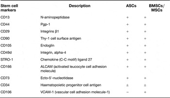
In culture, ASCs express cell-surface markers similar to those expressed by MSCs, including CD105, CD90 and CD44. Specifically, ASCs express CD49d, whereas this antigen was not observed in nonadipose MSC cultures (Table 1). In contrast to MSCs, no expression of CD106 was observed in ASCs (Refs Reference Katz4, Reference Matsumoto7). The abundance and yield of ASCs and MSCs might be different but their biology, although not identical, appears to be similar. ASCs may be a better source of multilineage mesodermal stem cells for gene therapy and tissue repair because of their copiousness and accessibility.
Markers of human ASCs

ASCs, adipose-derived stem cells; BMSCs, bone marrow-derived stem cells; MSCs, mesenchymal stem cells.
Adapted from Refs Reference Katz4, Reference Matsumoto7.
Origin and in vivo location of ASCs
Despite the enormous efforts in numerous laboratories, there are still significant gaps in our understanding of the origin and location of such cells. Indeed, although it has been shown that adipocytes develop in coordination with the vasculature, limited data is available on the identity and in vivo location of white adipocyte progenitors. From studies in rodents, it has been suggested that adipocytes descend from a pool of these proliferating progenitors that are already committed, either prenatally or early in postnatal life (Ref. Reference Tang18). These progenitors may reside in the mural cell compartment of the adipose vasculature, as a subset of both pericytes and capillary endothelial cells, but not in the vasculature of other tissues. Thus, the adipose vasculature appears to function as a progenitor niche and may provide signals for adipocyte development (Refs Reference Tang18, Reference Gupta19). In adipose tissue, perivascular stromal cells are organised in two discrete layers, the innermost consisting of CD146+/α-SMA+/CD34− pericytes, and the outermost represented by CD146−/α-SMA−/CD34+ supra-adventitial ASCs (SA-ASCs), both of which show adipogenic potential in vitro. Pericytes had the most potent adipogenic potential, followed by the more numerous SA-ASCs. Endothelial populations had significantly reduced adipogenic potential compared with unsorted expanded SVF cells (Ref. Reference Zimmerlin20). Moreover, only the α-SMA+ cells seem to show multilineage differentiation ability whereas the α-SMA− cells can only differentiate in an adipogenic direction (Ref. Reference Cai21).
Multilineage differentiation of ASCs in vitro
As mentioned above, adipose tissue is a major source of adult multipotent stem cells that can differentiate along the mesenchymal lineage to produce adipocytes, chondrocytes, osteocytes and myocytes (Refs Reference Zuk11, Reference Rodriguez12), and along the neuronal lineage (Refs Reference Mareschi22, Reference Ashjian23). They can also differentiate into pancreatic beta-cells (Ref. Reference Chandra24), myocardiocytes (Refs Reference Choi25, Reference Choi26) and endothelial cells (Refs Reference Auxenfans27, Reference Zhai28).
White adipocytes
Several multiphase events are involved in the adipogenic differentiation and ultimate maturation of ASCs. Kuri-Harchuch et al. demonstrated that adipogenic induction of ASCs resulted in a significant increase in glycerol-3-phosphate dehydrogenase (GPDH) activity, a lipogenic enzyme involved in triglyceride synthesis (Ref. Reference Kuri-Harcuch and Green29). In addition to possessing metabolic activity consistent with the formation of mature adipocytes, differentiating ASCs express several genes involved in lipid biosynthesis and storage, including (i) specific expression of PPARγ2, a fat-specific transcription factor that functions in preadipocyte commitment, (ii) increased expression of lipoprotein lipase (LPL), a lipid exchange enzyme upregulated during adipogenesis (Ref. Reference Ailhaud, Grimaldi and Négrel30), (iii) upregulation of aP2, a protein associated with lipid accumulation within mature adipocytes (Ref. Reference Bernlohr31), (iv) increased expression of both leptin and GLUT4 and restriction of these proteins to lipid-filled ASCs and (v) reduced expression of Pref-1, also known as Delta-like Protein 1 (DLK-1), an epidermal growth factor-like domain-containing trans-membrane protein involved in adipogenesis repression and cell fate decision (Refs Reference Kim32, Reference Jing33). Differentiation potential varies between clones of ASCs, raising the possibility that epigenetic differences account for this variability. Indeed, the in vitro differentiation towards various pathways maintains the overall methylation profiles of undifferentiated cells, suggesting that ASCs are epigenetically programmed for adipogenesis. Thus, the extent of DNA methylation of lineage-specific genes may predict the propensity for differentiation into a particular cell type. Specifically, hypomethylation of adipogenic loci in undifferentiated ASCs may reflect a commitment of these cells to adipogenic lineage (Ref. Reference Boquest34).
Brown adipocytes
The last decade has brought increasing interest in the study of brown adipose tissue which is equally specialised for thermogenic expenditure of energy and it is the focus of intense investigation. Although it was long thought that adult humans had only a modest amount of brown fat, studies using positron emission tomography to identify fluorodeoxyglucose uptake by metabolically active cells revealed a significant amount of brown adipose tissue located in the neck, supraclavicular, mediastinal, suprarenal and paravertebral regions (Ref. Reference Nedergaard35). Some authors have demonstrated a myogenic transcriptional signature suggesting a distinct origin than that of white adipocytes both by usage of lineage-tracing model in which expression of YFP was dependent on Myf5 (myogenic lineage specific)-promoter driven cre-recombinase expression (Ref. Reference Seale36) and by microarray analysis to examine global gene expression patterns in white versus brown preadipocytes (Ref. Reference Timmons37).
The possibility of achieving transdifferentiation of white adipose tissue into brown adipose tissue is of special interest. Transdifferentiation is a process whereby a mature differentiated cell transforms into a new phenotype with different morphology and physiology without going through dedifferentiation. This was indeed observed by several authors (Refs Reference Murano38, Reference Himms-Hagen39, Reference Granneman40); specifically, exposure of rodents to low temperature (Ref. Reference Murano38) or treatment with beta3-adrenergic agonists (Ref. Reference Himms-Hagen39, Reference Granneman40) was found to result in an increased number of multilocular cells expressing uncoupling protein-1 and a decreased abundance of unilocular adipocytes, whereas the total fat cell number remained constant (Ref. Reference Cinti41). Interestingly, studies by Nedergaard and coworkers (Ref. Reference Petrovic42) showed that even though these brown adipocytes are functionally thermogenic, they fail to express the classic brown adipocyte genes, including Zic1, Lhx8, Meox2 and PDRM16 and continue to express white adipocyte markers like Hoxc9. Thus, it is presumed that the brown adipocytes arose via transdifferentiation from white adipocytes, rather than brown-driven differentiation of ASCs.
Osteocytes
ASCs differentiate in vitro into osteoblast-like cells that are capable of depositing a mineralised extracellular matrix in a similar manner and under the same general conditions as MSCs. Thus, ASCs cultured between 14 and 21 days on low concentrations of ascorbic acid, beta-glycerophosphate and dexamethasone and/or vitamin D analogues will express multiple markers of osteocytes, including cbfa-1, alkaline phosphatase, osteopontin, osteocalcin and collagen I (Ref. Reference Halvorsen43). As with MSCs, transduction of ASCs with the gene encoding bone morphogenic protein 2 (BMP2) results in the generation of ectopic bone production in vivo. Thus, ASCs transduced with adenovirus-BMP2 and seeded into standard collagen type I sponges will form a radio-opaque, bone-like material when implanted, intramuscularly, into SCID mice (Ref. Reference Dragoo44). In addition, some groups showed that human ASCs were able to adhere to titanium scaffolds, proliferate, acquire an osteoblastic-like phenotype and produce a calcified extracellular matrix with protein, such as decorin, fibronectin, osteocalcin, osteonectin, osteopontin and type I collagen (Ref. Reference Gastaldi45).
Chondrocytes
Several groups have demonstrated the ability of ASCs to undergo chondrocytic differentiation (Refs Reference Dragoo44, Reference Nöth46, Reference Yoon47, Reference Winter48). These systems incorporate high cell-density (micromass) cultures supplemented with transforming growth factor-beta1 (TGF-beta1) to generate proteoglycan-rich spheroids that express collagen II, aggrecan, decorin and other markers consistent with chondrogenesis. Hyaluronic acid scaffold with chondrogenic medium is a promising chondrogenic culture system of ASCs and BMP-2 could potentially serve as a chondrogenic supplement for ASCs increasing the level of chondrogenic differentiation (Ref. Reference Yoon47). Winter et al. reported that the gene expression profile of ASCs under chondrogenic conditions is similar to that of MSCs (Ref. Reference Winter48). Following an in vitro differentiation process, Erickson et al. showed that human ASCs retain the chondrocyte phenotype and form cartilaginous tissue when implanted subcutaneously in vivo in immunodeficient mice for up to 12 weeks (Ref. Reference Erickson49).
Myocytes
The culture of ASCs in the presence of dexamethasone and hydrocortisone results in a time-dependent increase in the expression of muscle-specific markers (MyoD1 and myosin), expression of transcription factors (myf5, myf6 and myogenin) and the appearance of elongated, multinucleated structures that closely resemble myotubes (Ref. Reference Mizuno50). Furthermore, Bacou et al. showed that primary cultures of ASCs transduced with LacZ and injected into crush-injured anterior tibialis muscles were incorporated into the myofibres within 15 days (Ref. Reference Bacou51). Two months after the injection, the ASCs-treated muscles showed increased mass, myotube fibre diameter and force generation compared with the untreated controls. Moreover, other studies suggest that ASC transplantation modulates inflammation and improves muscle tissue regeneration, ameliorating the dystrophic phenotype in dystrophin-deficient mice (Refs Reference Vieira52, Reference da Justa Pinheiro53). ASC transplantation into the skeletal muscle of these mice led to improved muscle strength and resistance to fatigue. Increases in the fibre cross-sectional area, number of fibres with centralised nuclei and myogenin content were observed. In ASC-treated muscles, a decrease in muscle content of TNF-alpha, IL-6 and oxidative stress was also found. The level of TGF-beta1 expression was lower, whereas expression of VEGF, IL-10 and IL-4 was increased. An increase in markers of M1 macrophages (CD11 and F4-80) and a decrease in the T lymphocyte markers CD3 and arginase-1 was also observed in ASC-treated dystrophic muscles (Ref. Reference da Justa Pinheiro53).
Endothelial cells
Adipose tissue is an excellent source of autologous endothelial cells to reconstruct endothelialised tissue equivalents. Miranville et al. performed a series of studies using CD34+/CD31+ cells isolated from human adipose tissue and demonstrated the ability of these cells to undergo in vitro and in vivo endothelial differentiation (Ref. Reference Miranville54). In these studies, cultures of selected cells, grown in low serum medium supplemented with VEGF and insulin-like growth factor-I (IGF-I), formed network-like structures that expressed endothelial markers. In contrast, Rehman et al. showed that these structures can be also generated in the absence of growth factor supplements by eliminating the process of cell selection; for example, the culture of adipose-derived cells in alpha medium and 10% fetal calf serum results in the creation of a monolayer of ASCs, upon which network structures of cells expressing endothelial markers formed (Ref. Reference Rehman55). This difference might reflect the ability of the ASCs to express VEGF and other proangiogenic factors. Auxenfans et al. reconstructed an autologous endothelialised skin equivalent by incorporating human ASCs isolated from lipoaspirates and amplified to obtain endothelial progenitor cells, which were subsequently differentiated into endothelial cells (Ref. Reference Auxenfans27).
Neurons
Until recently, neural tissues have not been regarded as regenerative populations of cells. Thus, the identification of cell populations capable of neuronal differentiation has generated immense interest. Adipose tissue may represent an alternative source of cells that are capable of neuronal differentiation, potentially enhancing its use in the treatment of neurological disease. Even though studies indicating neuronal differentiation of ASCs need to be reviewed with particular care, the ability of either ASCs to acquire the key functional attributes of neuronal cells (formation of synaptic junctions and the generation or transmission of an action potential) (Refs Reference Liqing56, Reference Cardozo, Gómez and Argibay57, Reference Woodbury58) and to enhance or induce regeneration and repair of impaired neuronal axons after injury (Refs Reference Arboleda59, Reference Scholz60) has recently been reported. Following neural induction with bFGF and forskolin, human ASCs differentiated into various types of neural cells including neurons and glia in vitro and this resulted in apparent immunoreactivity for neural stem cell (nestin), neuronal (Tuj1, MAP2, NFL, NFM, NFH, NSE and NeuN), astrocyte (GFAP) and oligodendrocyte (CNPase) markers. Moreover, ASCs acquired neuron-like functions characterised by the display of voltage-dependent tetrodotoxin (TTX)-sensitive sodium currents, outward potassium currents and prominent negative resting membrane potentials under whole-cell patch clamp recordings (Ref. Reference Jang61). Interestingly, both Wnt and Notch signalling are present in proliferating ASCs and both cascades are downregulated when cells are differentiated into a neuronal phenotype (Ref. Reference Cardozo, Gómez and Argibay57).
Pancreatic beta-cells
Many studies have attempted to generate insulin-producing cells in vitro for the treatment of insulin-deficient type-1 diabetes. Although ESCs can be induced to differentiate into insulin-producing cells (Refs Reference Marchand62, Reference Nakanishi63, Reference D'Amour64, Reference Blyszczuk65, Reference Soria66, Reference Lumelsky67) concern exists that these cells, as well as the induced pluripotent stem cells, may form teratomas after transplantation. Therefore, somatic cells may represent a safer option for transplantation therapy. ASCs could be induced to differentiate into physiologically competent functional islet-like cell aggregates, which may provide a source of alternative islets for cell replacement therapy in type-1 diabetes. Chandra et al. showed that specific differentiation protocols permit the conversion of mesodermic human ASCs to definitive endoderm (with expression of Hnf3β, TCF2 and Sox17) and to PDX1+, Ngn3+, NeuroD+ and Pax4+ pancreatic endoderm, which further matures in vitro to secrete insulin. Transplantation of mature insulin-secreting cells, packed in immuno-isolatory biocompatible capsules to streptozotocin-induced diabetic mice restored near-normoglycemia within 3–4 weeks (Ref. Reference Chandra24). The results of this study form the basis of a promising new approach to the treatment of insulin-deficient diabetes mellitus.
Cardiomyocytes
Human ASCs may differentiate into cardiomyocytes and thus provide a source of donor cells for tissue engineering. Various types of stem cells have been shown to exhibit beneficial effects on cardiac function and cell-to-cell interaction seems to be a key inducer of cardiomyogenic differentiation. Human ASCs exhibit both stem cell (proliferation) and cardiomyocyte properties (action potential and spontaneous rhythmic beating) after fusion with rat cardiomyocytes, supporting the theory that fusion, even if artificially induced in vitro in a laboratory setting, could represent a mechanism for cardiomyocyte renewal in the heart in vivo (Ref. Reference Metzele68). ASCs co-cultured in direct contact with contracting cardiomyocytes exhibited a time-dependent increase in cardiac actin mRNA expression (up to 33-fold) between days 3 and 14. Immunocytochemistry revealed co-expression of the cardiomyoblast/cardiomyocyte markers GATA4, Nkx2.5, alpha-actin, troponin I and cardiac myosin heavy chain. Most importantly, many of these cells in culture showed spontaneous synchronous contractions (at approximately 100 beats/min) accompanied by changes in calcium flux. Thus, co-culture with contracting cardiomyocytes may provide a potential cardiac differentiation system to foster application of ASCs for cardiac cell therapy or tissue engineering (Ref. Reference Choi26).
Modulation of ASCs properties according to fat depot and age
Experimental evidence suggests that the fat cells located in the abdominal region possess distinct cellular features as compared with the fat cells from other fat depots such as the subcutaneous adipose tissue (Ref. Reference Giorgino, Laviola and Eriksson1). On the other hand, insulin resistance, type-2 diabetes, hypertension, dyslipidemia, sleep apnea and other complications of obesity are strongly linked with abdominal and, in particular, visceral adiposity rather than with peripheral or subcutaneous fat accumulation. It has been suggested that the distinct metabolic properties of visceral as compared with subcutaneous adipocytes may be due to inherent or intrinsic characteristics of the cells that are resident in this fat depot. This conclusion derives from the observation that human adipocytes differentiated in vitro from precursor stromal cells obtained from visceral and subcutaneous fat depots showed depot-specific differences in genetic, biochemical and metabolic endpoints (Refs Reference Tchkonia3, Reference Katz4, Reference Perrini5). Accordingly, several studies have reported fat depot-related differences in gene expression not only in whole adipose tissue biopsies (Refs Reference Vohl69, Reference Linder70, Reference Atzmon71) but also in adipocyte precursors or ASCs (Refs Reference Tchkonia3, Reference Tchkonia72). In these studies, the ASCs also showed differences in their proliferative and differentiation abilities according to the specific fat depots of origin (Refs Reference Van Harmelen73, Reference Baglioni74). Altogether, these findings suggest that genetic programming may differentially affect the development of specific adipose tissue depots through a specific commitment of the ASCs. To address this hypothesis, Gesta et al. performed a gene expression analysis of both adipocytes and SVF containing preadipocytes taken from mouse subcutaneous (flank) and intra-abdominal (epididymal) fat, using a microarray chip with 8017 probe sets representing 6174 different annotated genes (Ref. Reference Gesta75). This analysis revealed coordinated depot-specific differences in expression of multiple genes involved in embryonic development and pattern specification in mice, which were intrinsic and persisted during in vitro culture and differentiation. Similar depot-specific differences in the expression levels of these developmental genes were observed in subcutaneous and visceral adipose tissue in humans.
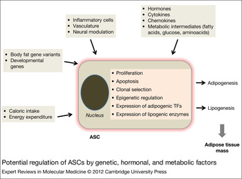
The regionally distinct ASCs characteristics could be epigenetically regulated, appearing during early development, to be set later by the particular microenvironment of a specific fat depot, or result from different populations of early progenitors, giving rise to ‘committed’ ASCs in distinct fat depots. In addition, sex, ageing, ASC replicative history and body mass index (BMI) might influence these properties through epigenetic, genomic or progenitor subpopulation selection mechanisms (Fig. 2). Since new fat cells continue to arise from ASCs in adulthood, regional differences in ASCs characteristics could contribute to differences in function among fat depots. Van Harmelen et al. (Ref. Reference Van Harmelen73) reported that subcutaneous ASCs proliferated at a greater rate than visceral cells. These authors also found that age correlated negatively with proliferation rates only in subcutaneous ASCs and not in visceral ASCs. This suggests that ageing may exert distinct effects on adipocyte precursors from different fat depots, providing a potential mechanism for the relative loss of subcutaneous fat that typically occurs with ageing (Ref. Reference Sepe76). The negative correlation between donor age and proliferation of subcutaneous but not visceral ASCs may potentially explain the greater capacity for adipose tissue growth in the visceral compared with the subcutaneous fat depot in aged individuals (Refs Reference Sepe76, Reference Schipper77, Reference Shahparaki, Grunder and Sorisky78). It should be noted, however, that there is no total agreement on the adipogenic differentiation capacity, since some studies showed apparently no significant differences in the in vitro differentiation capacity between subcutaneous and visceral ASCs (Refs Reference Van Harmelen73, Reference Shahparaki, Grunder and Sorisky78), whereas others provided evidence for greater adipocyte differentiation in cultures derived from subcutaneous compared with visceral ASCs (Refs Reference Baglioni74, Reference Montague79, Reference Adams80, Reference Tchoukalova81). Differences in the adipogenic media used and efficiency of adipogenesis obtained with various protocols could explain, at least in part, these discrepancies. Regulation of adipose tissue accumulation in various compartments in humans could also involve depot-specific differences in rates of ASC apoptosis, since apoptotic indices were found to be substantially greater in ASCs from the visceral compared with the subcutaneous depot (Refs Reference Tchkonia72, Reference Niesler, Siddel and Prins82). Since ASCs/preadipocytes appear to be distinct among different depots, it is conceivable that drugs could be developed to target specific fat cell populations: controlling the numbers of each of these ASCs and their depot-specific differentiation could potentially provide new treatments for obesity and its complications.
Potential regulation of adipose-derived stem cells (ASCs) by genetic, hormonal and metabolic factors. Several physiological and pathophysiological factors may regulate both adipogenesis and lipogenesis by affecting multiple cellular events in the ASC, including cell proliferation, apoptosis, clonal selection, epigenetic mechanisms and expression of adipogenic transcription factors (TFs) and lipogenic enzymes, ultimately leading to changes in fat mass.

ASCs in obesity
Obesity, especially central obesity, is a hereditable trait associated with a high risk for the development of diabetes and metabolic disorders, cardiovascular disease and cancer. It is quite evident that excessive adipose tissue may be detrimental through the adipocyte-mediated secretion of cytokines, which may exert relevant effects in the regulation of metabolic responses, leading to insulin resistance, beta-cell dysfunction and lipid abnormalities (Refs Reference Kern83, Reference Hotta84, Reference Friedman85), as well as of cell proliferation, potentially leading to tumourigenesis and/or cancer growth (Refs Reference Iyengar86, Reference D'Esposito87). However, not all obese patients have the same risk of developing these disorders. Individuals with peripheral obesity, i.e. fat distributed subcutaneously in the gluteo-femoral region, are at little or no risk of the common medical complications of obesity, whereas individuals with central obesity, i.e. fat accumulated in visceral depots, are prone to these complications. Although differentiation of adipocytes has been extensively characterised and there have been considerable recent insights into the control of appetite and energy expenditure as contributing factors to obesity, little is known about the genetic basis for determination of adipocyte number, differences in body fat distribution or their association with metabolic disorders. Twin and population studies have revealed that both BMI and waist/hip ratio (WHR) are heritable traits, with genetics accounting for 25–70% of the observed variability (Refs Reference Baker88, Reference Nelson89). In addition, it is known that some obese individuals, especially those with early-onset obesity, have increased adipocyte number, but how these are distributed and why this occurs is unknown. Potential differences between ASCs from normal and obese individuals could support the hypothesis that fat distribution and obesity itself may have a developmental genetic origin (Ref. Reference Gesta75). Depot-specific differences in the expression of genes involved in embryonic development and pattern specification have been reported to persist during in vitro culture and differentiation of mouse adipose precursors, as already recalled (Ref. Reference Gesta75). Specifically, the expression levels of HoxA5, Gpc4 and Tbx15 in human adipose tissue were found to be different between depots and to correlate significantly with BMI. No other biological parameter related to obesity or fat mass, including serum leptin, adiponectin or insulin, shows such a strong association, suggesting that it would be possible to define the overweight population based on the expression levels of these genes. For example, it has been suggested that HoxA5 expression levels in adipose tissue may represent a good marker for visceral fat expansion since HoxA5 expression is greater in visceral than in subcutaneous fat and correlates positively with BMI and WHR (Ref. Reference Gesta75). Furthermore, the expression of genes that regulate early development, including homeotic genes, differed extensively among undifferentiated preadipocytes isolated from different fat depots (Ref. Reference Tchkonia72) (e.g. HOXA4, A5, C8, C9, En1 and pregnancy associated plasma protein A are differentially expressed in abdominal subcutaneous and visceral ASCs). Thus, fat depot-dependent differences in developmental gene expression may contribute directly to regional variation in fat tissue mass and function, since these genes may affect the proliferation and differentiation capabilities of the ASCs. In addition, upstream mechanisms, such as histone acetylation or methylation patterns or other epigenetic processes, could cause or contribute to regional differences in both developmental gene expression and fat tissue function. It is to be expected that genetically programmed developmental differences in adipocytes and their precursors in different fat regions may play an important role in the functional differences between visceral and subcutaneous adipose tissues as well as in defining body fat excess and distribution.
Recently, by making use of the information based on the integration of 14C, derived from nuclear bomb tests, into genomic DNA, attempts to estimate the rates of adipocyte and lipid turnover in vivo in humans have been carried out. These studies have highlighted that approximately 10% of adipocytes are renewed annually at all adult ages and all levels of BMI (Ref. Reference Spalding90). In addition, obese individuals were found to have a significantly greater number of adipocytes added per year than lean individuals (Ref. Reference Spalding90). Thus, obesity appears to be characterised by an excess of adipocyte generation with preserved turnover of these cells once differentiated from the relevant precursors. In addition, human fat in obesity shows a slower triglyceride turnover resulting in higher triglyceride amounts stored annually (Ref. Reference Arner91). These findings suggest that an enhanced adipogenesis may occur in vivo in obese humans and that this could result from an enhanced ability of the ASCs from these individuals to undertake the adipogenic differentiation programme. Furthermore, they suggest that the mechanisms leading to excess fat accumulation in obesity may be multiple, involving changes in ‘adipogenesis’ (i.e. the number of newly formed adipocytes) as well as in ‘lipogenesis’ (i.e. the amount of triglyceride stored in adipose tissue).
Interestingly, obese subjects were found to have larger stromal and fat cell numbers when compared with lean subjects (Ref. Reference Van Harmelen73). However, in contrast to the expectation, the differentiation capacity of the ASCs was shown to decrease with increasing BMI. Indeed, BMI was found to correlate positively with the total number of adipocytes and stromal cells per body but negatively to the number of adipocytes and stromal cells per gram of adipose tissue and the capacity of preadipocytes to differentiate (Ref. Reference Van Harmelen73). In line with these findings, other studies have shown a reduced ability of ASCs from the subcutaneous fat depot of obese donors to differentiate in vitro into mature adipocytes (Refs Reference Permana92, Reference Tchoukalova, Koutsari and Jensen93, Reference Isakson94). It should be noted, however, that in these previous studies only ASCs from the subcutaneous adipose tissue have been investigated, specifically those collected from the SVF. As a consequence, investigation of the adipogenic and lipogenic potential of the ASCs from the visceral fat depot in humans as well as that of various subsets of ASCs, which were recently identified in addition to those in the SVF (see above, Refs Reference Miyazaki6, Reference Matsumoto7, Reference Perrini8), is very limited. Furthermore, information on adipogenesis and lipogenesis as related yet distinct endpoints is not available. The relationship between these in vitro cell phenotypes and the changes in developmental genes that occur in adipocytes and their precursors in a fat depot-dependent manner is also unexplored. Finally, different results may potentially emerge from evaluating the differentiation capabilities of the ASCs when examined in an in vivo setting, which is currently less feasible experimentally.
The effect of lifestyle modifications on the biological characteristics of ASCs in humans has also not been elucidated. Previous studies have shown that exercise training results in reduced white adipose tissue (WAT) mass and that the smaller fat mass contained fewer adipocytes; however, it is not clear whether exercise may affect the adipogenic/lipogenic potential of the ASCs. In a murine model, exercise training was shown to largely inhibit the ability of SVF cells to differentiate into adipocytes and that this response could be mediated via up-regulation of HIF-1α expression (Ref. Reference Sakurai95). Whether the exercise-mediated decrease in adipocytes in the WAT results from inhibition of ASC differentiation into mature adipocytes in humans remains to be established.
On the other hand, chronic overfeeding may affect the ASC population in different adipose tissue depots such that this could have sustained adverse health consequences. In a recent study, long-term high-fat intake was found to decrease the ASC number in BAT but not in WAT, indicating differential regulation of specific adipocyte progenitor populations in distinct adipose tissues in mice (Ref. Reference Xu96).
ASCs for cell-based therapy
Several observations suggest the therapeutic potential of ASCs, which has led to conducting of several clinical trials around the world (Ref. 97). In general, stem cells have been long looked at as possible therapeutic vehicles for different health related problems. As noted, ASCs have been gathering high attention in the last 10 years since they can be easily isolated although they provide simultaneously higher yields upon the processing of adipose tissue as compared with other stem cells. It was initially thought that the main potential of ASCs for regenerative medicine approaches was intimately related to their differentiation capabilities. Although this is true, there has been an increasing body of literature also describing the trophic effect of ASCs on the protection, survival and differentiation of a variety of endogenous cells/tissues. However, critical points remain to be addressed. As the stromal cells contained in adipose tissue represent a heterogeneous cell population containing several committed progenitors and their progenies, the evidence in favour of the occurrence of stem cells with a multi-lineage potential at the single cell level in vitro and in vivo remains to be demonstrated.
Cell therapies using ASCs are widely promising in various clinical fields based on in vitro and in vivo results. The most obvious prospective application of ASCs would be the prevention of metabolic diseases through modulation of adipogenesis and lipogenesis. Indeed, molecules that are expressed in a specific manner in differentiating ASCs and drive the maturation process of adipocytes may represent excellent drug targets for the prevention of obesity and associated diseases (Refs Reference Lee14, Reference Bacou51). The demonstration that adipose tissue derived cells have the capacity to participate in muscle regeneration (Ref. Reference Kim98) and to promote neovascularisation (Ref. Reference Miranville54) after transplantation into animal models highlights the concept that adipose tissue is a new source of stem cells that could be potentially directed towards alternative phenotypes. This concept includes the possibility of using ASCs to promote differentiation into brown adipocyte with associated metabolic improvements (Ref. Reference Tseng, Cypess and Kahn99).
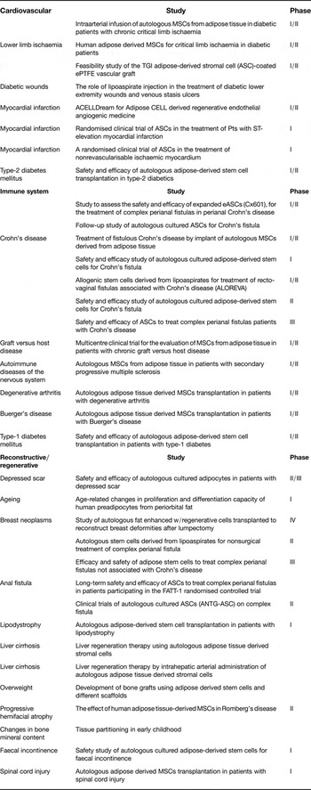
Adipose precursor cells may provide applications also in tissue reconstruction in humans. Purification of ASCs and enhanced reinjection could improve long-term survival of fat grafts by promoting adipogenesis and supporting blood vessel formation and growth, thus improving contour defects and wrinkles (Ref. Reference Kuhbier100). In addition, ASCs may have an important role in the treatment of HIV-associated lipodystrophy. Relevant to this concept, in an experimental study using a murine model, adipose transplantation was shown to revert the lipodystrophic condition (Ref. Reference Gavrilova101). Currently, several clinical trials for the regeneration of soft tissue, craniofacial tissue and cardiovascular tissue are being carried out (Table 2). In accordance with the potential of ASCs to differentiate into the mesenchymal lineage, ASCs could be potentially used to treat different aspects related to osteogenesis, chondrogenesis and myogenesis. Transplantation of human ASCs cells into mdx mouse, an animal model of Duchenne muscular dystrophy, resulted in substantial expression of human dystrophin on a long-term basis (Ref. Reference Liu102). ASCs have also been used to reossify large cranial defects (Ref. Reference Thesleff103). Taking into consideration the difficulties in stimulating cartilage regeneration in vivo, the ASC-base therapy shows promising future clinical applications in chondropathies (Ref. Reference Nathan104). Isolated ASCs express and secrete diverse haematopoietic cell cytokines, including macrophage colony-stimulating factor and granulocyte-macrophage colony-stimulating factor (Ref. Reference Gimble and Guilak9) and antiapoptotic and proangiogenic factors, including VEGF, hepatocyte growth factor (HGF) and TGF-β (Refs Reference Rehman55, Reference Cao105). Therefore, ASC-associated enhancement of angiogenesis is more likely the effect of secretion of angiogenic factors than the result of a true differentiation of ASCs into endothelial cells. The ASC-mediated proliferation of haematopoietic pluripotent stem cells was suggested in an animal study in which intraperitoneal injection of ASCs was able to reconstitute up to 40% of the bone marrow of lethally irradiated mice (Ref. Reference Cousin106). Therefore, ASCs appear to be an optimal tool for clinical applications because of their ability to act as a functional and vascular support for several tissues. Autologous adult stem cells are now considered for cell replacement therapy in diabetes as they have the potential to generate neo-islets which are genetically a part of the treated individual. Human ASCs are an ideal population of personal stem cells for cell replacement therapy, given that they are abundant, easily available and autologous in origin (Ref. Reference Chandra24). In addition, ASCs possess immunosuppressive properties in a dose-dependent and time-dependent manner and have the capacity to be used as immune-privileged universal donor cells (Refs Reference Shi107, Reference Tse108, Reference Puissant109). Encouraging evidence suggests that transplantation of ASCs can improve cardiac function of infarcted myocardium in rat models with a limitation of cardiac remodelling, improved wall thickness and increased neovascularisation with no cardiac side effects, such as electrical instability (Refs Reference Madonna, Geng and De Caterina110, Reference Hwangbo111, Reference Bai112). Currently, clinical trials are being carried out in patients with acute myocardial infarction (AMI) (Ref. Reference Sanz-Ruiz113). Freshly isolated ASCs are delivered through intracoronary infusion in patients with AMI and left ventricular ejection fraction impairment. For chronic myocardial ischaemia, a clinical trial is also being carried out in up to 36 patients (Ref. Reference Sanz-Ruiz113). The freshly isolated ASCs are delivered via transendocardial injections in patients with coronary artery disease. However, clinical trials of cardiac delivery of stem cells have yielded inconsistent results (Ref. Reference Bel114). A number of issues, including appropriate type and number of cells, timing and route of cell delivery and the detailed mechanism of action should be optimised for more consistent clinical results (Ref. Reference Danoviz115).
Ongoing clinical trials (Ref. 97) using human ASCs for the regeneration of soft tissue, craniofacial tissue and cardiovascular tissue

ASCs, adipose-derived stem cells; eASCs, allogenic adipose-derived stem cells; MSCs, mesenchymal stem cells.
Conclusions
Adipose tissue represents an interesting source of multipotent stem cells which represent a key aspect to be investigated for the understanding of adipose tissue biology under normal physiology and in disease states. In addition, these cells hold great promise for several clinical applications. The easily repeatable access to subcutaneous adipose tissue provides a clear advantage for the isolation of the ASCs. In addition, both isolation and culture techniques are easier to perform than BMSCs, making these cells readily available for cell therapy purposes.
In the past few years, there has been great progress in the understanding of the biology of ASCs and in defining ASC expansion, phenotypic characterisation, multipotency and different functional properties. Thus, evidence has emerged that there might be regionally distinct ASC characteristics, appearing during early development or being regulated at a later stage by the particular microenvironment of the specific fat depot potentially via epigenetic mechanisms. Alternatively or additionally, fat depot diversity may result from different subsets of ASCs, resulting in distinct committed adipocyte precursors within each fat depot. Evidence for changes in the functional features of ASCs in obesity and potentially other pathophysiological conditions is also starting to develop, even though limited information is available on visceral ASCs and the relationship between in vitro and in vivo adipogenesis. Nevertheless, ASCs are clearly at the interface between environmental and genetic factors involved in the pathogenesis of obesity and the development of metabolic and cardiovascular abnormalities (Ref. Reference Giorgino116). Thus, the possibility to modulate the differentiation potential and functional attitude of the ASCs represents a fascinating area of current research.
The potential use of ASCs for clinical applications is also of interest, although still in its early stages. It should be noted that standardisation regarding purification and characterisation of these cells is very limited. As previously noted, the SVF is a heterogeneous cell population and, even though only a minor proportion of these cells is able to adhere to the plastic surface of culture dishes, it cannot be excluded that a contamination of the ASCs with other cell types might occur during the isolation process. Hence, the identification of a specific pattern of cell surface markers appears to be critical to identify and isolate true ASCs, avoiding the use of impure cell populations. In addition, cell sorting analysis after isolation could potentially discriminate between markers of ASCs expressed in vivo and markers expressed after in vitro culture. A deeper understanding of ASC biology may thus represent a fundamental tool for the appropriate utilisation of ASCs in cell-based therapy applications.
Acknowledgements and funding
This work was supported in part by grants from the Ministero dell'Università e Ricerca (MIUR, Italy; MERIT Program), the COST Action BM0602, the Fondazione della Società Italiana di Diabetologia per la Ricerca in Diabetologia e Malattie Metaboliche (Fo.Ri.SID) and NovoNordisk to F. Giorgino.