Introduction
Species of the genus Entedon Dalman (Hymenoptera: Eulophidae: Entedoninae) are solitary or gregarious koinobiont endoparasitoids of eggs and larvae of various beetles. The most reliable host records concern weevils (Curculionidae: Graham, Reference Graham1971; Noyes, Reference Noyes2010) and bean beetles (Chrysomelidae, Bruchinae, mostly in Afrotropical region: Rasplus, Reference Rasplus1990). Other beetles (Anobiidae, Buprestidae, Cerambycidae, Mordellidae, Nitidulidae) are recorded as hosts, but these reports are not associated with biological essays. The non-coleopteran host records have not gained reliable proves so far and are regarded by some authors as doubtful (Bouček & Askew, Reference Bouček and Askew1968; Askew & Kopelke, Reference Askew and Kopelke1989).
The genus Erythrina (Fabaceae), or so called ‘coral trees’, comprise about 120 species which are spread throughout tropical and subtropical regions of the world (Mabberly, Reference Mabberly2008). Sixteen Erythrina species are critically endangered and placed on the World Conservation Union Red List of Threatened Species (IUCN, 2010). The coral trees are commonly used as ornamental trees in streets and city parks. They are also described as part of folk traditions and indigenous medicine (Graham et al., Reference Graham, Quinn, Fabricant and Farnsworth2000; List & Horhammer, Reference List and Horhammer1969–1979; Perry, Reference Perry1980; Rotar et al., Reference Rotar, Joy and Weissich1986; Yang et al., Reference Yang, Tung, La Salle and Wu2004).
The Hawaiian Islands (USA) house more than half of the world's Erythrina species at three botanical gardens. All are introduced, except for the wili wili, Erythrina sandwicensis O. Deg. Erythrina sandwicensis is the only endemic Erythrina species in dry land forests of the Hawaiian Islands, one of the most sensitive and threatened ecological systems on the planet due to the accidental introductions of invasive pests and to animal grazing (Medeiros et al., Reference Medeiros, von Allmen, Fukada, Samuelson and Lau2008). The wili wili is also a culturally significant tree and has been traditionally used for surfboards and canoes. The brightly colored orange-red seeds and flowers of the wili wili were used for botanical jewelry and leis by native Hawaiians (Medeiros et al., Reference Medeiros, von Allmen, Fukada, Samuelson and Lau2008).
The major damage to the coral trees in Hawaii is caused by two invasive species of African origin, the gall-inducing chalcidoid Quadrastichus erythrinae Kim (Hymenoptera: Eulophidae: Tetrastichinae), the so-called Erythrina Gall Wasp (EGW), and the bean beetle, Specularius impressithorax (Pic.) (Coleoptera: Chrysomelidae: Bruchinae). The EGW infestation has altered the reproductive capacity of wili wili populations, leaving merely one-third of the population reproductively active (Kim et al., Reference Kim, Delvare and La Salle2004; Yang et al., Reference Yang, Tung, La Salle and Wu2004; Heu et al., Reference Heu, Tsuda, Nagamine, Yalemar and Suh2006). The beetle arrived on the Hawaiian Islands in January 2001 and has caused the loss of over two-thirds of the annual wili wili seed crop (Medeiros et al., Reference Medeiros, von Allmen, Fukada, Samuelson and Lau2008). The pathway of infestation on Hawaii could be the imported decorative seeds from Africa. Seeds imported for ornamental purposes may also serve as the source of seed beetles. In the Netherlands, S. impressithorax sustained several generations indoors, after having been introduced from South Africa along with seeds of Erythrina used for decoration, but eventually was not established (Heetman & Beenen, Reference Heetman and Beenen2008). Adult beetles oviposit directly onto seeds, and the hatching larvae tunnel into them, rendering the seeds unviable (Medeiros et al., Reference Medeiros, von Allmen, Fukada, Samuelson and Lau2008). Specularius impressithorax is also reported as feeding on seeds of seven introduced Erythrina species including the native wili wili. Mean wili wili seed infestation during the first three years of introduction was 77.4% seed crop loss in six of the Hawaiian Islands (Samuelson & Medeiros, Reference Samuelson and Medeiros2006; Medeiros et al., Reference Medeiros, von Allmen, Fukada, Samuelson and Lau2008). The development of S. impressithorax in the seed may impair seed viability, depending on the size and type of seed. Infestation by a single larva can halt germination of 97.5% of wili wili seeds (Medeiros et al., Reference Medeiros, von Allmen, Fukada, Samuelson and Lau2008). The beetle also destroys stored Erythrina seeds and expensive botanical jewelry, if not stored properly (Ernst, Reference Ernst1993; Nápoles et al., Reference Nápoles, Kingsolver and Hernández2009). Specularius impressithorax also caused havoc in Erythrina seeds in Mexico in 2007, with 28.7% seed infestation of Erythrina coralloides D.C., a common native tree in Mexico, USA and Central America (Nápoles et al., Reference Nápoles, Kingsolver and Hernández2009). The EGW and seed damage from S. impressithorax is now presenting a further threat to the reproductive fitness of the wili wili populations. Efforts are underway to create a seed bank of various wili wili stands as a safeguard in case the trees die out completely (HEAR, 2010).
Preliminary surveys in Africa to discover new biological control agents of S. impressithorax revealed that a species of Entedon is a major mortality factor of the beetle's larvae. This species is probably the dominant parasitoid naturally occurring in infested seeds of several Erythrina species in tropical and subtropical Africa (M. Ramadan, personal observations). Before initiating plans for its introduction for a biological control program, recent surveys on Oahu and Maui Islands showed that the same parasitoid has been accidentally introduced there, apparently with its host in infested Erythrina seeds (Medeiros et al., Reference Medeiros, von Allmen, Fukada, Samuelson and Lau2008).
This study provides a description, detailed life history and observations on host-parasitoid relationships of this species of Entedon, which appeared to be new. We also discuss the potential of this parasitoid as a biological control agent against S. impresithorax in Hawaii and elsewhere where this beetle is considered a pest.
Materials and methods
Materials
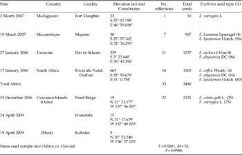
At least 5000 seeds of seven species of Erythrina were collected from various places in Hawaii and Africa and were examined for rates of infestation and parasitism (table 1). Seed samples were held for parasitoid emergence and afterward dissected to determine the fate of parasitoids, immature mortalities and numbers of parasitoids per host. Entedon specimens and other unidentified parasitoids of S. impressithorax were preserved as voucher specimens at the Hawaii Department of Agriculture (HDOA, Hawaii, USA). The beetles and parasitoids reared from the seeds collected in Hawaii were used to start a laboratory colony during April–May, 2009.
Erythrina seed samples from Africa and Hawaii examined for the presence of Specularius impressithorax and associated parasitoids.

The following acronyms of depositaries were used: BMNH (The Natural History Museum, London, UK), HBM (Bishop Museum, Honolulu, Hawaii, USA), HDOA (Hawaii Department of Agriculture, Honolulu, USA), MNHN (Muséum National d'Histoire Naturelle, Paris, France), SIZK (Schmalhausen Institute of Zoology, Kiev, Ukraine), ZSM (Zoologische Staatssammlung München), SANC (the South African National Collection of Insects, Plant Protection Research Institute, Pretoria, South Africa), UHM (University of Hawaii at Manoa, Honolulu, Hawaii, USA), USNM (United States National Museum of Natural History, Washington, DC, USA).
Laboratory studies
The development of the new species was observed in a laboratory culture of the host maintained on seeds of Erythrina variegata L., E. crista-galli L., E. sandwicensis O. Deg. and E. abyssinica Lam. ex DC (ZSM: 18.0±1.0°C at night and 25.0±2°C during the day, 50–70% RH and 14L:10D photoperiod; and HDOA: 22.0±1.0°C at night and 34.0±2°C during the day, 60–80% RH and 13L:11D photoperiod). The infested host plant seeds were collected from Oahu and Maui Islands. When adult beetles emerged from the collected seeds, the sound host-plant seeds were exposed to them for oviposition. The seeds bearing freshly laid eggs of beetles were then introduced to the females of the parasitoid. The potential fecundity (in mated females, 3–7 days old) and immature mortality were determined for the parasitoids emerged from S. impressithorax reared on E. crista-galli seeds in Hawaii. The parasitized hosts at various stages of development were dissected either immediately or 1–3 h after detected oviposition. The young parasitized host larvae were dissected in 1–2 day intervals. The older host larvae were dissected at week intervals because the development was found to be slower. Adult emergence was calculated by counting the number of emerged parasitoids and also the number of adults trapped inside the host pupation chamber.
DNA studies
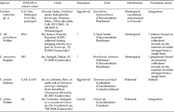
DNA extraction and sequencing were undertaken in the DNA-Tax Laboratory of ZSM. The holotype female of E. erythrinae sp. n. and four other species of Entedon, which share certain characteristics with the new species, were studied (table 2).
Species of Entedon used for molecular studies and attributes shared with Entedon erythrinae sp. n.

Genomic DNA was extracted from ethanol-preserved individuals using a protocol largely based on those described in the DNeasy Tissue Handbook provided by Qiagen (Hilden, Germany) but modified for the non-destructive DNA extraction. Each ethanol-preserved specimen was allowed to air dry briefly. Then the specimen was immersed into 180 μl ATL buffer (DNeasy Kit provided by Qiagen) in a 1.5 ml Eppendorf tube and frozen at –20°C for eight hours or overnight. The freezing is thought to improve the lysis by the formation of minute ice crystals, which damage intact cells to provide better penetration of extraction chemicals. Then, the mixture was thawed, 20 μl of proteinase K was added, and this digestive mix was incubated at 55°C for eight hours or overnight. Then, 20 μl of fresh ATL buffer were added, and the mix was incubated at 55°C for two hours. The liquid was carefully removed by a pipette into a clean 1.5 ml Eppendorf tube, and further DNA extraction followed the protocol described in the kit. The DNA extracts were stored in ZSM DNA bank under the codes listed in table 2. We amplified partial sequences of the nuclear 28S D2 rDNA, mitochondrial cytochrome oxidase subunit I and mitochondrial cytochrome B genes, for all studied samples, using the following primers: 28S D1 and D2 rDNA (nuclear), D1F (ACCCGCTGAATT TAAGCATAT) (Harry et al., Reference Harry, Solignac and Lachaise1996), D2R (TTGG TCCGTGTTTCAAGACGG) (Campbell et al., Reference Campbell, Steffen-Campbell and Werren1993); cytochrome oxidase 1 mtDNA (mitochondrial, COI), COI-Jerry (CAACATTTATTTTGATTTTTTGG), COI-2613 (ATTGCAAATACTGCACCTAT) (both: Simon et al., Reference Simon, Frati, Beckenbach, Crespi, Liu and Flook1994); cytochrome B mtDNA (mitochondrial, CytB), CB3 (GAGGAGCAACTGTAATTACTAA), CB4 (AAAAGAAA(AG)TATCATTCAGGTTGAAT) (Barraclough et al., Reference Barraclough, Hogan and Vogler1999). Standard polymerase chain reactions (PCR) were carried out in 25 μl reaction mixtures consisting of 0.25 μl Taq Polymerase, 2.5 μl PCR Buffer, 1.5 μl 25 mM MgCl2, 2.5 μl dNTP, 0.25 μl of each, forward and reverse primers, 16.75 μl water and 1.0 μl DNA extract. DNA fragments were sequenced in one direction (with forward primer). The obtained PCR products were cleaned using the QIAquick PCR Purification Kit. DNA fragments were sequenced in one direction (with forward primer) in the Sequencing Service of the Department Biology Genomics Service Unit (GSU, Ludwig-Maximilians-Univesität, Munich, Germany). The obtained sequences were manually corrected, if obvious mistakes in the reading of nucleotides were observed. Finally, the sequences were aligned using the ClustalW algorithm in BioEdit software version 5.0.0 (Hall, Reference Hall1999). Identities or similarities for two given aligned sequences were calculated using the corresponding option of BioEdit with gaps treated as characters if there was a residue in one sequence and a gap in the other. The sequences obtained in the same laboratory (ZSM) were compared.
Imaging
The dissections of parasitized host eggs and first-instars were made in the PBS buffer. Dissections conducted in water caused explosion and distortion of early parasitoid embryos. Some of the isolated embryos and larvae of the parasitoid were fixed in Boin fixative and 70% ethanol. The early embryos were stained best using methylene followed by eosin dyes and then partial de-coloration by formaldehyde. The stained embryos were kept in glycerol. Some early embryos and parasitoid eggs were studied without staining.
For the Scanning Electron Microscopy studies (ZSM), the ethanol preserved specimens were initially treated with proteinase K in ATL buffer (likewise in the non-destructive method of DNA extraction described above). Then, these specimens were washed out with distilled water and transferred to 70% ethanol for one day, then to 100% ethanol for one day and, finally, in 100% molecular sieved ethanol for maximal dehydration (also for one day). After dehydration, the specimens were critical point dried, transferred to SEM stubs, coated with gold and observed using a scanning electronic microscope.
Seed infestation and parasitism data analysis
Results of seed infestation and parasitism rates from seeds collected in Africa and Hawaii, in addition to immature mortality, adult emergence and sex ratios in field and laboratory reared seed samples, were analyzed using Student's t-test at P<0.05 (SAS Institute, 2001). Percentage data were arcsine-square root transformed before statistical analysis, but untransformed mean values are reported in the text and charts.
Results
DNA sequences
The fragments of the 28S D2, COI and CytB loci were successfully amplified for the holotype and male paratype of Entedon erythrinae and other studied herein species (tables 2 and 3). New sequences were deposited in GenBank (accession numbers JF262143–JF262156).
Similarity of the DNA sequences of obtained series of Entedon erythrinae sp. n. with four congeneric species (1.0 means 100% similarity).
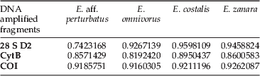
The 28S D2 sequences were of 421–424 bp long, the CytB sequences were of 343 bp long, the COI sequences were of 393 bp long. The similarity of the obtained sequences is shown in table 3.
Taxonomy
Entedon erythrinae Gumovsky & Ramadan sp. n
Type material
Holotype ♀ (voucher number GA1), Hawaii, Oahu, Erythrina seeds, Kaluakauila, lower site, Waianae Mtns, 100 m elevation, Coll. 051123001, 16.xii.2005 (L. Weisenberger) (BMNH); Paratypes: 6 ♀, 8 ♂, ibid. (BMNH, HBM); 9 ♀, 11 ♂, Hawaii, Oahu, Erythrina seeds [series γ] (USNM, HBM); 123 ♀; 75 ♂, Hawaii, laboratory culture started on S. impressithorax on Erythrina crista-galli seeds, v.2009 (M. Ramadan & A. Gumovsky) (HBM, ZSM, SIZK, BMNH); 9 ♀, 123 ♂, Hawaii, Maui, Ulupalakua, Kanaio, ex E. sandwicensis, vi.2008 (L. Kaufman, UHM); 7 ♀, 8 ♂, Hawaii, Oahu, Waimea Arboretum and Botanical Garden, Erythrina folkersii Krukoff & Moldenke, Kaluakauila, Coll. ‘Mexico’ 839528, coll. 05.iv.2009, r. vi.2009 (L. Kaufmann, UHM); 3 ♀, 1 ♂, RSA: ‘USA [Union of South Africa], San Francisco POE [Port of Entry], July 18, 1948, Parasite Specularius erythrinae Brd.’; 2 ♀, Union S. Afr., in mail, N.4, #89780, III-14-41, with Bruchid in Erythrina seeds (USNM).
Non-type material
75 ♂, 3 ♀, Tanzania, Dar es Salaam, ex Erythrina sacleuxii Hua, and Erythrina abyssinica Lam. ex DC., 27.i.2006 (M. Ramadan); 35 ♂, 13 ♀, Tanzania, Arusha, Masai village, ex E. abyssinica, 27.i.2007 (M. Ramadan); 65 ♂, 14 ♀, Mozambique, Maputo, 19.iii. 2007, ex Erythrina humeana Spreng., and Erythrina lysistemon Hutch. (M. Ramadan); 34 ♂, 56 ♀, Republic of South Africa, Kwazulu Natal, Durban, ex Erythrina caffra Thunb., E. abyssinica, E. lysistemon (M. Ramadan) (HDOA).
Diagnosis
Body robust, dark, with weak green or blue tint, more metallic green or bronze in males; female gaster short ovate, attached with a short elongate petiole to mesosoma, frontal sulcus missing, propodeum with median carina surrounded by channels, lateral propodeum with complete antero-lateral sulcus and smooth nearly flat submedian areas; lateral lobes of prepectus with thin processes oriented inwards; whole legs predominantly darkened, just knees and posterior one-quarter of hind tibia and one-fifth of fore and mid tibiae, first three tarsi of hind and mid legs, pale; axillular protrusion in shape of a callus with short irregular spines; costal cell bare; female flagellum with three-segmented funicle and two-segmented clava, male flagellum with five free segments (first segment elongate) separated by distinct peduncles; male gaster without a pale subbasal spot.
Female. Length 1.2–1.6 mm
Body dark with green or blue tint; legs predominantly dark, except for posterior tips of all tibiae and first three tarsi of hind and mid legs; wings transparent, venation pale brown; oval membranous areas dark, traced mainly by sculpture.
Head in dorsal view 2.2× as broad as long; ocelli moderate in size; POL≈3.7× as long as OOL. Occipital margin marked off by sharp carina, very weakly raised laterally. MDO:OOL:OCL in ratio 4:5:3. Dorsal surface of head evenly reticulate. Eye and occiput moderately pubescent.
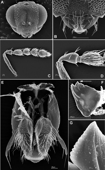
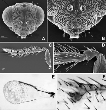
Head in frontal view 1.3× as broad as high (fig. 1a). Frons without frontal sulcus. Eye height slightly shorter than interocular distance. Eye merely pubescent, ≈3.0× as long as malar space. Distance between lower eye margin and antennal torulus about as long as diameter of the torulus. Face with raised border along eye margin and with row of short setae along this border. Surface between scrobal depressions not raised, without projection.
Female of Entedon erythrinae: (a) head in frontal view; (b) lower face; (c) antenna; (d) articulation of scape, pedicel and F1; (e) labio-maxillary complex; (f) mandible (view from inside); (g) indentation of inner tooth (enlarged portion arrowed on (f)).

Breadth of mouth opening ≈2.0× as wide as malar space (fig. 1b). Gena weakly convex, with light sculpture. Clypeus not delimited by sutures, its surface with reticulation somewhat finer than rest of face. Anterior margin of clypeus weakly produced and evenly rounded, with four protruding setae (fig. 1b). Mandibles bidentate, with elongate striation on outer tooth and anteriorly on inner tooth; cutting margin of the teeth with rather minute indentation (fig. 1f, g). Labial and maxillar palpi one-segmented, bearing two short setae, galea moderately pubescent (fig. 1e).
Antennal scape (fig. 1c) cylindrical, slightly more than 6.0× as long as broad in the middle, nearly (≈0.9×) as long as eye height; combined length of pedicel and flagellum ≈0.7× as long as breadth of head; pedicel nearly 2.0× as long as broad, ≈0.8× as long as F1 (fig. 1d); F1 nearly 2.0× as long as broad, F2 ≈1.2× as long as broad, length of F3 slightly exceeds its breadth; clava two-segmented, ≈2.0× as long as broad (fig. 1c).
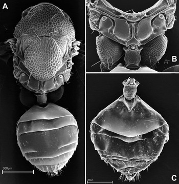
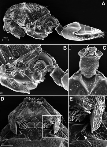
Mesosoma 1.6× as long as broad (fig. 2a). Pronotal collar not carinate (fig. 3a). Prosternum with paired subparallel carinae somewhat continued posteriorly, forming a Y-shaped pattern (fig. 3d), propleuron not bent posteriorly (fig. 3e, ppl). Lateral lobes of prepectus with thin processes oriented inwards (fig. 3e, arrow). Mesoscutum ≈2.0× as broad as long, length of scutellum slightly exceeds its own breadth and ≈1.3–1.4× as long as mesoscutum (fig. 2a). Axilla with one seta. Axillula present as a callus with irregular short spines (fig. 3b, axl). Dorsellum narrow, present as half-circular area bearing coriaceous sculpture (fig. 2b); each lateral panel of metanotum with one seta near the dorsellum (fig. 3b).
Female of Entedon erythrinae: (a) body in dorsal view; (b) propodeum and metasomal petiole in dorsal view; (c) metasoma in dorsal view.

Female of Entedon erythrinae: (a) body in lateral view; (b) propodeum and metasomal petiole in lareral view; (c) metasomal petiole enlarged; (d) anterior part of mesosoma. (e) Inset framed on (d) (a process on prepectus is arrowed); ppl, propleural margin.

Propodeum (figs 2b and 3b) with complete median carina continued to a wide nucha and surrounded at both sides by longitudinal channels bearing alutaceous sculpture. The channels connected with complete lateral propodeal sulcus, which spreading around spiracular elevation and further along relatively convex submedian areas of propodeum. The submedian areas are nearly smooth, with weak alutaceous sculpture posteriorly. Each spiracular elevation bearing short sharp projection beneath; supracoxal flange narrow. Propodeal callus with two large and 15–17 smaller setae.
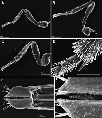
Spur of fore tibia (calcar) short and straight (fig. 4a), spur of mid tibia 1.8× as long as breadth of its tibia (fig. 4b), spur of hind tibia 0.8–0.9× as long as breadth of its tibia (fig. 4c, d).
Female of Entedon erythrinae, legs: (a) fore leg; (b) mid leg; (c) hind leg; (d) end of hind tibia. (e) Isolated ovipositor with ovipositorial sheaths; (f) ovipositorial saw enlarged, dorsal view.

Fore wing (fig. 5e, f) 2.1–2.2× as long as broad, costal cell bare, 5.6× as long as broad, subcosta of submarginal vein with two dorsal setae; marginal vein ≈0.6× as long as costal cell, postmarginal vein 1.5× as long as stigmal vein (fig. 5f); speculum open below; fringe of apical margin ≈0.3× as wide as breadth of parastigma in its widest part.
Male of Entedon erythrinae: (a) head in frontal view; (b) lower face; (c) antenna; (d) articulation of scape, pedicel and F1. (e, f) Fore wing of the female of E. erythrinae.

Metasoma. Petiole ≈1.3× as long as broad, with distinct collar and weak alutaceous sculpture dorsally (figs 2b, c and 3b, c). Gaster about as long as broad (fig. 2c). Ovipositorial sheaths with stout setae (fig. 4e), ovipositorial saw with short irregular crests (fig. 4f).
Male. Similar to female, but differs as follows: more bright metallic (generally bronze) body color, gaster without a pale subbasal spot. Male antenna (fig. 5c) with five separate segments (clava one-segmented); antennal scape moderately wide,≈2.7× as long as broad, with notable reticulation, pedicel 1.4× as long as broad (fig. 5d), F1–F5 separated by distinct peduncles, F1 3.3× as long as broad, nearly 2.0× as long as F2 and nearly 4.0× as long as pedicel, F2 1.8, F3 and F4 1.5, F5 (without long terminal spine) 2.0× as long as broad; combined length of pedicel plus flagellum equal to the breadth of head; head in dorsal view 2.4–2.5× as wide as broad, breadth of mouth opening 1.8–2.0× malar space (fig. 5b). Prosternum with distinct depressions in place of contact with propleura laterally and nearly bilobed anteriorly. Petiole 1.31–1.4× as long as broad, also with a collar as in female.
Host. A bruchine beetle Specularius impressithorax in pods of Erythrina spp.
Biology. Koinobiont, gregarious egg-larval parasitoid.
Distribution. Afrotropical region: Republic of South Africa, Tanzania, Mozambique, adventive to the Hawaiian Islands.
Etymology. The species epithet derives from the generic name of the host plant.
Comparative notes. The new species may be assigned to the perturbatus group sensu Gumovsky & Boyadzhiev (Reference Gumovsky and Boyadzhiev2003) due to the possession of the complete lateral propodeal sulcus. Entedon erythrinae is close to E. delvarei Rasplus, but differs in longer and free flagellar segments of male (two terminal flagellomeres are fused into a clava in males of E. delvarei). Females of these two species are difficult to separate, but pedicel somewhat longer than or as long as F1 in E. delvarei, whereas in E. erythrinae F1 is ≈1.2× as long as F2. They also differ in their known host plants: Erythrina spp. in E. erythrinae and Acacia senegal (L.) Willd. in E. delvarei.
Ecology
The host, Specularius impressithorax
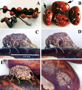
Specularius impressithorax damages wide spectrum of Erythrina plants (Kingsolver & Decele, Reference Kingsolver and Decele1979) and has also been associated with E. sandwicensis in Hawaii since its introduction in 2001 (Medeiros et al., Reference Medeiros, von Allmen, Fukada, Samuelson and Lau2008). The life history of S. impressithorax is typical for the subfamily Bruchinae and entirely associated with the Erythrina seeds. As was suggested by Medeiros et al. (Reference Medeiros, von Allmen, Fukada, Samuelson and Lau2008) and confirmed by our experiments, the females of the beetle lay eggs on a variety of seeds and beans or even on gelatin capsules filled with pinto bean flour in no-choice oviposition tests under laboratory conditions. However, the hatching larvae never complete their development beyond first instar in any non-Erythrina hosts. Female of S. impressithorax lays her eggs onto the surface of the host plant bean (fig. 6c–e). Field eggs were always collected from open pods where eggs are laid directly on the seed (fig. 6a, b). Eggs were never found on seed pods as the habit of other bruchine species. The female first makes circular movements by her partly erected telescopic ovipositor around the oviposition site directly on the seed integument. The movements last for ≥40 s. Afterwards, the beetle adjusts its fully erected ovipositor onto the seed's surface and pulsing movements of the ovipositor start. Eventually, the egg is ejected and glued onto the surface. The eggs are of a peculiar ovoid shape, with short forks on their caudal ends. Freshly laid eggs are pale, nearly transparent and, later, with notable fat granules inside. Mean (±SEM) duration of egg development was 5.1±0.1 days (n=7, HDOA).
Erythrina seeds damaged by Specularius impressithorax: (a) infested fruit of E. folkersii; (b) heavily damaged seeds of E. sandwicensis (black arrows point to exit holes of the beetle, white arrows point to exit holes of Entedon erythrinae; (c–e) female S. impressithorax laying eggs on a seed of E. crista-galli: (c, d) steps of oviposition; (e) female prepares a place before ovipositing; (f) female near a freshly laid egg.

At 8–9 days after oviposition (ZSM, laboratory conditions), the beetle first instar starts drilling into the seed integument and starts filling, subsequently, the egg shell with frass, so that the shell is getting whitish or yellowish. The entry hole made by first instar bruchine is discernible under the hatched egg shell as a circular pin hole, Ø 0.18±0.01 mm (n=7). The creamy egg shell is generally an indication of the larva's successful penetration into the host-plant seed.
The first instar larva burrows through the peeling into the cotyledons during the second week after oviposition and molts into the second instar. During this time, the larva drills into the mid part of the seed and, afterwards, generally does not move much, but rather eats the seed tissues around. We encountered five larval instars based on the number of larval skins with head capsules (Pfaffenberger, Reference Pfaffenberger1990). The final instars are found 3–4 weeks after the egg was laid. Mean duration of larval instars was 31.7±1.2 days (n=3, HDOA). Larval development is strongly dependent on food availability; and, in case of mutual feeding of some larvae in the same seed, the first emerged ones continue their development earlier then the step-followers, which occasionally die due to the drying of the seed. The mature beetle larva pupates in a chewed oval chamber, the walls of which are constructed by its glued characteristic frass. The mine leading to the pupation chamber is also filled with this frass. Pupal stage takes a mean of 8.5±0.9 days (n=4, HDOA) and the total lifespan (egg to adult duration) is 47.5±0.3 days (n=132, HDOA). One seed of Erythrina may support the development of 4–15 adults, depending on the seed size. Adults emerge by gnawing a rounded opening in the seed peeling (fig. 6a, b). The opening appears in advance as a convex circle (Ø 1.9±0.04 mm, n=8), prepared and pushed out afterwards by the emerging adult. The characteristic gun-shot circular or ovoid hole of S. impressithorax (fig. 6b) may be confused with other seed feeders. The anobiid cigarette beetle, Lasioderma serricorne (F.), may also be found in infested seeds but emerge through an unevenly edged hole of Ø 1.6–1.8 mm.
The parasitoid, Entedon erythrinae
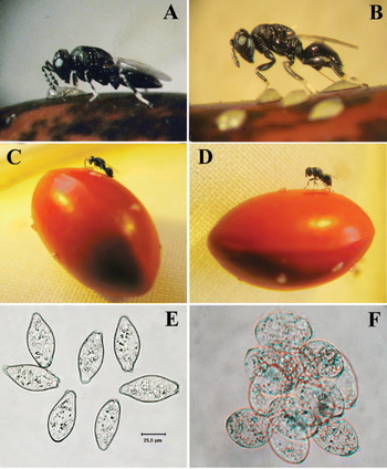
Ovipositing behaviour of the parasitoid (fig. 7a–d). Females of E. erythrinae are proovigenic: they emerge with a full capacity of mature eggs in their ovaries. The mature ovarian egg measures 61.0±0.7 μm long and 26.5±0.4 μm maximum wide (n=10) (fig. 7e). Mean number (±SEM) of ovarioles per ovary of mated female was 35.8±0.6 (n=5), and mean number of mature eggs in both ovaries was 712±48.8 eggs (n=25). Generally, no differences were found between the numbers of mature eggs in each ovary (t=1.434, df=12, P=0.177). However, on one occasion, a female had 331 eggs in one ovary and no mature eggs in the second ovary. Egg resorption also was observed in some freshly eclosed females, but 10-day-old females retained a full load of mature eggs with no sign of resorption. The maximum recorded number of mature eggs was 894 eggs in both ovaries.
Female Entedon erythrinae observing a freshly laid egg of Specularius impressithorax on Erythrina seeds (b, d). Female E. erythrinae ovipositing: (a, b) E. crista-galli; (c, d) E. abyssinica; (e) ovarian eggs of E. erythrinae; (f) eggs of E. erythrinae isolated from the egg of S. impressithorax.

The females of E. erythrinae start ovipositing only about the next day after emergence, generally after mating and feeding on honey. Mating lasts ≈16 s, immediately as the female emerges from the seed. The parasitoid female searches along the Erythrina bean's surface by drumming it with her antennal tips (fig. 7a, c). Once the female has located an egg, she starts more frequent drumming and walks back and forth around the position where the egg is located, continuing antennal drumming of the egg. After these examinations, she starts probing the egg with her ovipositor. She bends her gaster downwards, briefly hooks the ovipositor saw into the egg shell and then releases the gaster so that it strengthens in a position perpendicular to the ovipositor (fig. 7b, d). Shortly afterwards the ovipositor's needle appears within the egg yolk, the female carries out rhythmic, twisting movements of her gaster, similarly to other species of the genus (Gumovsky, Reference Gumovsky2007, Reference Gumovsky2008). Oviposition lasts 1–3 min or longer. Quite often, after oviposition or simply ovipositor probing, the host egg bears some air bubbles inside. These bubbles are likely artifacts of ovipositor withdrawal. All the beetle eggs containing bubbles dry afterwards, likewise unfertilized eggs. In the laboratory, the number of the host eggs damaged this way occasionally prevailed over the number of the successfully survived eggs (parasitized and unparasitized) altogether.
Parasitoid ecology and parasitoid-host relations
The parasitoid female lays a clutch of eggs (5–14) during a single oviposition episode (fig. 7f). The parasitoid eggs are free floating as a single mass within the host's egg yolk and are not discernible, unless the egg is dissected and the yolk and fat granules are rinsed. No obvious external marks of oviposition were shown on the beetle's egg chorion or on the hatching larvae.
The parasitoid egg clutches are passively encompassed into the developing weevil's embryos, but do not demonstrate any evident marks of early development. The hatched first instar of the host still contains the parasitoid eggs. However, the parasitoid first instars develop as peculiar embryonic structures, which we call further, the ‘soccer-ball chambers’.
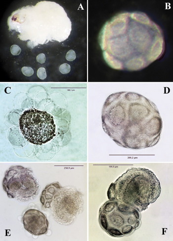
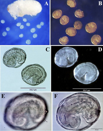
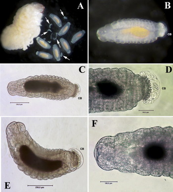
The first evident forms of the parasitoid development are the spherical embryos represented by masses of proliferating cells with larger cellular formations on the periphery (fig. 8). Smaller embryos (≈Ø 45.0 μm) are found mostly in the actively feeding host first instars at about the 10th day after the parasitoid's oviposition (fig. 8c, d). The larger embryos (≈Ø 115.0 μm) are found in the second instar host larva (fig. 8e, f). The fully-grown second instars and early third instars of the beetle contain the parasitoid embryos in the so-called ‘soccer ball’ chambers (figs 9 and 10). If just freshly molted, parasitized second instar of S. impressithorax is dissected, these ‘embryonic chambers’ emerge from the host's body and look like just small milky granules (Ø 43.0–50.0 μm) that may be confused with the granules of the host's fat body (fig. 8a). If the mature, parasitized second instar of the host is dissected, the embryos are larger (≈Ø 115.0 μm) and bear distinct penta- or hexagonal ‘soccer ball’ pattern on their surfaces (fig. 9b, d). The ‘soccer ball’ chambers change over time in size. They are rounded in shape and ≈Ø 200.0 μm in the beginning, when they contain undifferentiated cell masses or worm-like bent germs (fig. 10e, f). Then, each of the embryos becomes oval shaped, reaching up to 500.0 μm long when containing the fully formed larva, which is ready to emerge (fig. 11a–c). About 20 days after the parasitoid's oviposition, the ‘soccer ball’ chamber breaks and reveals the first active parasitoid larva (figs 11d and 12a). This generally happens inside the third host instar, but occasionally in second or fourth instars of the beetle, too. These larvae are usually concentrated near the host's guts, float free in the host's haemolymph and start feeding soon after vacating the chamber (figs 13 and 14). Young final instars (fig. 15) are found in the fourth and fifth host instars. They look much similar to the second instar, but differ in their larger size (growing from 1.3 mm in length after molt to 2.0 mm when mature), have seven pairs of spiracles and sharp, bent mandibles (fig. 15c, e).
Early development of Entedon erythrinae in early instars of Specularius impressithorax (‘germ stage’): (a) dissected freshly molted second instar of S. impressithorax with isolated embryos of E. erythrinae (enlarged on b, e, f); (c, d) early developing embryos (at about tenth day after oviposition, the first-instar host larva just emerged into the seed tissue), treated by Eozyne Dye; (e, f) mature embryos (shown as a group on (a, b) 14th day after oviposition), treated by Eozyne Dye.

Early development of Entedon erythrinae in early instars of Specularius impressithorax (‘soccer-ball stage’): (a) dissected mature second instar of S. impressithorax (≈4.0 mm deep burrowed in seed) with isolated ‘soccer ball’ embryos of E. erythrinae (enlarged on b, d): (b) reflected light, (d) transmissing light; (c) artificial view of the embryo, immersed in water: with numerous swellings of tiny internal membrane, caused by internal osmotic pressure and prevailing explosion of the embryo; (e) broken ‘soccer ball’ embryos revealing somewhat formed larval embryos. (f) One of the embryos shown in (e), enlarged.

Development of Entedon erythrinae in mid instars of Specularius impressithorax (‘soccer-ball stage’): (a) dissected mature second instar of S. impressithorax with isolated ‘soccer ball’ embryos of E. erythrinae (enlarged on b–f): (b, d) reflected light, (c–f) transmissing light, (c, d) same pair of embryos, (e, f) same embryo. (e) Demonstrates the granulate surface of the ‘soccer-ball chamber’; (f) demonstrates the nearly formed second-instar larva inside (the caudal bladder is not everted although).

The larvae of Entedon erythrinae leaving the ‘soccer ball’ chambers: (a) the dissected larva of Specularius impressithorax with the isolated parasitoid larvae inside or emerging from the ‘soccer ball’ chambers (enlarged in b); (c, e) the fully formed larva inside the ‘soccer ball’ chamber; (d, f) the fully formed larva just emerged from the ‘soccer ball’ chamber (its remnants are visible caudad). (c, d) View in reflected light; (e, f) viewed in transmitted light.

Larva of Entedon erythrinae emerged from the ‘soccer ball’ chamber: (a) larva leaving the ‘soccer ball’ chamber; (b) the dorsal view of the emerged larva encapsulated into the external transparent membrane (enlarged in fig. 13), with remnants of the ‘soccer ball’ chamber at its caudal end; (c) the ventral view of the larva.

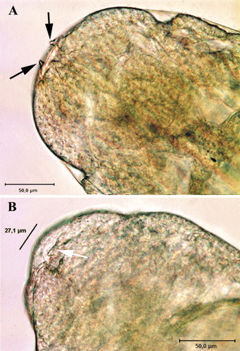
The head of the larva of Entedon erythrinae emerged from the ‘soccer ball’ chamber (a) dorsal view: the extra-embryonic membrane bearing remnants of the early instar mandibles is visible; the poorly sclerotized mandibles of the formed larva inside are visible behind); (b) ventral view: poorly sclerotized mandibles of the formed larva inside are visible.

The first active larva of Entedon erythrinae isolated from the mature second instar of Specularius impressithorax: (a) dissected host with larvae and remnants of the ‘soccer ball’ chambers (white arrows); (b, c, e) habitus of the parasitoid larva ((b) reflected light, (c, e) transmission light); (d) caudal end of the larva; (f) cranial end of the larva. CB, caudal bladder.

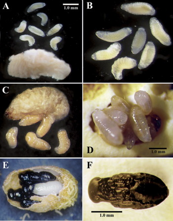
The morphology of the final instar larva of Entedon erythrinae isolated from the mature larva of Specularius impressithorax: (a) habitus; (b, d) cranial end; (c, e) head ventrally with visible mandibles (MD). SP, spiracle.

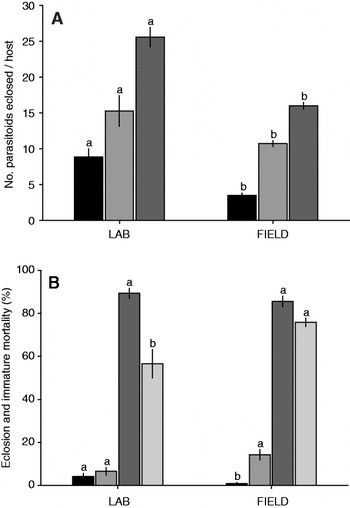
Both parasitized and unparasitized beetle larvae actively tunnel and feed on host-plant seeds and successfully construct pupation chambers. Subsequently, development of a parasitized beetle larva is arrested after creation of the pupation chamber; so, it never pupates but is consumed internally by the final parasitoid instars. The fully fed parasitoid larvae leave the host's remnants (fig. 16d), expel their meconia and transit into the prepupal stage, which lasts 3–6 days: 2.9±0.1 days, n=42 (HDOA) or 5.4±0.1 days, n=26 (ZSM). Then, they pupate inside the host's pupation chamber, but the pupation is asynchronous in siblings (fig. 16e). Adult parasitoids emerge at different intervals from two weeks to one month. Adults emerge from the chamber through a circular exit hole (Ø 0.63±0.02 mm, n=6) on the Erythrina seed (fig. 6b). This hole is ≈0.3× as large as the diameter of a circular hole made by the bruchine beetle and 3.0× larger than the entry hole made by the first instar bruchine. However, the parasitoid may also emerge through an adjacent beetle pupation chamber or through another parasitoid exit hole. The time lapsed from the parasitoid eclosion from the pupation chamber placed in a gelatin capsule and its flee to a rearing vial was 5.2±1.4 days (n=6). This suggests that the adult parasitoids may survive without honey and water for ≈5 days, using this time to gnaw an opening and get out of the seed. Some adult parasitoids do not manage to escape from the host-plant seeds due to the damage caused by the Grain Itch Mite, Pyemotes tritici (LaGrèze-Fossat & Montagné) (Acari: Pyemotidae) or simply because of overall weakness. Mean percentage of the parasitoid emergence from field collected samples (85.5±2.2%, n=165, HDOA) and from laboratory reared samples (89.3±3.8%, n=56, HDOA) were not significantly different (t=–0.168, df=219, P=0.8666, graph 1). Mating was observed outside the seed within 16 s post-eclosion. Adult parasitoids live for ≥10 days when provided with honey, but males normally die the next day after mating. The total average lifespan for the parasitoid is 59.4 days (5.1 days egg+31.7 days larva+2.9 days prepupa+19.7 days pupa, HDOA).
The final instar larvae of Entedon erythrinae (a–c) isolated or (d) emerged from the mature larva of Specularius impressithorax: (e, f) pupae of E. erythrinae: (e) parasitoid pupae in the host cell isolated from the seed of Erythrina variegata; (f) habitus of the pupa.

Entedon erythrinae sp. n. (a) progeny per host larva (▪, males;  , females;
 , total Entedon) and (b) adult emergence, sex ratio, and immature mortality from laboratory and field colonies (HDOA, Hawaii) (▪, larval mortality; , pupal mortality; , adult emergence;
 , sex ratio (%female)). Bars of the same parameter from laboratory and field grouping (mean±SEM) topped by the same letters are not significantly different (P>0.05; t-test).

Parasitism and sex ratio
The percentages of parasitism and immature mortality were not the same under laboratory and field conditions (graph 1). The proportion of males of E. erythrinae among the laboratory-reared offsprings (8.9±0.83 ♂ per host, n=23, HDOA) was significantly higher than of the field-reared male offspring (3.4±0.34 ♂ per host, n=134) (t=–5.989, df=156, P<0.0001: Oahu and Maui populations). The number of females emerged from the laboratory colony was significantly higher (15.3±1.2 ♀ per host, n=23) than the number of the females emerged from the field samples (10.7±0.5 ♀ per host, n=135) (t=–3.404, df=156, P=0.0008: Oahu, Maui populations). The total number of parasitoid's progeny produced per host larva was significantly higher in the laboratory than in the field collected samples: 25.6±1.0 parasitoid/host, n=56 (HDOA) vs. 15.9±0.6 parasitoid/host, n=167 (Oahu and Maui populations) (t=–8.736, df=221, P<0.0001). The females significantly prevailed in the field collected samples (75.9±2.0% ♀, n=16: Hawaii: Oahu, Maui) than in laboratory colonies (56.6±4.8% ♀, n=230 (HDOA), (t=3.741, df=157, P=0.0003). This may be attributed to the lack of adequate mating under laboratory conditions, as noticed when propagating several hymenopterous parasitoids under laboratory conditions (Kobayashi & Shimada, Reference Kobayashi and Shimada2000).
The percentage of larval mortality was significantly higher in the laboratory culture (4.2±1.0% dead larvae, n=56, HDOA) than in the field (0.85±0.6% dead larvae, n=165: Hawaii: Oahu, Maui) (t=–3.481, df=219, P=0.0006). This difference may be caused by the lower host quality due to higher occurrence of superparasitism under long exposure and biased parasitoid-host ratios under laboratory conditions. The mortality of the parasitoid pupae appeared higher in the field-collected seeds (14.2±2.1% dead pupae, n=165: Hawaii: Oahu, Maui) than in the seeds from the laboratory culture (6.5±3.6% dead pupae, n=56, HDOA), but differences were not significant (t=1.057, df=219, P=0.2917).
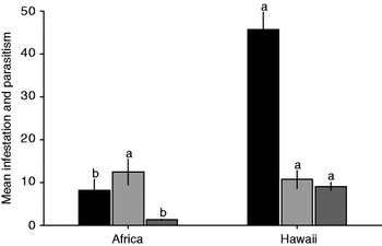
Field observations from the surveys conducted in Africa (table 1, graph 2) suggest much lower degree of infestation of Erythrina seeds by S. impressithorax and lower parasitism rate of E. erythrinae. For example, only five infested seeds and two seeds with parasitoids were present among one hundred seeds of Erythrina abyssinica sampled from the Durban Botanical Garden (Durban, Kwazulu Natal province, RSA, 07.i.2006). Also, just eight seeds infested by S. impressithorax and two with E. erythrinae were present among 100 seeds of Erythrina lysistemon collected at Elysium, South Coast of Durban (KZN province, RSA, 16.i.2006). Also, both, the beetle and the parasitoid, were present in low numbers in the collections of the seeds of Erythrina abyssinica in Tanzania (Arusha, Masai village, 27.i.2007) and the seeds of E. humeana and E. lysistemon in Mozambique (Maputo, 19.iii.2007).
Mean infestation and parasitism rates for the parasitoid-host system of Specularius impressithorax and Entedon erythrinae sp. n. (generalized materials from RSA, Tanzania and Hawaii). Bars of the same parameter from Africa and Hawaii (mean±SEM) topped by the same letters are not significantly different (P>0.05; t-test) (▪, %infested seeds; ,%parasitism; , Specularius eggs per seed).

Immature stages of parasitoid
Egg
The egg isolated from ovaries is mostly transparent, spindle-shaped, 61.0±0.7 μm long and 26.5±0.4 μm wide and lacks any discernible chorionic sculpture in reflected light (fig. 7e). The egg's fore end is somewhat narrowed, its hind end is blunt and bearing a micropyle. The latter is discernible as a small conical invagination (fig. 7e). The developing eggs (usually gathered in a clutch), isolated from the host egg and freshly hatched host first-instar, are more evenly rounded throughout, oval in shape, ≈50 μm long and 30 μm wide (fig. 7f). Apart from the shape, the developed eggs differ from ones isolated from ovaries in the possession of higher number of discernible cells (with distinct cell walls and nuclei) inside.
The ‘soccer ball’ chamber's embryo
The early development of the parasitoid eggs results in the formation of the embryos (fig. 8a, b), which are generally found within the host first instar. The individual embryo is of oval or spherical shape, Ø 43.0–50.0 μm (fig. 8c, d). It consists just of a mass of undifferentiated proliferating cells covered externally by a thin single-cell layer of larger cells (fig. 8d–f). The difference in shape becomes more obvious about 14–15 days after the parasitoid's oviposition, when the host molts into the second instar and the embryos gain a size of ≈Ø115 μm; the elongate peripherial cells are nearly 10.0× longer than the cells of the inner mass (fig. 8e, f).
While the second instar larva of the host grows to its third instar, the parasitoid's embryos also grow (fig. 9). The external cells become more rough and separate, as a membrane from the rest of the proliferating embryonic cell masses (fig. 9e), forming the characteristic ‘soccer ball’ pattern (fig. 9b, d). When the ‘soccer ball’ pattern is formed, the chamber does not change in size anymore. The first embryos on the ‘soccer ball’ stage are generally of Ø 200.0 μm (fig. 9d), although smaller or larger embryos also may be found. If the embryos are immersed in water, they appear in an artificial view, i.e. with numerous peripherial bladders (fig. 9c), which appear likely to be due to the pressure caused by the difference in internal and external concentrations. The bladders are formed by the evertions of another internal membrane, which retracts back when the embryo is placed into the PBS buffer.
The cell masses inside the ‘soccer-ball chamber’ are undifferentiated initially; however, when isolated from older host instars, these masses are organized into worm-like germs (fig. 9e, f). These germs have distinct outlines of a hymenopteriform larva later (fig. 10c–f). The internal organs are not differentiated at this stage; however, it is possible to recognize the tracheae, which are already filled with gas. The hind gut is also discernible as an invagination at the caudal end of the embryo (fig. 10f).
Free-floating instar
At about the third week after the parasitoid's oviposition, the ‘soccer ball chambers’ become more elongate and reach ≈500.0 μm (fig. 11a). The individual meshes of the ‘soccer ball’ pattern look more evidently like widened cells (with nuclei inside) at this stage (figs 11e and 12a). Each of these chambers contains a fully formed hymenopteriform larva. As soon as the larva is fully developed, the chamber breaks and reveals it (figs 11d, f and 12A). The newly emerged larvae (fig. 12b, c) are ≈600.0 μm long and 200.0 μm wide, and are initially enclosed into a thin transparent membrane, which bears a pair of minute projections resembling mandible tips at its cranial end (fig. 13a). This membrane breaks soon after the break of the ‘soccer-ball chamber’, releasing the larva. The larva is bearing poorly sclerotized triangular mandibles of the length ≈27.0 μm (fig. 13b), mid gut, the developed tracheal system (filled with gas bubbles), everted caudal bladder and a dense formation adjusted to it (figs 13c and 14). The bladder is bearing short filaments around its proximal part (fig. 14c, d) and contains numerous small, actively moving symbionts (likely bacteria judging from their minute size and peculiar motility). The peristalting membrane of the mid gut of the parasitoid larva may be observed in living preparations in the PBS buffer.
When the larva starts feeding, it approaches the size of 750.0 μm, its mid gut obtains a characteristic pale-yellowish color, likely due to the consumption of the host's fat bodies and haemolymph (fig. 14a, b). Occasionally, the remnants of the ‘soccer ball’ chambers may be found nearby the parasitoid larvae, when isolated from the host (fig. 14a, arrow).
Final instar (figs 15 and 16a–d)
The final instar (the follower of the instar emerging from the ‘soccer ball’ chamber) is easily distinguishable from the two previous larvae in having a developed tracheal system with one pair of spiracles on the 2nd thoracic segment and six pairs on the 6th to the 11th abdominal segments (fig. 15D). The spiracles are closed until the larva is mature and most host fluid is consumed. The length of the newly molted final instar is ≈1.3 mm, its width is ≈0.6 mm (fig. 15a). It has lightly sclerotized mandibles (fig. 15c, e) and relatively large spherical caudal bladder with traceable filaments around its proximal portion (fig. 15a). The mature final instar (fig. 16a–d) is ≈2.0–2.5 mm long and has no caudal bladder. The bladder breaks before the eclosion of the larva, but the remnants of its membrane are visible in the larvae, which are still inside the host remnants. The sensory structures of the head are not very distinct, but epistome, pleurostomal and maxillar palpi are recognizable.
Pupa (fig. 11e, f)
The pupa is black, with no metallic tint, obtect in shape, with distinct outlines of head, mesosoma, metasoma, wings, legs and antennae. The average length is 3.0 mm, the width of the head is ≈0.8 mm, of mesosoma ≈1.0 mm, of metasoma ≈1.3 mm.
Discussion
Erythrina production and biocontrol needs
The newly described Entedon erythrinae is a parasitoid of an invasive pest Specularius impressitorax specialized only to Erythrina seeds and damaging endemic Hawaiian wili wili trees. This beetle may happen to be of global concern in damaging non-African seeds of the New World Erythrina. Therefore, E. erythrinae is considered a potential biological control agent in Hawaii and possibly elsewhere where S. impressitorax is a pest.
As it was mentioned by other authors and supported here, S. impressithorax causes significantly less damage in its native Africa than in other newly invaded regions (Hawaiian Islands: 75–97%, Medeiros et al., Reference Medeiros, von Allmen, Fukada, Samuelson and Lau2008; California: up to 100%, Gulmahamad, Reference Gulmahamad2006; Mexico: 29%, Nápoles et al., Reference Nápoles, Kingsolver and Hernández2009), which may be explained by the suppression of the beetle populations by parasitoids in the native region (graph 2). In Africa, Erythrina seed infestation was just ≈0.18× the infestation rate in Hawaii. Entedon erythrinae was able to maintain the bruchine populations in the native region and average ratio of beetle eggs per seed was 0.14× the infestation rate in Hawaii. There is a short infestation period by the bruchine during the wili wili fruiting season in Hawaii (August–October), but other nearby Erythrina species may act as a reservoir for the bruchine and also for the parasitoid. The beetle may also reproduce on fallen seeds.
So far, E. erythrinae is the best prospective biological control agent for managing the Erythrina bruchine populations in Hawaii. It may prove to be an effective alternative to reduce the beetle populations in the field. Apparently, this parasitoid was accidently introduced to Hawaii with the pest but remained at low levels since 2003. The earliest evidence of parasitism came from an Erythrina variegata infested seed with a typical parasitoid exit hole and remnants of pupal exuviae in the empty host chamber. It was the only parasitized host (5.9% parasitism) out of 17 pupal chambers with normal bruchine exit holes (Oahu Island, Honolulu Civic Center, 9.viii.2003, J. Harada, coll.).
Entedon erythrinae seems to be the most dominant biological control agent in its native region of South and East Africa. It is persistent though in low numbers during low population levels in the field when most of the host bruchine and its favorable Erythrina seeds are rare. This is an important character of a successful biological control agent that synchronizes with its primary host at low levels of host populations. Host range of S. impressithorax and E. erythrinae are still unknown under Hawaii's conditions. It is not clear yet if the bruchine beetle has shifted to attack any new hosts in Hawaii. In literature, there are records of alternative Fabaceae host seeds based on laboratory oviposition studies, but there are no field records to confirm any new host plants beside mature seeds of Erythrina (Kingslover & Decele, 1979; Johnson & Romero, Reference Johnson and Romero2004; Medeiros et al., Reference Medeiros, von Allmen, Fukada, Samuelson and Lau2008). Besides, it is unknown if E. erythrinae can survive on other bruchine species infesting other Fabaceae seeds in Hawaii.
The wili wili seed production in Hawaii is currently in jeopardy (Medeiros et al., Reference Medeiros, von Allmen, Fukada, Samuelson and Lau2008). The Hawaiian Islands have had serious damage to wili wili trees by Quadrastichus erythrinae (EGW) in recent years. Actually, EGW has killed the vast majority of coral trees and wili wili in many areas. Recent market value of the native wili wili seed lei of 74 cm reached $US 500 because the wili wili seeds are harder to come by since the infestation of Quadrastichus erythrinae (EGW). However, in 2010 the new flush and abundant flowers on wili wili trees appeared for the first time after three years of devastating infestation by the EGW. It may be expected, that with care for the trees and the augmentation of the effective biological control agent of EGW (Gates & Delvare, Reference Gates and Delvare2008), these trees are coming back and starting to create new harvests of the wili wili flowers and seeds. Thus, the problem of seed infestations may resurface and new surveys will be conducted on the islands to ensure that the parasitoid is well established on all infested fields.
Entedon erythrinae has been recorded at new sites, Kamuela, Big Island (Hai On samples) in April 2009, and the wili wili seeds seemed less infested and free of bruchine infestation at many sites in 2010 (M. Ramadan, unreported data). However, the infested seed samples of several wili wili stands on three islands (Hawaii, Molokai and Lanai) are still lacking the Entedon parasitoids (M. Ramadan, unreported data 2010). Those areas can be targeted for inoculate release of the parasitoids. Augmentative release of the parasitoid can reduce the bruchine populations to low levels similar to what has been shown from the African Erythrina seed samples.
Natural enemies of S. impressithorax
Two other unidentified parasitoids (Hymenoptera: Braconidae and Eurytomidae) and a Pyemotes sp. mite were also found attacking the bruchine larvae in Africa, but E. erythrinae was the dominant species (M. Ramadan, personal observations). In addition to E. erythrinae, there are three local hymenopterous parasitoids reported to attack S. impressithorax on Oahu, Maui and Kauai Islands since 2003. These are Stenocorse bruchivora (Crawford) (Braconidae), Eupelmus cushmani (Crawford) (Eupelmidae) and Goniozus emigratus (Rohwer) (Bethylidae) (M. Ramadan, Mach Fukada, HDOA, personal communication). Medeiros et al. (Reference Medeiros, von Allmen, Fukada, Samuelson and Lau2008) also reported three unidentified larval parasitoids (Bethylidae and Eulophidae) from the samples collected on Maui and Molokai Islands. The pyemotes mite, Pyemotes tritici, is also an important biotic factor on Oahu Island, as it kills the bruchine and the parasitoid reducing rates of parasitoid emergence and increasing immature mortality as shown from field collections.
In 2004, S. impressithorax has been attacking Erythrina seeds in northern America, with records of four hymenoperous parasitoids (Gulmahamad, Reference Gulmahamad2006). One of the parasitoids associated with S. impressithorax in California is an eulophid new to North America, which appears to be the predominant species attacking the bruchine in south California. That could be the E. erythrinae, described above. The other wasps are unidentified larval parasitoids, a bethylid wasp, an eupelmid wasp (Brasema sp.) and an eurytomid (Eurytoma sp.). However, these parasitoids are generalists and apparently not adequate in reducing the beetle larval populations, unlike E. erythrinae which should enhance biological control of S. impressithorax on the Hawaiian Islands. The parasitoid is advancing on its own to new sites on all islands. A survey is in progress to quantify recent levels of seed infestation and parasitism.
Eulophid parasitoids are documented to attack important bruchine pests and, thus, may be considered prospective biocontrol agents. In Brazil, eulophid parasitoids are known to regulate the seed bruchine Ctenocolum crotonae (Fahraeus) in native leguminous plants. Infestation was recorded as low as 4.9% attributed to the eulophid larval parasitoid, Horismenus missouriensis Ashmead (Sari et al., Reference Sari, Ribeiro-Costa and Medeiros2002). A successful biological control program against Acanthoscelides obtectus (Say), a major bruchine pest of stored beans in Colombia, Latin America, was achieved by releasing the eulophid wasp Horismenus ashmeadii (Dalla Torre). The parasitoid was reported to reduce the bruchine infestation in the field (18% average parasitism) but failed to develop under storage conditions and was not considered as a postharvest control agent (Schmale et al., Reference Schmale, Wackers, Cardona and Dorn2002).
Parasitoid development
The data reported herein are the first report on the life history of the Entedon species, which is of African origin and associated with bruchine beetles. Previously, just host records were available for this group of Entedon (Rasplus, Reference Rasplus1990). Apart from nominal novelty, the preimaginal development of this species is rather peculiar and differs from the observations published on other species of the genus (Gumovsky, Reference Gumovsky2006, Reference Gumovsky2007, Reference Gumovsky2008). The biology of E. erythrinae is remarkable in that this is the first recorded egg-larval gregarious endoparasitoid in its genus. The other known gregarious parasitoids of Entedon (the cioni group) attack the larvae of their hosts, Cionus weevils (Curculionidae) (Gumovsky, Reference Gumovsky1997). The other known egg-larval species of genus are all solitary parasitoids (Gumovsky, Reference Gumovsky2007, Reference Gumovsky2008). Entedon erythrinae is proovigenic species. Its egg is ≈0.1× the size of the egg of E. thomsonianus Erdös (≈450 μm long: Gumovsky, Reference Gumovsky2007), ≈0.2–0.3× the size of the egg of E. sparetus Walker (≈250 μm long: Gumovsky, Reference Gumovsky2007), ≈0.2–0.3× the size of the egg of E. sylvestris Szelényi (260–300 μm long: Gumovsky, Reference Gumovsky2006), ≈0.2× the size of the eggs of E. costalis Dalman (≈300 μm long: Gumovsky, Reference Gumovsky2008) and of the species of the cioni-group (≈310 μm long: Gumovsky, unpublished data). The small size of the eggs of E. erythrinae is likely associated with the relatively small size of the eggs of the host beetle (if compared with the eggs of most weevils) and the number of the eggs laid by the parasitoid female. Further studies are needed to find out whether these peculiarities of E. erythrinae are shared with other congeneric species.
The early development of E. erythrinae differs from all other species of the genus and is peculiar among known life histories of chalcidoid wasps. Unlike other non-polyembryonic chalcidoids, the early development of the egg is not associated with organogenesis, but simply results in multiplication of proliferating cells. Instead, the cells differentiate to the external layer (formed by the larger elongate cells) and the inner cell mass (formed by the smaller relatively rounded cells). The external layer gives start to an extraembryonic membrane (‘soccer ball’ chamber), while the internal cell aggregation develops further into the larva. This larva shares all the morphological characters with the second instar of other Entedon species, for which immature stages are described. The second-instar larvae of Entedon species (Gumovsky, Reference Gumovsky2006, Reference Gumovsky2007, Reference Gumovsky2008) are distinguishable from the first instars in having:
1. the rounded head with poorly visible labrum and sensoria (‘beak’-shaped, with protruding labrum and clearly outlined sensoria in the first instar);
2. the smooth body segments (dorsal posterior margins of IV–XIII segments bear fine but distinct indentation and the terminal sclerite is bearing a peculiar ‘caudal crown’ consisting of elongate denticles in the first instar); and
3. the poorly sclerotized subtriangular mandibles (sharp elongate and sickle-shaped and distinctly sclerotized in the first instar).
Also, likewise other second-instar larvae of the genus, the young larva of E. erythrinae has tracheae filled with gas bubbles, despite no spiracular openings being present. This character is also shared with other second-instar larvae of Entedon, although mechanism of the gas diffusion is not clear. One can assume that oxygen transits from the host's haemolymph to the parasitoid's haemolymph through the parasitoid's integument (cutaneous respiration) and then condenses in the closed tracheae of the parasitoid.
Another character which may support the statement that the young larva of E. erythrinae is morphologically the second instar is the possession of the small peak-shaped mandibles on the membrane covering the fully-formed young parasitoid larva before its eclosion (fig. 13a). We assume that these projections may happen to be the vestigial mandibles of the first instar (the only morphological evidence of this undifferentiated morphologically immature stage).
So, we conclude that the peculiarity of the onthogenesis of E. erythrinae is that the true first instar is embryonized and substituted by multicellular embryo covered by an extraembryonic membrane. The first active parasitoid instar corresponds morphologically to the second instar of other Entedon species. This embryonization of the first instar is unique and has not been recorded for Chalcidoidea so far. Such loss of the first instar may be an adaptation preventing siblicide among gregarious offsprings, since the first instars are responsible for the elimination of rival siblings in solitary parasitoids of the genus. Further studies on gregarious parasitoids of Entedon and related genera are needed to verify the presumption about such embryological feature.
The extraembryonic (‘soccer ball’) membrane described here is unique among Eulophidae. A similar extraembryonic membrane was reported by Pedata et al. (Reference Pedata, Garonna, Zabatta, Zeppa, Romani and Isidoro2003) for Encarsia berlesei Howard (Hymenoptera: Aphelinidae). However, the extraembryonic membrane of E. berlesei dissociates into teratocytes, whereas the membrane of E. erythrinae remains solid, its cells are toughly aggregated and do not undergo any further transformations.
Jackson (Reference Jackson1964) and Gumovsky (Reference Gumovsky2008) mentioned the embryonic cuticle between the developing first-instar embryo and egg chorion for the developing larvae of entedonines Mestocharis bimacularis (Dalman) and Entedon costalis Dalman, correspondingly. This membrane is probably homological to the thin membrane of the embryo of E. erythrinae, which everts from inside (in appearance of numerous bladders; fig. 9c), when the ‘soccer ball’ chamber is put in the water (not into a PBS buffer). Later, the membrane likely adjusts to the ‘soccer ball’ extraembryonic membrane; and the newly formed larva of E. erythrinae is also enclosed into another membrane (fig. 13a), which is a probably rudimentary integument of the first instar. So, in total we can count three extraembryonic membranes in E. erythrinae: (i) the ‘soccer ball’ membrane (fig. 9d–f); (ii) the thin everting membrane (fig. 9c); and (iii) the membrane bearing two processes on head end (probable rudimentary integument of the first instar; fig. 13a).
DNA sequences
The obtained sequences of nuclear and mitochondrial genes of E. erythrinae proved to be sufficient enough to distinguish E. erythrinae from those known species where a molecular characterization has been undertaken (table 3). It is remarkable that E. erythrinae did not demonstrate high similarity with its congeneric Afrotropical bruchine-associates (namely with E. omnivorus and E. aff. perturbatus, which may also be assigned to the perturbatus group sensu Gumovsky & Boyadzhiev (Reference Gumovsky and Boyadzhiev2003) based on their morphology). Otherwise, the DNA sequences of E. erythrinae proved to be more similar to the Palearctic E. costalis and E. zanara, the curculionid associates. The biological characteristics shared by E. erythrinae with these species are egg-larval parasitism (shared with E. costalis) and gregarious parasitism (shared with E. zanara). This illustrates the compound pattern of life history traits in the genus Entedon and emphasizes the necessity of studies on phylogeny and evolution in this and allied genera.
Acknowledgements
The studies, results of which are reported in this paper, were fully supported by AVG's research grant of the Alexander von Humboldt Foundation (Germany) and partly by the grant of the State Fund of Fundamental Research, Ukraine (SFFR, F35/002). AVG thanks Stephan Schmidt, Eva Lodde and Isabella Stöger for their help during his work in ZSM and also is grateful to John Noyes, Alfried Vogler (The Natural History Museum, London) and Chao-Dong Zhu (Institute of Zoology, Chinese Academy of Sciences, Beijing) for their advices on non-destructive DNA-extraction. The authors express their thanks to Jeffrey Harvey (Netherlands Institute of Ecology, NIOO-KNAW, Heteren) for the reading the manuscript and to Paul Krushelnicky, Stephanie Jo, Leyla Kaufman (University of Hawaii at Manoa, Honolulu, USA), Bernarr Kumashiro, Cynthia King, Juliana Yalemar (Hawaii Department of Agriculture, Honolulu, USA) and other colleagues, who submitted the materials mentioned in this paper.