Introduction
Benign paroxysmal positional vertigo (BPPV) is a disorder characterised by recurrent, brief episodes of vertigo and distinctive nystagmus, triggered by changes in head position relative to gravity, with an annual incidence of approximately 1.6 per cent.Reference Bhattacharyya, Gubbels, Schwartz, Edlow, El-Kashlan and Fife1 Posterior canal BPPV (PC-BPPV) is the most common type, accounting for approximately 80–90 per cent of cases.Reference Power, Murray and Szmulewicz2 With advances in vestibular function testing, the diagnosis and treatment of PC-BPPV have become increasingly refined, with current clinical treatment success rates as high as 90 to 96 per cent.Reference Li, Li, Zhang, Yang and Tan3 While otolith repositioning therapy can quickly relieve vertigo symptoms, a subset of patients continues to experience residual dizziness (RD), including sensations of instability, lightheadedness, floating and non-rotational dizziness after treatment. The incidence of RD has been reported to be as high as 61 per cent.Reference Seok, Lee, Yoo and Lee4
The factors associated with the occurrence and development of BPPV have been a key area of research. According to published studies, these influencing factors can be categorised into four groups: (1) factors that trigger BPPV, such as vitamin D deficiency, traumatic brain injury and aging; (2) factors contributing to BPPV recurrence, such as otolith dysfunction, orthostatic hypotension and improper sleeping posture; (3) factors that exacerbate BPPV symptoms; and (4) medications that alleviate its symptoms. The most extensively studied are the factors that trigger BPPV and those that may lead to its recurrence.Reference Hu, Lu, Wang, Quan, Ren and Rong5 Additionally, several studies suggest that RD may be associated with factors such as age, duration of vertigo prior to repositioning, anxiety disorders, sympathetic nervous system dysfunction and utricular dysfunction. Whereas, current studies have not fully replicated previous findings, indicating that further research is needed to clarify these influencing factors.Reference Ke, Ma, Jing, Diao and Yu6
According to the previous report, many patients with BPPV experience sleep disorders, which can affect treatment outcomes to some extent.Reference Zhu and Peng7 Sleep disorders may lead to ischemia, hypoxia and recurrent hypoxemia. Prolonged chronic hypoxia can cause damage and aging of the otolithic organs, resulting in the detachment of otoconia, which can trigger the onset of BPPV.Reference Shu, Liao, Fang, Shi, Yan and Hu8 Yet, there is limited research on the impact of sleep quality on vertigo-related symptoms before and after repositioning in PC-BPPV, particularly in patients with cupulolithiasis. Further studies on the effectiveness of otolith repositioning in this patient population are needed.
Hence, this study aims to observe the characteristics of nystagmus during the Dix-Hallpike test in patients with PC-BPPV with varying sleep quality and explore the impact of sleep quality on the effectiveness of repositioning in patients with PC-BPPV. Additionally, the changes in the scores of different dimensions of the Dizziness Handicap Inventory (DHI) before and after successful repositioning in both patient groups and the correlation between sleep quality and the occurrence of residual dizziness after repositioning were also investigated.
Materials and methods
This study was a prospective cohort study, which planned to enroll patients diagnosed with posterior canal BPPV between February 2020 and February 2023.
Grouping and diagnosis
The Pittsburgh Sleep Quality Index (PSQI) was used to assess the patients’ sleep quality. Based on the PSQI scores, patients with a PSQI score greater than or equal to 7 were classified into the low sleep quality group, while those with a PSQI score less than 7 were classified into the high sleep quality group.Reference Yuanyuan, Jing, Yan and Huifang9 The diagnoses of PC-BPPV-canalolithiasis (PC-BPPV-ca) and PC-BPPV-cupulolithiasis (PC-BPPV-cu) were according to the latest diagnostic criteria for BPPV established by the International Bárány Society in 2015. Diagnosis was based on the patient’s typical nystagmus and clinical manifestations.Reference Yuanyuan, Jing, Yan and Huifang9, Reference von Brevern, Bertholon, Brandt, Fife, Imai and Nuti10 Patients who met the diagnostic criteria were included in the study. The exclusion criteria were as follows: (1) age greater than 80 years; (2) presence of severe central nervous system or other systemic organ diseases; (3) severe cardiac, hepatic or renal diseases.
Clinical assessment indicators
All patients were able to cooperate with the required data collection, laboratory tests and questionnaire surveys. Clinical baseline data were collected from both groups, including age, gender, affected side, disease duration, proportion of atherosclerosis, rates of smoking, alcohol consumption, osteoporosis, peripheral vestibular and otological diseases and immune dysfunction. Based on the patient’s cerebrovascular ultrasound and vascular imaging results, the presence or absence of intra-cranial arterial atherosclerosis could be determined. Immune function abnormalities were identified through relevant laboratory tests and medical history associated with autoimmune diseases.
The nystagmus characteristics during the Dix-Hallpike test and reverse nystagmus upon sitting up and the efficacy of the repositioning treatment at the initial treatment, and after one week and after one month were compared between the two sleep quality groups. Changes in DHI, Hamilton Depression Rating Scale (HAMD-24) and Hamilton Anxiety Rating Scale (HAMA-14) scores before and after repositioning were also assessed. Additionally, a correlation analysis was conducted to examine factors influencing the efficacy of otolith repositioning.
All patients underwent simulated positional testing using the G-Force BPPV diagnostic and therapeutic device (Product model: GYZ-ZLY-I; China Medical [TianJin] Group, China), and the VisualEyes 525 system was used to video record nystagmus in each position. Meanwhile, simulated Epley and Semont repositioning techniques were administered for treatment. Please refer to our previous study for detailed operation methods.Reference Wang, Yan, Zhang, Han, Li and Liu11
Residual symptoms after otolith repositioning were defined as persistent or intermittent sensations of dizziness, unsteadiness or floating, without visual spinning or nystagmus.Reference Shuai, Fu, Lei and Hui12–Reference Martellucci, Pagliuca, de Vincentiis, Greco, De and Nobili Benedetti14 The duration of residual symptoms was measured from the time of the last repositioning treatment.
Efficacy evaluation
Following repositioning, a positional test was conducted to reassess the results. The efficacy was evaluated based on the BPPV treatment outcome criteria published by the Chinese Medical Association in 2017:15 (1) cured, positional vertigo disappears; (2) improved, positional vertigo and/or positional nystagmus were reduced but not fully resolved; (3) ineffective, positional vertigo and/or nystagmus showed no improvement or even worsened. The initial repositioning outcomes were compared between the two groups. For patients whose initial repositioning was classified as “improved” or “ineffective,” multiple repositioning sessions were performed. The number of sessions required to achieve complete resolution of nystagmus or its reduction (no further improvement in nystagmus after additional repositioning) was compared between the two groups.Reference Liu, Zheng, Dong, Xiang, He and Wen16 Follow-up was conducted every other day, with repositioning efficacy assessed one week and one month after the initial treatment. Additionally, changes in the scores of different dimensions of the DHI before and one week after repositioning were compared.
Statistical analysis
SPSS 25.0 software was used for statistical analysis. Categorical data were expressed as frequency (per cent), and continuous data were presented as mean ± standard deviation (SD) for normally distributed variables, or as median (interquartile range [IQR]) for non-normally distributed variables. Group comparisons of categorical data were performed using the chi-squared test, with Yates’ continuity correction or Fisher’s exact test applied when necessary. For continuous data, the independent t-test was used for comparisons between groups if the data were normally distributed; otherwise, the Mann–Whitney U test (a non-parametric test) was employed. A significance level of α = 0.05 was set for hypothesis testing, and for pairwise comparisons, the significance level was adjusted to α′ = 0.017.
Results
Clinical characteristics
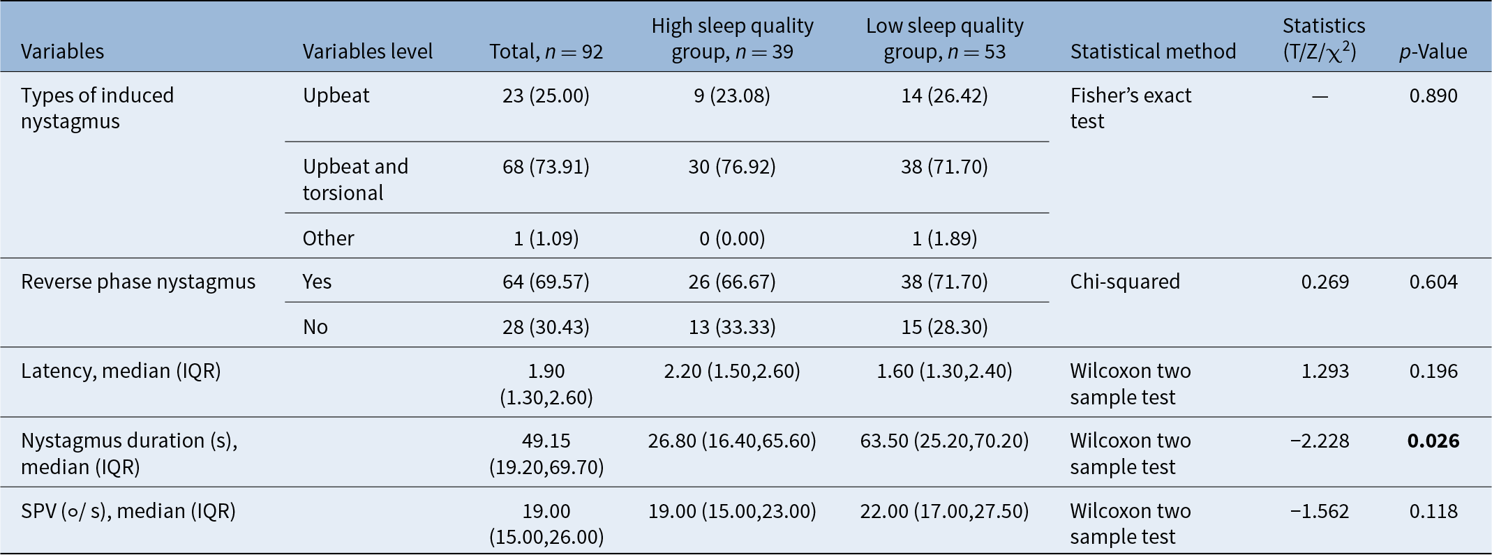
A total of 92 patients with PC-BPPV were included in the study, with 53 in the low sleep quality group and 39 in the high sleep quality group. The two groups were comparable in terms of age, gender, affected side, disease duration, proportion of atherosclerosis, smoking and alcohol consumption rates, osteoporosis rates, prevalence of peripheral vestibular and otological diseases, migraines or immune dysfunction. However, the proportion of patients with PC-BPPV-cu was significantly higher in the low sleep quality group compared to the high sleep quality group (60.38 per cent vs. 35.90 per cent; p = 0.020), as shown in Table 1. Regarding nystagmus characteristics, the median duration of upbeat nystagmus during the Dix-Hallpike test was significantly longer in the low sleep quality group (63.50 seconds vs. 26.80 seconds; p = 0.026). Other indicators were similar between the two groups, as displayed in Table 2.
General conditions of patients in both groups

Characteristics of Dix-Hallpike test in both groups

Repositioning efficacy
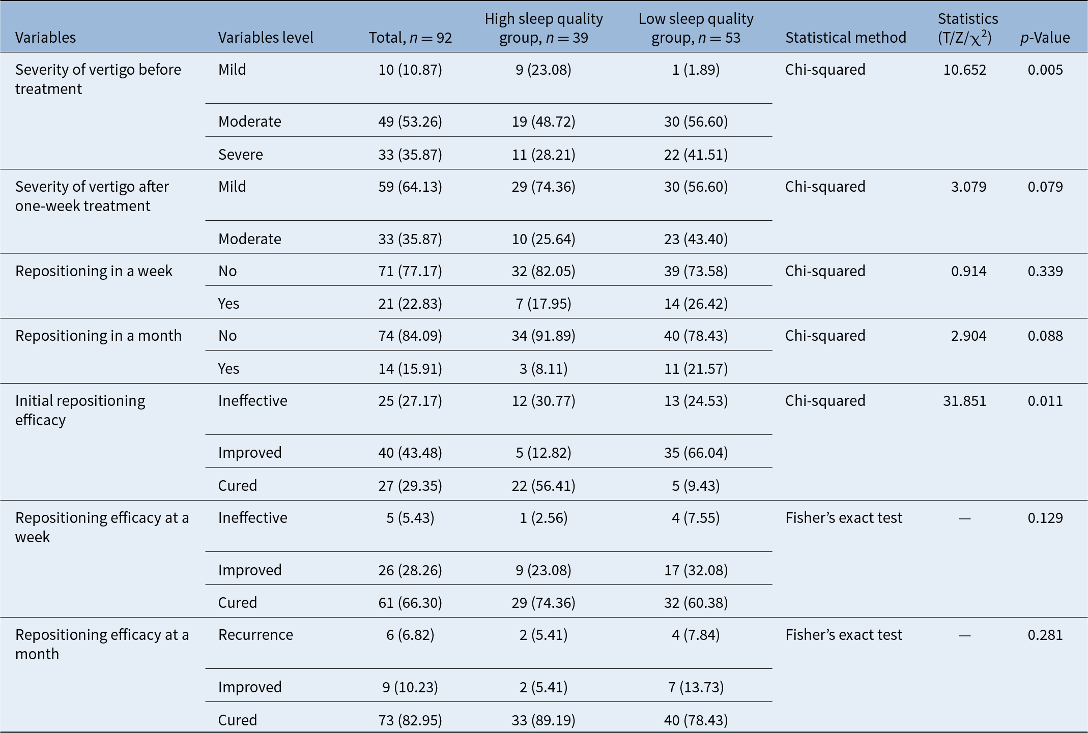
Before treatment, the severity of vertigo was significantly higher in the low sleep quality group compared to the high sleep quality group (p = 0.005), with the proportion of mild, moderate and severe being 23.08, 48.72 and 28.21 per cent in the high sleep quality group versus 1.89, 56.60 and 41.51 per cent in the low sleep quality group, respectively. After one week of repositioning treatment, the severity of vertigo between the two groups became comparable (p = 0.079), vertigo severity was predominantly mild and moderate, accounting for 74.36 per cent and 24.64 per cent in high sleep quality group and 56.60 per cent and 43.40 per cent in low sleep quality group accordingly, as demonstrated in Table 3. The initial short-term efficacy of repositioning was poorer in the low sleep quality group, with a higher recurrence rate (p = 0.011). In the high sleep quality group, the proportions of ineffective, effective and cured cases were 30.77, 12.82 and 56.41 per cent, respectively. In contrast, the group with low sleep quality had proportions of 24.53, 66.04 and 9.43 per cent, respectively, as illustrated in Figure 1. One week and one month after repositioning, there were no statistically significant differences in overall efficacy between the two groups (both p > 0.05), as shown in Table 3. After adjusting for confounding factors related to the different proportions of otolith types in the two groups, the success rate of the initial repositioning was significantly lower in the low sleep quality group (odds ratio [OR] = 0.372; 95 per cent confidence interval [CI]: 0.163–0.848; p < 0.05).
Results of repositioning treatment in both groups

Comparison of efficacy of first maneuvers in both groups.

Comparison of vertigo disability before and after repositioning
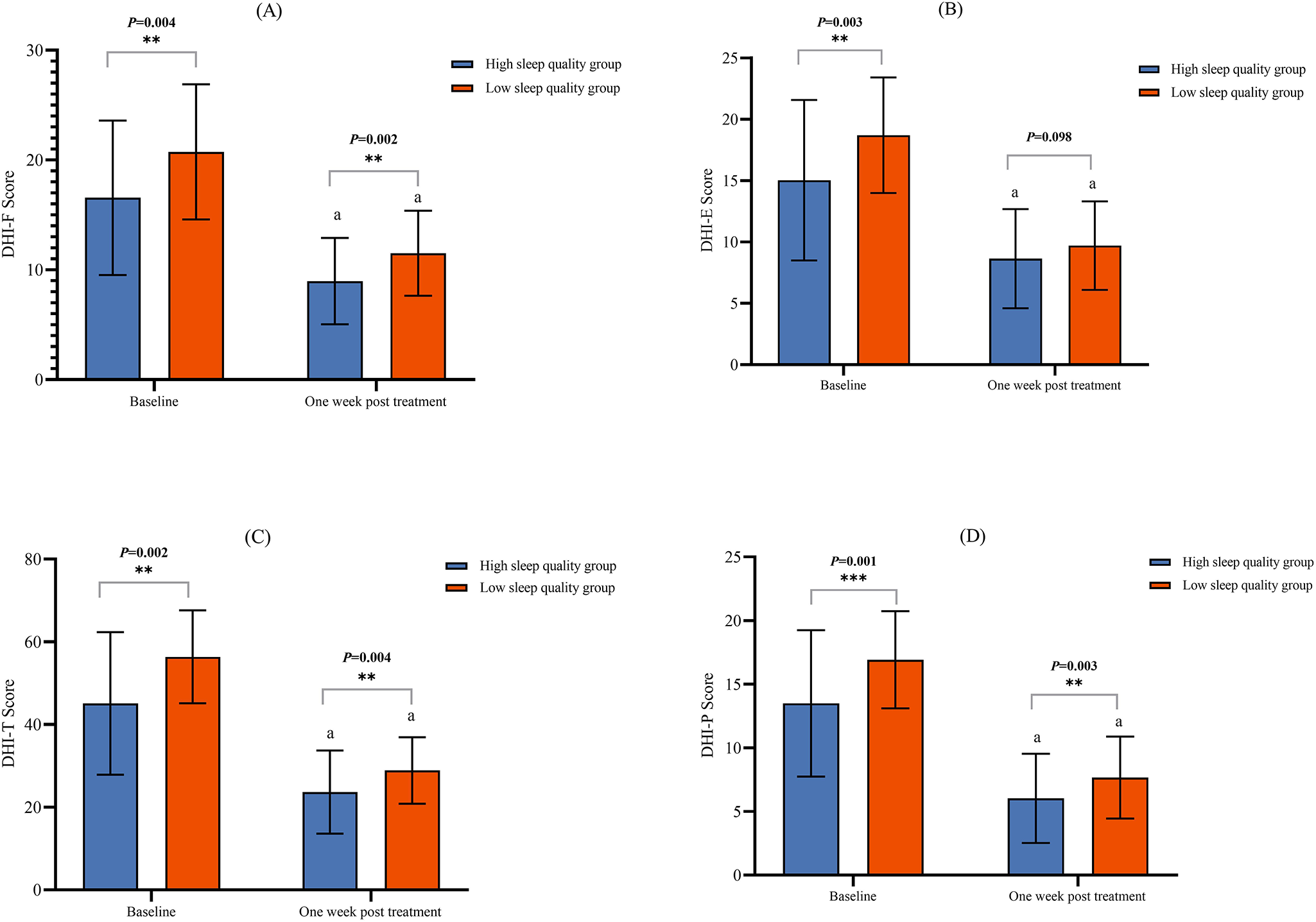
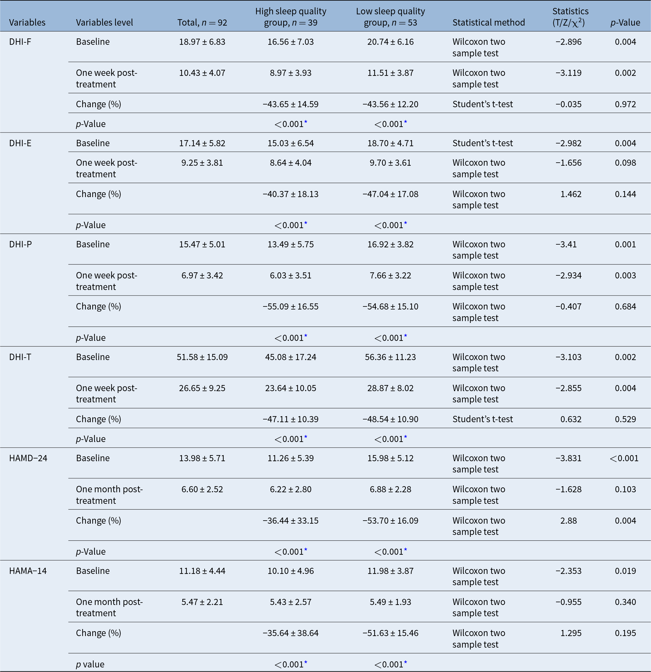
Before repositioning treatment, patients with PC-BPPV in both the low and high sleep quality groups exhibited moderate to severe vertigo disability, as indicated by the DHI total score. The low sleep quality group had significantly higher scores across all dimensions, including the DHI-T (total), DHI-P (physical), DHI-E (emaotional) and DHI-F (functional), compared to the high sleep quality group (Figure 2). One week after repositioning treatment, both groups showed a significant reduction in DHI scores compared to pre-treatment levels. The average reductions in DHI-F, DHI-E, DHI-P and DHI-T scores were 43.65 per cent vs. 43.56 per cent, 40.37 per cent vs. 47.04 per cent, 55.09 per cent vs. 54.68 per cent and 47.11 per cent vs. 48.54 per cent, respectively, with no significant differences in the average reduction between the two groups (all p > 0.05). However, even after repositioning, significant differences remained between the two groups in terms of DHI-T, DHI-P and DHI-F scores, with the low sleep quality group continuing to exhibit more severe symptoms (all p < 0.05), with the DHI-T scores of 28.87 ± 8.02 vs. 23.64 ± 10.05, DHI-P scores of 7.66 ± 3.22 vs. 6.03 ± 3.51 and DHI-F scores of 11.51 ± 3.87 vs. 8.97 ± 3.93, respectively. However, there was no significant difference between the two groups in the DHI-E scores after repositioning (9.70 ± 3.61 vs. 8.64 ± 4.04; p = 0.098), as shown in Table 4 and Figure 2.
(A–D) Change of DHI scores before and after maneuvers in both groups. “P” stands for comparison between groups at the same time point; “a” stands for comparison (p < 0.05) before and after treatment in the same group.

Results of DHI, HAMD-24 and HAMA-14 score before and after repositioning treatment in both groups

* Represents the p value compared within the group before and after treatment.
Comparison of depression and anxiety before and after repositioning
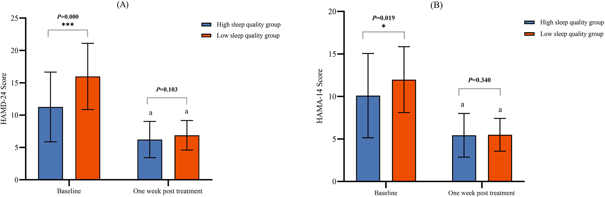
Before repositioning, the HAMD-24 scores for patients in the low and high sleep quality groups with PC-BPPV were 15.98 ± 5.12 and 11.26 ± 5.39, respectively, with the low sleep quality group showing significantly higher scores (p < 0.001), indicating more pronounced depressive symptoms in patients with low sleep quality. After repositioning treatment, both groups experienced significant reductions in HAMD-24 scores. The average reduction was 36.44 per cent in the high sleep quality group compared to 53.70 per cent in the low sleep quality group, with the low sleep quality group showing a significantly greater improvement (p = 0.004). One month after repositioning, in HAMD-24 scores were comparable between the two groups (6.88 ± 2.28 vs. 6.22 ± 2.80; p = 0.103), indicating that repositioning treatment significantly improved depressive symptoms in patients with PC-BPPV, especially in those with low sleep quality, as shown in Table 4 and Figure 3A.
Change of HAMD-24 (A) and HAMA-14 (B) scores before and after maneuvers in both groups. “P” stands for comparison between groups at the same time point; “a” stands for comparison (p < 0.05) before after treatment in the same group.

Similarly, the HAMA-14 score improvement followed the same pattern as HAMD-24. Before repositioning, the HAMA-14 scores in the low and high sleep quality groups were 11.98 ± 3.87 and 10.10 ± 4.96, respectively, with the low sleep quality group having significantly higher anxiety levels (p = 0.019). After repositioning, both groups showed significant reductions in HAMA-14 scores, with an average reduction of 35.64 per cent in the high sleep quality group and 51.63 per cent in the low sleep quality group (p = 0.195). After repositioning treatment, the HAMA-14 scores of the two groups were similar (5.49 ± 1.93 vs. 5.43 ± 2.57; p = 0.340), as shown in Table 4 and Figure 3B.
Discussion
Patients with sleep disorders, such as insomnia, sleep apnoea and sleep deprivation, often experience a decline in sleep quality, which is one of the most common symptoms among hospitalised patients with chronic dizziness. BPPV is one of the most common types of peripheral vertigo in clinical practice, and patients frequently suffer from sleep disorders such as insomnia.Reference Yuanyuan, Jing, Yan and Huifang9 As a result, the relationship between sleep and BPPV has received increasing attention. Most clinical studies have evaluated the relationship between vertigo and sleep using subjective rating scales. In this study, the PSQI was used to assess sleep quality, which has been widely used in both domestic and international research, showing high reliability and validity.Reference Zhu and Peng7 A case–control study demonstrated that the prevalence of sleep disorders in patients with BPPV was significantly higher than in healthy controls.Reference Xue, Wang, Meng, Zhao, Wang and Zhang17 Consistent with previous research, this study found that 57.6 per cent of the 92 patients with PC-BPPV had poor sleep quality. Another population-based cohort study revealed that individuals with chronic sleep disorders were at the highest risk of developing BPPV compared to those with other types of non-apnoea sleep disorders.Reference Shih, Wang, Chung, Lin, Chen and Lee18 Furthermore, Tsai et al. Reference Tsai, Lee, Tsai, Yang, Liu and Lin19 found that an adjusted Cox proportional hazards model demonstrated a significant association between sleep apnoea and an increased incidence of vertigo, strongly supporting the idea that sleep disorders are an independent risk factor for vertigo. In terms of the severity of vertigo, this study found that patients with sleep disorders experienced more severe overall vertigo, suggesting that the presence of sleep disturbances may exacerbate disease progression and lead to more adverse outcomes. Additionally, according to Ichijo’s report, the etiology of posterior cupulolithiasis is closely related to sleep, and he proposed a micro-otoconia accumulation hypothesis;Reference Ichijo20 our study found that the proportion of patients with cupulolithiasis was higher in the low sleep quality group, which suggests that low sleep quality might have more influence on cupulolithiasis type.
Currently, research on the impact of sleep quality on BPPV is still limited. Sleep disorders may contribute to the onset and progression of BPPV through the following mechanisms: First, the main components of otoliths are calcium and carbonate, which are susceptible to dissolution and detachment under changes in the surrounding pH. During nighttime sleep, mild respiratory acidosis and a decrease in arterial blood oxygen levels typically occur, placing the body in a mildly acidic and hypoxic environment. This condition may promote structural changes in the otoliths, making them more prone to dissolution and detachment.Reference Han and Kim21 Valli et al. proposed a hypothesis suggesting that a single detached otolith particle may not be sufficient to induce endolymphatic flow or stimulate nerve cells. However, the continuous daily shedding of otolith particles could accumulate, and once reaching a certain mass, they start moving under their own weight. At this point, the movement of the endolymph is triggered, leading to vertigo and nystagmus.Reference Valli, Botta, Zucca, Valli and Buizza22 Patients with sleep apnoea, who experience more pronounced nocturnal ischemia and hypoxia, are at a higher risk of developing BPPV.Reference Li and Li23 Moreover, BPPV often occurs suddenly without warning, causing asymmetry in bilateral vestibular function and triggering clinical symptoms of acute vestibular syndrome, such as vertigo and nystagmus. Central vestibular compensation may play a significant role in the alleviation of BPPV symptoms.Reference Deveze, Bernard-Demanze, Xavier, Lavieille and Elziere24, Reference Vats, Kothari and Biswas25 Some studies suggest that sleep deprivation can cause changes in posterior parietal cortical function, leading to abnormal vestibular-evoked myogenic potentials, indicating functional or structural alterations in the vestibulospinal pathway. This could result in impaired spatial orientation and diminished capacity for spatial information storage.Reference Wang, Zhang, Lei, Zeng and Cai26 Additionally, both sleep disorder and vestibular dysfunction models in animals have shown increased histamine levels, suggesting that histamine may be involved in the process of vestibular compensation.Reference Qian, Wang and Zhang27 The regulation of sleep is closely related to certain structures within the central nervous system, such as the raphe nuclei in the brainstem, the solitary tract nucleus, the locus coeruleus in the dorsomedial pontine tegmentum, the suprachiasmatic nucleus and the thalamus. The anatomical pathways and neuroendocrine signaling between the vestibular nuclei and these structures may represent potential mechanisms underlying the interplay between BPPV and sleep disorders.Reference Wang, Zhang, Lei, Zeng and Cai26
Otolith repositioning is the preferred method for treating BPPV. Several studies have identified factors that may contribute to poor treatment outcomes, including patient age, gender, disease duration, the affected semicircular canal, psychological factors and hyperlipidemia.Reference Wang and Wang28 However, there is no conclusive evidence on the impact of sleep disorders on BPPV repositioning efficacy. Yuanyuan et al. Reference Yuanyuan, Jing, Yan and Huifang9 explored the effects of sleep on primary PC-BPPV and found that sleep disorders do have some impact on PC-BPPV, with patients experiencing sleep disturbances generally showing poorer short-term outcomes following repositioning. In this study, the group with low sleep quality demonstrated worse short-term efficacy after the initial repositioning and had higher recurrence rates. Meanwhile, two meta-analyses evaluated risk factors for BPPV recurrence after repositioning and concluded that sleep disorders were not among the factors affecting recurrence. However, due to the limited number of included studies, no definitive conclusion could be drawn.Reference Sfakianaki, Binos, Karkos, Dimas and Psillas29, Reference Li, Wang, Liu, Cao, Zheng and Jing30 Further exploration is needed to better understand the influence of sleep quality on the efficacy of BPPV repositioning and the potential underlying mechanisms.
After successful repositioning in patients with BPPV, although vertigo and nystagmus may be relieved, some patients still experience residual symptoms such as persistent dizziness, lightheadedness or unsteadiness.Reference Gu, Chen, Zhang, Shen, Ma and Wang31 The mechanisms leading to these residual symptoms remain unclear, but they may be related to the following factors: (1) co-morbid mechanisms, such as functional abnormalities in the peripheral otolithic apparatus, its conduction pathways or the vestibular system itself;Reference Tian, Zhang, Ma and Liu32, Reference Pollak, Davies and Luxon33 (2) residual otolith fragments that remain after repositioning, though insufficient to cause significant positional vertigo or nystagmus;Reference Dispenza, Mazzucco, Mazzola and Martines34 (3) the central nervous system requires time to readapt after otolith repositioning;Reference Faralli, Lapenna, Giommetti, Pellegrino and Ricci35 (4) delayed diagnosis and repositioning of otoliths;Reference Teggi, Quaglieri, Gatti, Benazzo and Bussi36 (5) subjective dizziness caused by psychological factors.Reference Wei, Sayyid, Ma, Wang and Dong37 Currently, there is a lack of tools specifically designed to assess residual symptoms following BPPV repositioning. Some studies have applied the DHI to evaluate the effects of repositioning therapy in patients with BPPV before and after treatment. The Chinese version of the DHI demonstrates good internal consistency and can comprehensively assess the quality of life of patients with BPPV in physical, emotional and psychological aspects. Therefore, it is considered an effective tool for evaluating the presence and severity of residual symptoms after otolith repositioning.Reference Gu, Chen, Zhang, Shen, Ma and Wang31 There is still no conclusive evidence on the impact of sleep quality on residual symptoms after BPPV repositioning. A meta-analysis by Ke et al. evaluated risk factors for residual dizziness in patients with BPPV after repositioning and found no association between sleep disorders and the occurrence of residual symptoms.Reference Ke, Ma, Jing, Diao and Yu38 In this study, residual symptoms were assessed across three aspects of the DHI after repositioning treatment. The low sleep quality group showed higher scores in DHI-T, DHI-P, DHI-E and DHI-F compared to the high sleep quality group, although the DHI-E scores were similar between the two groups after repositioning. This suggests that special attention should be given to the physical and social functioning of patients with PC-BPPV and low sleep quality, providing appropriate and reasonable support and guidance to achieve better disease outcomes.
The factors influencing the development of BPPV are quite complex, and sleep quality may be just one contributing factor. Many patients also experience depression, anxiety and physical symptoms, which can further exacerbate sleep disorders. Preventing the onset and recurrence of BPPV is emerging as a new research direction in neurotology. Since sleep quality may also be related to other forms of otogenic vertigo, further studies are needed to determine the impact of sleep disorders on BPPV and the underlying mechanisms involved.
• Previously, there were limited researches on the impact of sleep quality on vertigo-related symptoms in posterior canal benign paroxysmal positional vertigo (PC-BPPV), particularly in patients with cupulolithiasis
• Patients with PC-BPPV and poor quality sleep have a higher proportion of cupulolithiasis, longer nystagmus duration during positional tests and lower efficacy of initial repositioning
• After repositioning, residual dizziness symptoms are primarily reflected in more pronounced physical symptoms and impaired social functioning
• Additionally, repositioning therapy results in a more significant improvement in depressive symptoms for patients with PC-BPPV and poor sleep quality
Conclusion
Patients with PC-BPPV and low sleep quality tend to have a higher proportion of cupulolithiasis, longer nystagmus duration and more severe vertigo-related symptoms as well as heightened anxiety and depression. These patients also acquire poorer initial outcomes from repositioning treatment, with more pronounced residual dizziness symptoms, particularly in terms of physical symptoms and reduced social functioning. Notably, repositioning treatment led to a more significant improvement in depressive symptoms in patients with PC-BPPV and low sleep quality.
Data availability statement
The datasets generated for this study are available on request to the corresponding author.
Funding statement
This study was supported by Hebei Provincial Health Commission (No. 20220174), “Spark” Research Project Cultivation Fund, the First hospital of Hebei Medical University (No. XH202410) and the National Natural Science Foundation of China (U23A20431).
Ethical standards
The research protocol was approved by the Ethics Committee of the First Hospital of Hebei Medical University.