Introduction
Extensive nasal polyposis is the protrusion of benign oedematous polyps into the nasal cavity. It is caused by various mechanisms, including chronic inflammation, aspirin intolerance, epithelial gene defects, aeroallergens or food allergy.Reference Norlander, Brönnegård and Stierna1 Histologically, polyp tissue consists of inflammatory cells such as eosinophils and neutrophils and contains an increased level of inflammatory mediators, a sign of a persistent chronic inflammation.Reference Pawankar2
Melatonin is a neuromodulatory hormone that is rhythmically released from the pineal gland into the plasma during darkness.Reference Reiter3 It has been shown in viral and bacterial infections that melatonin protects against functional disorders in various organs. In addition, melatonin is effective within the antioxidative defense system and in the anti-inflammatory pathway.Reference Reiter and Maestroni4
Keeping in mind the pathogenesis of nasal polyposis and the effect of melatonin on the immune system, this study aims to establish if melatonin in the tissue or serum plays any aetiological role in nasal polyposis.
Material and methods
This study included 29 patients (16 female and 13 male; mean age: 44 years) who had presented to our clinic between June 2013 and June 2014 because of nasal polyposis and underwent functional endoscopic sinus surgery. As control group, we enrolled 26 patients (13 female and 13 male; mean age: 39 years) who did not have nasal polyps but were operated on for a deviated nasal septum and concha bullosa.
Patients were excluded if they: had another chronic infection history; had a diagnosis of obstructive sleep apnoea; had asthma; had aspirin sensitivity; had been using hormone therapy for other reasons; or had been using other drugs (including antidepressants, antipsychotic medications, benzodiazepines, calcium channel blockers, betablockers, anticoagulants, interleukin-2 or non-steroidal anti-inflammatory drugs) that could influence melatonin levels.
The study protocol was approved by the Clinical Trials Ethics Committee of Bezmialem Vakif University (approval number: 050.01.04/556).
The surgical procedures were all performed during the same hours of the day (10 am to 12 noon). Samples were retained from the nasal polyp tissue and from the mucosa of the middle concha. Five millilitre venous blood samples were collected from volunteers’ peripheral veins and put into tubes containing disodium ethylene diamine triacetic acid (EDTA). These were centrifuged at 4500 × g for 10 minutes. The resultant plasma samples were stored at −20°C. The tissues removed from the volunteers were stored at −80°C until analysis.
Chemicals
Melatonin was purchased from Sigma-Aldrich (St. Louis, Missouri, USA). Acetonitrile, chloroform and acetic acid were of high-performance liquid chromatography grade. All other solvents and chemicals were analytical grade. Stock solutions of melatonin standard were prepared in water and stored at 4°C. Water was purified by Human Ultra Water Purification System (Human Science, Seoul, Korea).
Melatonin extraction
To extract melatonin from the plasma samples, 200 µl of plasma was acidified with 200 µl of 0.1 M acetic acid (pH 4.6). The solution was then extracted into 2 ml of chloroform. The contents were mixed using a vortex mixer at moderate speed for 5 minutes and centrifuged at 4500 × g for 3 minutes. The aqueous layer was discarded. The organic phase was alkalinised with 500 µl of 0.1 M sodium hydroxide and mixed with a vortex mixer. After centrifugation at 4500 × g for 2 minutes, the organic phase was removed and evaporated to dryness under a stream of nitrogen at 40°C. Next, 100 µl of the mobile phase was added to the residue and the solution was filtered; 20 µl of the filtrate was injected into the high-performance liquid chromatography system. In order to extract melatonin from each type of tissue, 20 mg of the tissue sample was homogenised, and 2 ml of 0.1 M acetic acid (pH 4.6) was added to the samples. The contents were mixed with a vortex mixer at moderate speed for 5 minutes and centrifuged at 4500 × g for 3 minutes. The supernatant was taken and extracted into 2 ml of chloroform. The extract was processed with the same procedures as the plasma extracts after the extraction step.
Analytical procedure
In order to determine the presence of melatonin in plasma and tissue samples, a high-performance liquid chromatographic method was developed and validated. The high-performance liquid chromatography analyses were performed on a Shimadzu (Kyoto, Japan) LC 20 liquid chromatograph, consisting of an LC-20AT pump, SIL AT-HT auto sampler part and an SPD-20A HT fluorimetric detector set to 285 nm for excitation and 360 nm for emission. A CTO 10 AC column oven was used. Chromatographic separation was achieved isocratically at 30°C on a GL Sciences (Tokyo, Japan) carbon 18 (octadecyl silane) column with dimensions of 4.6 mm inner diameter, 100 mm length and 3 µm particle size. The mobile phase consisted of 85 mM acetic acetate buffer and 0.1 M EDTA-disodium mixture and acetonitrile (86:14) with a flow rate of 1.0 ml per minute. Quantification of melatonin peaks was carried out by using a calibration curve prepared using standard melatonin solutions.
Summary statistics
Statistical analysis was carried out using SPSS® (version 13) statistical software. All quantitative variables were estimated using measures of central location (i.e. mean and median) and measures of dispersion (i.e. standard deviation). Data normality was controlled using the Kolmogorov–Smirnov tests of normality. Student's t-test was used for comparing between group variable data. Repeated analysis of variance test was used for within-group variability analysis. The Bonferroni test was used to compare analyses of the groups. A value of p < 0.05 was accepted as statistically significant.
Results
There were 29 patients (16 female and 13 male; mean age: 44 years) in the nasal polyposis study group. The control group included 26 patients (13 female and 13 male; mean age: 39). There was no statistically significant difference between the groups regarding gender distribution.
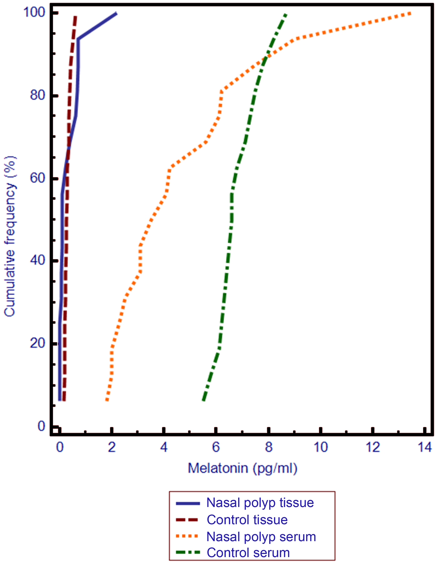
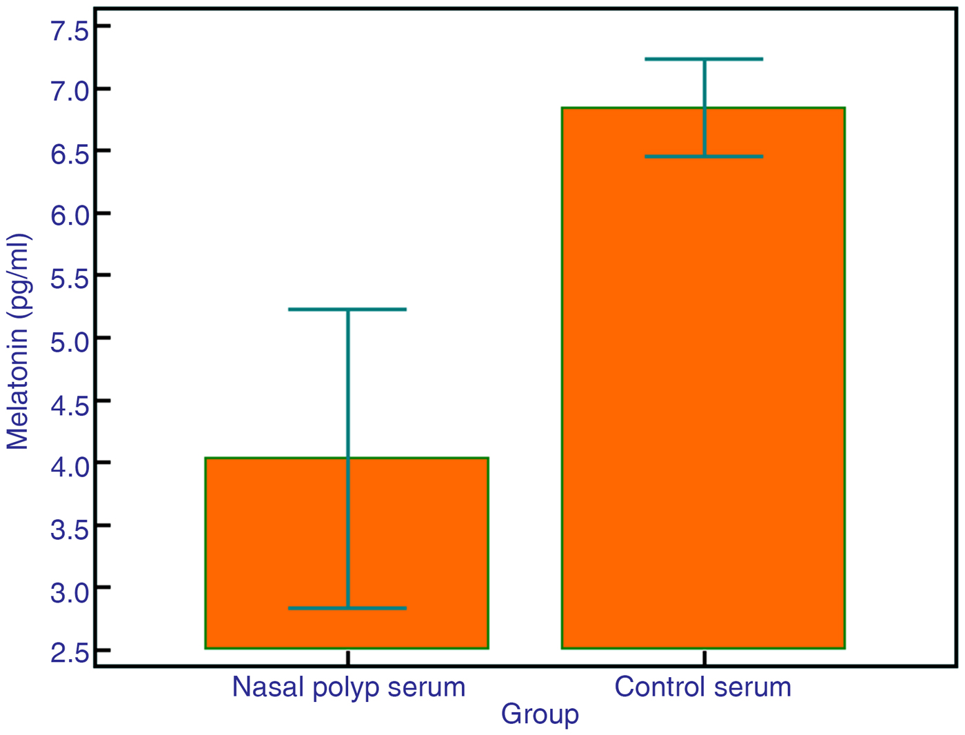
It was found that the tissue melatonin levels in the nasal polyp group were significantly lower compared with the tissue melatonin levels in the control group (p = 0.0129) (Figure 1). The serum melatonin levels in the nasal polyp group were significantly lower compared with the serum melatonin levels in the control group (p = 0.0003) (Figure 2).
Graph showing the tissue and serum melatonin levels in the nasal polyp group and control group.

Graph showing the serum melatonin levels in the nasal polyp group and control group.

In both groups, the tissue melatonin levels were statistically significantly lower than the serum melatonin levels (p = 0.0001) (Table 1).
Melatonin level comparison between nasal polyp and control group

*n = 29; †n = 26.
Discussion
A nasal polyp is a chronic inflammatory disease of the mucous membranes in the nose and the paranasal sinuses. Among the list of symptoms are a blocked nose, reduced sense of smell, runny nose, headache and pain in the face. It can be a local condition but may also be a result of systemic diseases such as cystic fibrosis, aspirin intolerance or primary ciliary dyskinesia. Prevalence in the population ranges between 1 and 4 per cent. There is no single factor in the aetiology of nasal polyps, but inflammation plays a major role.Reference Pawankar2
There are a number of studies that have researched the role of melatonin in inflammation. Melatonin affects the immune system directly and indirectly.Reference Cardinali and Freire5 It has been shown to increase both specific and non-specific immunity.Reference Ravindra, Lakshmi and Ahuja6,Reference Jung and Ahmad7 It binds to receptors on T helper type 1 and T helper type 2 lymphocytesReference Maestroni and Conti8–Reference Gonzalez-Haba, García-Mauriño, Calvo, Goberna and Guerrero10 and enhances cytokine production.Reference García-Mauriño, Pozo, Carrillo-Vico, Calvo and Guerrero11 It also lengthens the lifespan of B lymphocyte precursors, which are the main cells responsible for humoral immunity, inhibiting their apoptosis and thus increasing the number of mature B lymphocytes.Reference Yu, Miller and Osmond12 By inhibiting the release of interleukin-8 and tumour necrosis factor-α from neutrophils, it can limit acute and chronic inflammation.Reference Silva, Rodrigues, Carvalho, Catalani, Campa and Ximenes13
Some studies show that during inflammation, melatonin levels are reduced and, in a vicious cycle, this state can increase inflammation.Reference Kos-Kudla, Ostrowska, Marek, Ciesielska-Kopacz, Kajdaniuk and Kudla14,Reference Fei, Liu, Zhang and Zhou15 It has been suggested that this reduction can result from pinealocytes being inhibited by cortisone, which is raised during inflammation, or from the faster metabolism of melatonin.Reference Barriga, Martin, Tabla, Ortega and Rodríguez16 However, there are very few studies that have researched the role of melatonin in nasal polyposis, where the primary aetiology is that of a chronic inflammation. Fidan et al. showed that melatonin levels in serum and mucus were significantly lower in their nasal polyposis group compared with the control, but the release rhythm did not change. As a treatment, they proposed to give melatonin and chronotherapy alongside steroids.Reference Fidan, Alp, Kalkandelen and Cingi17 Contrary to this, another study showed that melatonin significantly increased in polyp tissue in polypous rhinosinusopathy.Reference Evsiukova, Okuneva, Zubzhitskaia and Kvetnoĭ18 As with the study by Fidan et al.,Reference Fidan, Alp, Kalkandelen and Cingi17 our study showed that melatonin serum and tissue levels in the nasal polyposis group were also significantly lower than in the control group.
The melatonin concentration in nasal polyp tissue has not been considered before. Our study is therefore the first of its kind. Compared to the healthy controls, we found that the melatonin concentration in the mucosa of the polyp group was significantly lower. It is possible that if the mucosa does not have a sufficient concentration of melatonin, it can turn into polyps due to chronic inflammation. In a chronically inflamed mucosa, melatonin concentration can also be reduced by a faster metabolism.
• Chronic inflammation is an important factor in the aetiology of nasal polyps
• Melatonin is effective at limiting chronic inflammation
• Melatonin deficiency in both serum and tissue may play a role in the formation of nasal polyposis
Conclusion
The most important factor in the aetiology of nasal polyps is chronic inflammation. Melatonin is effective in limiting chronic inflammation in a number of body systems. In the formation of nasal polyposis, melatonin deficiency, both in the serum and tissue, may play a role. Future studies need to establish if melatonin can be used for treatment alongside steroids.
Acknowledgements
This research was supported by Bezmialem Vakif University.
Competing interests
None declared