Dilated cardiomyopathy is the most common form of cardiac muscle disease defined by the presence of left ventricular dilatation and left ventricular systolic dysfunction in the absence of abnormal loading conditions – hypertension, valve disease – or coronary artery disease sufficient to cause global systolic impairment. Right ventricular dilatation and dysfunction may be present but are not necessary for diagnosis.Reference Elliott, Andersson and Arbustini1 The aetiology is unclear in about half of the cases – idiopathic dilated cardiomyopathy – but may be genetic, viral, metabolic, or toxic. The clinical presentation usually involves cardiac failure, which is often progressive and correlates to the degree of myocardial dysfunction.Reference Soler, Rodríguez, Remuiñán, Bello and Díaz2 Although dilated cardiomyopathy is prognostically important and is a common indication for cardiac transplantation in all age groups, the incidence and age distribution of dilated cardiomyopathy in a well-defined paediatric population have been poorly characterized. Marked variability was seen to occur among the different age groups of children with dilated cardiomyopathy, suggesting that different pathophysiologic mechanisms, and possibly aetiologies, may exist in different age groups.Reference Arola, Jokinen and Ruuskanen3
Paediatric cardiomyopathy is a rare but serious and often life-threatening condition. The estimated incidence is 0.34–0.58 cases per 100,000 children per year, with rates that are 8 to 12 times higher in infants than in older children. Nearly 40% of children with symptomatic cardiomyopathy receive a transplant or die within 2 years, and outcomes have not improved substantially, despite advances in medical care and technology.Reference Cox, Sleeper and Lowe4
Natural history and prognostic factors of dilated cardiomyopathy in paediatric age are not well identified so far and are difficult to predict due to heterogeneous diseases that can result in cardiomyopathy.
Published data on dilated cardiomyopathy in children are sparse and little is known about the long-term clinical course and the factors that may influence prognosis for better or worse. It is necessary to identify the group of patients having a poor outcome in order to define the prognostic factors of impending death so that a cardiac transplant could be offered to them as a reasonable therapeutic choice.Reference Caso, Cioppa, Musto, De Leva, Vitale and Calabrò5
The purpose of this study was to review our experience with children diagnosed with dilated cardiomyopathy and attempt to discover prognostic factors.
Materials and methods
A retrospective study was carried out, collecting the data of 41 patients under 14 years of age who presented with dilated cardiomyopathy at our unit from 1993 to 2008. Patients with cardiomyopathy related to anomalous origin of the left coronary artery were excluded. No patients had underlying congenital cardiac defects. We reviewed the medical history of the patients with dilated cardiomyopathy to determine the age at diagnosis, gender, family history, preceding viral illness, aetiology, symptoms and signs at presentation, treatment, and outcome. The patients were categorised according to the guidelines of the Task Force on Cardiomyopathies Working Group on Myocardial and Pericardial Disease 2007Reference Elliott, Andersson and Arbustini1: seven children (17%) had a family disease while 34 patients (83%) had a non-familial form.
The diagnosis of cardiomyopathy was based on a paediatric cardiologic examination plus ECG and echocardiography.
In echocardiography, we consider the left ventricular end-diastolic dimension, posterior wall and septal thickness, and fractional shortening. All parameters were measured and expressed conditionally on the body surface area. Standard echocardiography was perfomed with pulse wave Doppler to record mitral – and tricuspid – inflow pattern, while mitral regurgitation was demonstrated by colour-flow Doppler imaging.
Patients underwent a metabolic evaluation including blood lactate, pyruvate, carnitine, amino acids, and urine organic acids, a skeletal muscle biopsy with ultrastructural analysis – the tissue was snap-frozen and submitted to standard histopathology examination.
Viral culture with polymerase chain reaction and serology was useful in establishing a diagnosis of viral myocarditis by demonstrating rising titres of neutralising antibodies, or virus-specific IgM class antibodies, as indicative of recent infection. Although viral identification from endomyocardial biopsy was not routinely undertaken during the study period, diagnosis has been greatly facilitated by the introduction of endomyocardial biopsy. In all, 18 patients underwent endomyocardial biopsy with microbiological and histological investigations.
Therapy was a combination of diuretics, angiotensin-converting enzyme inhibitors, digoxin, β-adrenergic receptor blockers, spironolactone, and intravenous inotropic support in resistant patients or immunoglobulin and cortisone infusion, respectively, in patients with viral or autoimmune myocarditis.
The patients were stratified by age at diagnosis, presenting symptoms, and therapy administered at onset and aetiology. During follow-up, the patients were examined frequently to evaluate cardiac function.
The primary endpoint of death/cardiac transplantation was associated with the need for intravenous inotropic support.
Data are presented as mean plus or minus standard deviation. Survival curves were plotted with Kaplan–Meier survival estimates and the differences between the groups were tested using a Log Rank test. Any p-value less than 0.05 was considered significant.
Informed consent was obtained from parents or legal guardians for all diagnostic procedures and molecular studies.
Results
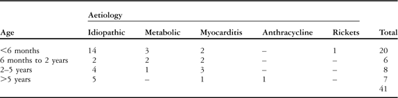
In all, 41 patients were identified with dilated cardiomyopathy from 1993 to 2008. Median age at diagnosis was 33.4 months – plus or minus 49.25 standard deviation – ranging from 0 to 14 years. There were 20 (49%) male and 21 (51%) female patients. At diagnosis, 20 patients (49%) were under 6 months, six patients (15%) were between 6 months and 2 years, eight patients (19%) were from 2 to 5 years, and seven patients (17%) were above the age of 5 years.
There were 28 (68.3%) patients with signs of congestive cardiac failure. The first cardiac examination in 13 (31.7%) asymptomatic patients was due to systolic murmur and/or abnormal ECG findings, pre-natal ultrasonography, or familial screening. Of the 28 patients with severe cardiac failure at presentation, 12 (42.9%) needed intravenous inotropic support while 16 (57.1%) received conventional treatment for cardiac failure.
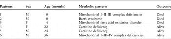
Intracardiac thrombus formation was present in three patients: they received anticoagulant therapy. The cause of dilated cardiomyopathy was not identified in 25 patients (61%). Of the 16 patients with a known cause, six (37.5%) had a metabolic form: one with Barth syndrome, two with mitochondrial disease, one with inborn error of fatty acid beta-oxidation, and two with acetyl-l-carnitine deficiency.
Among the remaining 10 patients, seven had a diagnosis of viral myocarditis, one had an autoimmune myocarditis, one had a dilated cardiomyopathy due to anthracycline therapy, and one had cardiomyopathy related to rickets.
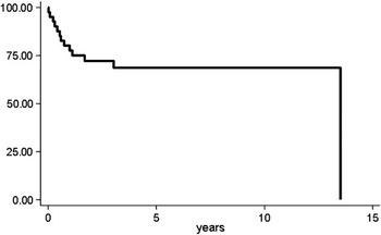
Median follow-up was 45.25 plus or minus 41.15 months, ranging from 10 days, death shortly after presentation, to 162 months. Freedom from death or cardiac transplantation was 68.3% at 5 years. Only one patient was lost to follow-up (Fig 1).
Survival curve: 5-year survival 68%.

Of the 20 patients under 6 months at presentation, three (15%) died, two (10%) underwent cardiac transplantation, whereas 15 (75%) were followed – of these, one had a Berlin Heart as a bridge to transplant. Of the six patients between 6 months and 2 years, five were followed – of them, one underwent Extra Corporeal Membrane Oxygenation – and one underwent cardiac transplantation.
Within the group of 2–5 years of age (eight patients), one patient underwent cardiac transplantation and seven were followed.
Of seven patients above the age of 5 years, one died at presentation, while three patients underwent successful cardiac transplantation and three were followed (Table 1).
Age at diagnosis and aetiology.

Symptoms were present in most patients at presentation and were usually severe. Congestive cardiac failure was the initial symptom in 68.3% (28) of patients, 42.9% (12) of whom needed intravenous inotropic support; two (16.7%) of these patients died, three (25%) patients underwent cardiac transplantation while ambulatory monitoring was performed in the remaining seven patients (58.3%): one of these has a Berlin Heart and is waiting for a cardiac transplant.
The other 16 patients with cardiac failure received conventional therapy and had significant improvement in cardiac function: 14 (87.5%) were followed while two (12.5%) underwent cardiac transplantation.
There was a significant difference in survival with respect to aetiology: three (50%) of six patients with a metabolic form died (Table 2).
Metabolic pattern.
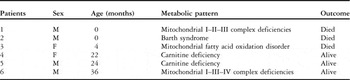
M = male; F = female
Discussion
Dilated cardiomyopathy represents a heterogeneous group of diseases with multiple aetiologies united by a common presentation of a dilated, poorly contractile heart, usually accompanied by cardiac failure.Reference Bostan and Cil6 Little is known about the long-tem functional outcome of children with dilated cardiomyopathy and the survival rates remain dismal.Reference Helton, Darragh and Francis7 The uniform diagnostic features of dilated cardiomyopathy fail to reflect the multitude of aetiologic factors that can lead to this disorder. Infectious, metabolic, toxic, and hereditary factors have been implicated in the disease pathogenesis.Reference Allen, Gutgesell, Clark and Driscoll8 The course of the disease often presents “odd” characteristics: some of the patients show clinical improvements, some remain unchanged, and some of them have a negative evolution in a very short time.Reference Caso, Cioppa, Musto, De Leva, Vitale and Calabrò5 The recent success with ventricular assist devices and cardiac transplantation and the limited number of donors for transplantation make it important for the clinician to develop an accurate means of risk stratification of children with dilated cardiomyopathyReference McMahon, Nagueh and Eapen9 and to identify the group of patients having a poor outcome in order to define prognostic factors of impending death so that a cardiac transplant could be offered to them as a reasonable therapeutic choice.Reference Caso, Cioppa, Musto, De Leva, Vitale and Calabrò5
This study aimed to determine the outcome of dilated cardiomyopathy presenting in childhood and the features that might be useful for prognostic stratification. Despite the highly variable cause of dilated cardiomyopathy in children, this study has identified some indices that can be used to rationalise the management of these patients. In children, dilated cardiomyopathy is a diverse disorder with outcomes that depend on cause, age, and cardiac failure status at presentation.
The Kaplan–Meier curves illustrate the difference in survival among different patients’ subgroups. Age at presentation has been a prognostic factor as stated in some previous studies.Reference Talercio, Seward, Driscoll, Fisher, Gersh and Tajik10–Reference Burch, Siddiqi, Celemajer, Scott, Bull and Deanfield12 In these studies, children who are younger than 2 years of age were found to have higher recovery rates than older children.Reference Bostan and Cil6 Towbin et alReference Towbin, Lowe and Colan13 have demonstrated that dilated cardiomyopathy is significantly more likely to present in the first year of life than at older paediatric ages but dilated cardiomyopathy presenting at older paediatric ages is associated with worse outcomes.
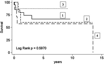
In our study, infants presenting their first symptoms over 5 years of age have a twofold increased risk of mortality/cardiac transplant during the observation period compared with those presenting earlier (Fig 2).
Survival curve for age categories; 1 = 0–6 months; 2 = 6 months to 2 years; 3 = 2–5 years; 4 = older than 5 years.

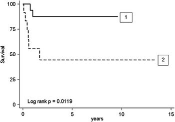
Cardiac failure was a frequent reason for the first consultation in our cohort. Cardiac failure was present in 28 of 41 patients and required intravenous inotropic support in 12 of 28 patients. Intractable cardiac failure has been found to be a statistically significant predictor of the outcome (p less than 0.005; Fig 3). Cardiac failure is an important prognostic indicator in dilated cardiomyopathy because it is the most common cause of death and therefore the most important indication for cardiac transplantation.Reference Bostan and Cil6 Our data showed that intravenous inotropic support at first symptoms has a highly increased risk of mortality. Finding the aetiology of dilated cardiomyopathy can be time-consuming and frustrating. The success rate is 39% in our study.
Relationship between clinical onset and outcome; 1 = conventional cardiac failure therapy; 2 = intravenous inotropic support.

Nevertheless, it is important to undertake every effort to establish the aetiology of the cardiomyopathy because there may be a causative therapy available. In addition, specific screening of other members of the family and meaningful counselling of the parents regarding the risk of recurrence are possible only if the aetiology of the cardiomyopathy is known.Reference Badertscher, Bauersfeld, Arbenz, Baumgartner, Schinzel and Balmer14
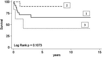
In our study, there was a significant difference between aetiology and outcome (Fig 4). Inherited metabolic diseases represent a vast group of disorders of energy metabolism with a wide range of symptoms, presentation, and severity.Reference Debray, Lambert and Chevalier15 Dilated cardiomyopathy has been reported to be frequent in metabolic disorders.Reference Scaglia, Towbin and Craigen16 In our study, for the children affected by DCM as a part of a multi-system involvement, the mortality was 50%. However, in common with other retrospective studies,Reference Bostan and Cil6, Reference Burch, Siddiqi, Celemajer, Scott, Bull and Deanfield12, Reference Daubeney, Nugent and Chondros17 we have reported good outcomes in children with dilated cardiomyopathy secondary to lymphocytic myocarditis treated with immunosuppressive medication. Despite high initial mortality, patients with lymphocytic myocarditis had better survival (one out of eight children died; Fig 4). This may reflect the rapid evolution of paediatric lymphocytic myocarditis, a more favourable natural history of this condition, and the benefit of the various therapies employed.Reference Daubeney, Nugent and Chondros17 Besides, the cardiovascular system of infants can cope better with severe myocardial damage as infant myocytes retain the propensity to regenerate or afterload may be lower due to more compliant peripheral arteries.Reference Bostan and Cil6, Reference Burch, Siddiqi, Celemajer, Scott, Bull and Deanfield12 All but one patient with myocarditis recovered completely.
Relationship between aetiology and outcome; 1 = familial/non-familial idiopathic; 2 = acquired; 3 = metabolic aetiology.

Endomyocardial biopsy has a diagnostic field in the assessment of myocarditis in children. It is not without hazard but procedural safety is higher in children greater than 5 kilogram of weight. Biopsy may have an important role in defining treatment.Reference Webber, Boyle, Jaffe, Pickering, Beerman and Fricker18 In case of detection of a viral genome in our specimens using polymerase chain reaction, we did not use immunosuppressive agents or steroids. Anyway, we treated all myocarditis with intravenous immunoglobulin.Reference Webber, Boyle, Jaffe, Pickering, Beerman and Fricker18
The patients of this study illustrate the need for a high level of suspicion for metabolic disease in children with dilated cardiomyopathy.
Inherited disorders of energy metabolism should be considered in any child with unexplained dilated cardiomyopathy.Reference Debray, Lambert and Chevalier15 On the basis of these considerations, we propose an algorithm of investigations for patients with (presumed) metabolic dilated cardiomyopathy. Since most of these conditions are currently untreatable, we suggest starting with the least invasive techniques – blood DNA testing for specific aetiologies – before performing muscle and/or endomyocardial biopsy.
To conclude, because of the small number of patients and the retrospective study design, we are not able to make further statements about the risk factors for death or transplant, but we observed the following trends, which concord with previously described seriesReference Towbin, Lowe and Colan13: overt cardiac failure is the only predictive factor of the global functional outcome (p = 0.01); metabolic status and age at presentation are not identified as independent predictors of the functional outcome of patients even if associated with a poorer prognosis.
Acknowledgements
We thank “Laboratorio della Sezione Malattie Metaboliche e Neuromuscolari Ereditarie, Istituto Meyer, Firenze” and “Unità Operativa di Neurogenetica Molecolare, Istituto Nazionale Neurologico Carlo Besta, Milano” for collaboration.