Disconnection of the pulmonary artery is a congenital defect characterised by discontinuity between the proximal part of a pulmonary artery and the main pulmonary trunk. This malformation can be isolated or associated with other CHDs.Reference Kim, Ban and Bae 1 Early surgical treatment is mandatory in order to support the growth of the disconnected pulmonary artery. However, surgery is associated with a high rate of re-stenosis.Reference Kim, Ban and Bae 1 – Reference Pfefferkorn, Loser and Pech 3 This paper describes a new hybrid approach that involves the reconstruction of the pulmonary artery and hybrid implantation of a stent for support.
Case series
Case 1
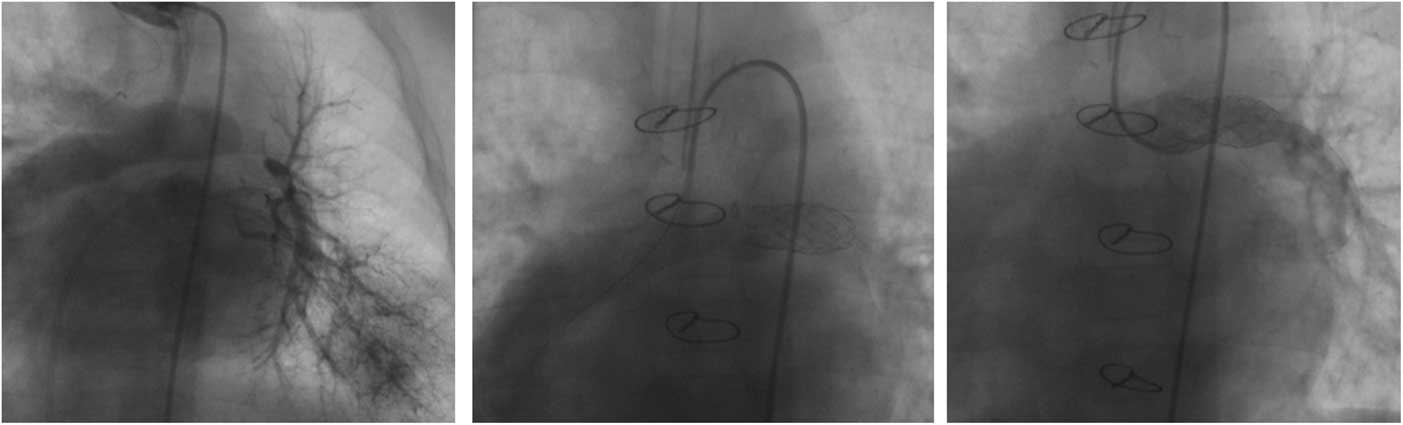
An 8-month-old boy weighing 7 kg was admitted with a diagnosis of dextrocardia, pulmonary atresia with ventricular septal defect, and congenitally corrected transposition of the great arteries. He was treated with a Blalock–Taussig shunt at birth. During clinical follow-up, he underwent cardiac catheterisation with evidence of a patent Blalock–Taussig shunt with normal flow and complete obstruction of the left pulmonary artery. Retrograde injection in the left pulmonary veins showed a 3-mm distal left pulmonary artery (Fig 1, left), whereas the right pulmonary artery measured 9 mm.
(Patient #1): Left: retrograde injection in the left pulmonary veins showing the disconnected left pulmonary artery. Middle: after intervention, an anterograde injection in the systemic-to-pulmonary artery shunt showed preferential flow towards the right pulmonary artery. Right: selective anterograde injection in the left pulmonary artery demonstrates a developed vessel.

An end–end anastomosis was performed between the inferior surface of the pulmonary trunk and the distal segment of the “atretic” pulmonary branch. This was accompanied by a pulmonary arterioplasty with patch augmentation up to the bifurcation of lobar pulmonary branches using a patch of heterologous pericardium (Edwards Lifescience Corp., Irvine, California, United States of America). Two genesis stents (Cordis, Belgium) dilated up to 8 mm and with a length of 19 mm were implanted to support the newly created connection. At the age of 28 months, the patient was assessed for complete physiological correction. Cardiac catheterisation showed preferential flow towards the right pulmonary artery (Fig 1, middle). However, despite this, the left pulmonary artery was patent and nicely developed with satisfactory flow (Fig 1, right). Surgery included ventricular septal defect closure, a left-ventricular-to-pulmonary artery conduit using a 14-mm Edwards conduit (Edwards Lifescience Corp.), and stent re-dilation to up to 10 mm. At 5 years of follow-up the left pulmonary artery remains widely patent.
Case 2
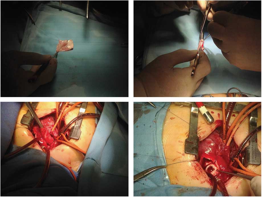
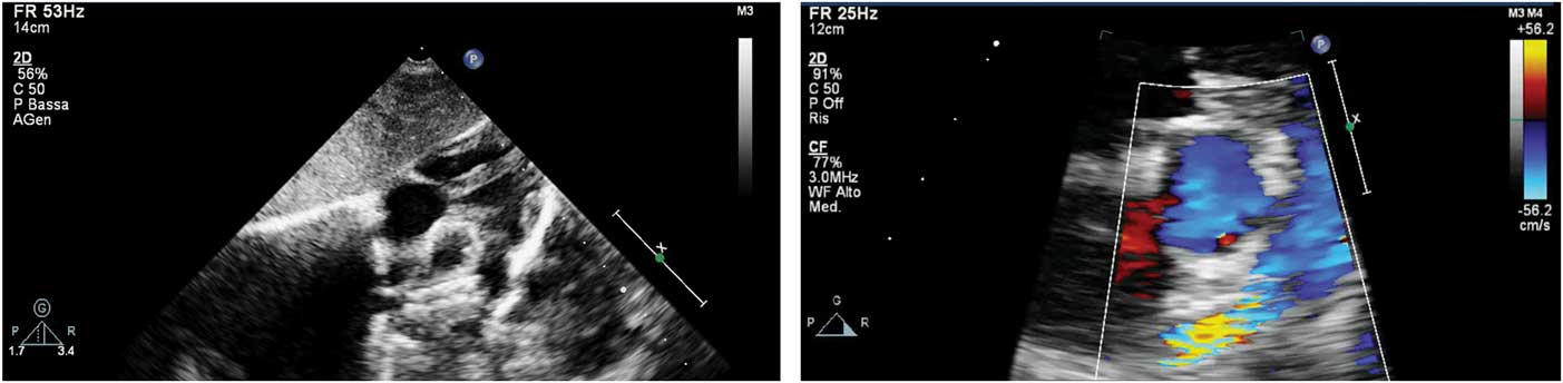
A 28-day-old boy weighing 3.3 kg was admitted because of respiratory distress and echocardiographic suspicion of a disconnected right pulmonary artery. At cardiac catheterisation, disconnection of the right pulmonary artery from the pulmonary trunk with a residual ductal origin was found. Patent ductus arteriosus stabilisation with coronary stent implantation (Multilink Vision 3×18 mm; Abbott, Santa Clara, California, United States of America) was performed. At 5 months of age, with a weight of 6.3 kg, he was electively re-admitted and underwent cardiac catheterisation and surgical correction. The complete unilateral primitive agenesis of the pulmonary branch was treated with a de novo-built conduit. Figure 2 and legend describes all of the details of the procedure. At 2 years of follow-up the pulmonary artery is widely patent with no signs of stenosis (Fig 3, left).
Steps of surgical reconstruction of the atretic pulmonary arterial branch. The complete unilateral primitive agenesis of the pulmonary branch was treated with a de novo-built conduit using a segment of homologous tissue. ( a ) Homologous tissue was obtained from the contralateral dilated pulmonary artery and autologous pericardial tissue. ( b ) A cylindrical conduit measuring 7 mm in diameter was created over an appropriate Hegar probe. ( c ) The biological conduit was connected with an end–end anastomosis between the pulmonary artery and the distal portion of the “atretic” pulmonary artery. ( d ) Finally, a Valeo stent 8×26 mm was implanted to support the surgical arterioplasty. The proximal end of the stent is seen through the “conduit”.

Echocardiographic views in subcostal views showing patent pulmonary artery stent in patient #2 (left) and in patient #3 (right) during follow-up.

Case 3
A 3-month-old boy, weighing 6 kg, was admitted in order to perform diagnostic cardiac catheterisation because of pulmonary arterial hypertension. Examination found disconnection of the right pulmonary artery from the pulmonary artery trunk. Surgical correction was performed similarly to case #2 with Valeo stent implantation (8×26 mm; Bard Peripheral Vascular Inc., Tempe, Arizona, United States of America). At 3 years of follow-up the right pulmonary artery is widely patent (Fig 3, right) and pulmonary pressures are normal.
Discussion
One of the possible embryologic causes of a disconnected pulmonary artery is involution of the proximal sixth aortic arch, destined to become the pulmonary artery, and the persistence of distal sixth aortic arch. After birth, the ductal origin of the disconnected pulmonary artery tends to close, leaving a diverticulum from the innominate artery.Reference Pfefferkorn, Loser and Pech 3 Disconnected pulmonary arteries can be further associated with other cardiac defects or be isolated. These patients may remain asymptomatic but many develop recurrent pulmonary infections, pulmonary arterial hypertension, respiratory distress, haemoptysis, pneumonitis, or bronchiectasis and can stay undetected into their adulthood.Reference Pfefferkorn, Loser and Pech 3
The diagnosis of a disconnected pulmonary artery may be suggested by chest radiography revealing asymmetric lung fields and mediastinal shift. In our cohort, echocardiography was the most sensitive diagnostic tool through the demonstration of the absence of the pulmonary artery bifurcation or pathological Doppler flows. Surgical approaches are based either on the creation of an aorto-pulmonary shunt or on the restoration of continuity between the disconnected vessel to the main pulmonary artery. The choice of strategy is determined by the size of the pulmonary artery. However, despite successful pulmonary artery surgical reconstruction, stenosis of the affected pulmonary artery frequently occurs.Reference Agnoletti, Boudjemline and Bonnet 2 , Reference Stamm, Friehs and Zurakowski 4 Agnoletti et alReference Agnoletti, Boudjemline and Bonnet 2 reported that all the patients in their series underwent cardiac catheterisation and interventional procedures. Stamm et alReference Stamm, Friehs and Zurakowski 4 studied 102 patients and reported that, when both surgical and catheter-based procedures are combined, the freedom from pulmonary angioplasty after construction of pulmonary artery continuity was 54±10% at 1 year and 31±11% at 5 years.Reference Stamm, Friehs and Zurakowski 4 In total, 67% of patients underwent up to five percutaneous balloon dilatations. Because of the high rate of re-stenosis after surgical reconstruction, we developed the different approach here. This included the reconstruction of pulmonary arterial continuity by using autologous tissue (Fig 2). In two cases, a de novo “conduit” was created in theatre. Furthermore, we then supported the arterioplasty by implanting a stent in a hybrid way during the same surgical procedure. The length of the stent was chosen to straddle the arterioplasty, whereas the diameter was chosen 2–3 mm larger than the distal disconnected arterial diameter. All of the procedures were performed in an on-pump normothermic beating heart and were uneventful. All patients showed no signs of stenosis, with stable results at a follow-up ranging from 2 to 6 years. These patients will need stent re-dilation during follow-up because of somatic growth. However, because of open-heart stent implantation, it was easy to implant a large stent or a stent that can then be dilated up to an adult-size diameter.Reference Butera, Giugno and Basile 5
There are several limitations to our report. Primarily this is a small patient cohort. In addition, as for known surgical treatments, the very long-term outcome of patients with reconstructed pulmonary arteries, including the success of transcatheter interventions, are still unknown, with limited reports addressing this issue.
In conclusion, we present a new strategy for disconnected pulmonary arteries, which includes single-stage surgical reconstruction with hybrid stent implantation to support the arterioplasty. Medium-term follow-up results are highly encouraging.
Financial Support
This research received no specific grant from any funding agency, commercial or not-for-profit sectors.
Conflicts of Interest
None.
Ethical Standards
All procedures performed in the study involving human patients were in accordance with the ethical standards of our institution and of the national research committee and with the 1964 Helsinki declaration and its later amendments or comparable ethical standards. Informed consent to the procedure and to the use of personal data for scientific purposes was obtained from all individual patients included in the study.
Acknowledgements
None.