Introduction
Aneurysmal bone cysts are rare benign tumours characterised by rapid growth and compressive destruction of local bone. The most common sites of aneurysmal bone cysts are the limbs.Reference Mankin, Hornicek, Ortiz-Cruz, Villafuerte and Gebhardt1 In recent years, aneurysmal bone cysts of the temporal bone have been reported occasionally; however, few cases of aneurysmal bone cysts with internal auditory canal involvement have been described.
This paper presents a rare case of aneurysmal bone cyst with compressive damage to the internal auditory canal, with clinical presentations of facial palsy, hearing loss and impaired vestibular function. The patient underwent tumour resection and recovered well without recurrence.
Case report
A 51-year-old man presented to our out-patient department with facial nerve palsy symptoms experienced for 2 weeks, as well as a 6-month history of hearing loss and a 1-year period of tinnitus in his right ear. The patient denied any vertigo, otalgia, headache or otorrhoea. On physical examination, the right external auditory canal was clean and the tympanic membrane was thickened.
Pure-tone audiometry of the right ear showed severe sensorineural hearing loss, with an auditory brainstem evoked response threshold of 95 dBnHL and a type B tympanogram curve for acoustic impedance. Vestibular-evoked myogenic potential testing revealed that both cervical and ocular vestibular-evoked myogenic potentials were unresponsive in the right ear, indicating a loss of function affecting saccular, utricular, inferior and superior vestibular nerves. No spontaneous nystagmus was detected. Caloric testing showed reduced right-sided horizontal semicircular canal function. Facial nerve electroneurography showed 100 per cent degeneration. The Burres–Fisch scores for facial nerve function were: static parameters = 6; raised eyebrows = 0; closed eyes = 0; teeth showing = 9; and blowing air = 3.
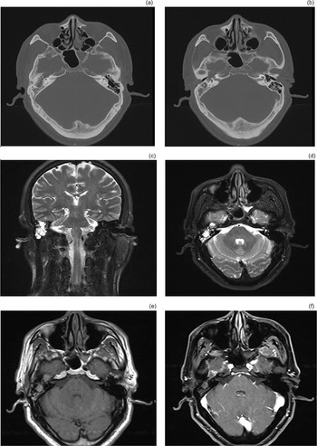
Computed tomography (CT) demonstrated a mass involving the apical and middle turn of the right cochlea, tympanic cavity, and genu of the facial nerve, with localised compressive bone defects and thinning of the middle cranial fossa plate (Figure 1a and b). Magnetic resonance imaging (MRI) showed a cystic lesion with peripheral heterogeneous enhancement involving the skull base, and a ‘soap bubble’ appearance of the fluid level visible within the lesion (Figure 1c–f).
(a & b) Axial computed tomography images: a mass could be seen on the right skull base with compressive bone defects, involving the apical and middle turn of the right cochlea, tympanic cavity, and genu of the facial nerve. (c) Coronal and (d) axial T2-weighted magnetic resonance imaging (MRI) scans. (e) Axial T1-weighted MRI scan. (f) Axial T1-weighted MRI scan with contrast. The mass presented medium signals on T1-weighted MRI and high mixed signals on T2-weighted MRI with peripheral heterogeneous enhancement, presenting a ‘soap bubble’ pattern.

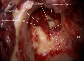
The patient underwent tumour resection, facial nerve decompression and external auditory canal occlusion by a right-sided infratemporal fossa approach. Intra-operatively, the tumour was found to be unencapsulated with an appearance of ‘fish flesh’. It appeared to be insinuating into various foramina, with erosion of the vestibular labyrinth, the cochlear apex, the dura of the skull base and the internal auditory canal (Figure 2), where the vestibulocochlear and facial nerves were surrounded and compressed by the lesion. The facial nerve was wrapped by the tumour, and was highly swollen but intact.
The tumour has invaded the cochlea and the inner auditory canal. FN = facial nerve

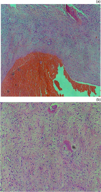
Histopathological evaluation of the bone and soft tissue specimens revealed characteristic features of an aneurysmal bone cyst, including scattered blood-filled cystic spaces without endothelial cells. The fibrous septa contained spindle cells, giant polynuclear cells and osteoclastic giant cells. Some cells were mildly heterogeneous, and scattered bone tissue and bone-like tissue were seen (Figure 3).
Microscopic sections demonstrate features of aneurysmal bone cyst. (a) Blood-filled cystic cavity (H&E; ×100). (b) Fibrous tissue septa surrounding the cavity with osteoid formation in sheet-like patterns, with no cellular atypia (H&E; ×100).

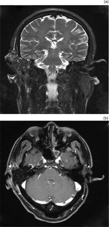
Post-operatively, the patient complained of vertigo and nausea 3 days post-operation, and displayed a horizontal (-torsional) spontaneous nystagmus toward the affected ear. His nystagmus persisted. Facial nerve function partially recovered, with Burres–Fisch scores as follows: static parameters = 14; raised eyebrows = 0; closed eyes = 9; exposed teeth = 21; and blowing air = 7. However, the exact extent of vestibular function recovery could not be determined because tamponade of the surgical cavity and closure of the external auditory canal intra-operatively prevented further vestibular-evoked myogenic potential testing to assess vestibular function. The MRI scan did not reveal any residual neoplasms (Figure 4).
(a) Coronal T2-weighted magnetic resonance imaging (MRI) scan. (b) Axial T1-weighted MRI scan with contrast. The tumour was removed without residue.

The patient was kept under close follow up, with no recurrence at five years of follow up. His vertigo resolved after three months of vestibular rehabilitation.
Discussion
Aneurysmal bone cysts were first described in 1942 by Henry L Jaffe and Louis Lichtenstein, and were defined as locally aggressive, blood-filled cystic lesions of the skeletal system.Reference Jaffe and Lichtenstein2 The pathogenesis of aneurysmal bone cysts remains unclear. Edling believed that aneurysmal bone cysts are a manifestation of fibrous dysplasia of bones or a defect in epiphyseal plate development, but this does not explain their occurrence in mature bone.Reference Edling3 Lichtenstein suggested that aneurysmal bone cysts may be caused by local circulatory disturbances due to sudden vascular occlusion of the venous drainage or the occurrence of arteriovenous shunts.Reference Lichtenstein4 Aneurysmal bone cysts are also thought to be secondary to an underlying vascular malformation due to a pre-existing primary bone lesion, such as osteoblastoma, chondroblastoma, chondromyxoid fibroma and giant cell tumour.Reference Wong, Chantharasamee, Nelson, Eckardt, Motamedi and Hornicek5,Reference Gutierrez, Link, Horvai, Joseph, O'Donnell and Motamedi6 Trauma is additionally considered to be a cause of aneurysmal bone cyst development.Reference Gutierrez, Link, Horvai, Joseph, O'Donnell and Motamedi6 Our reported patient denied having any pre-existing lesion of the temporal area or any history of trauma. Thus, our case can be considered as a primary aneurysmal bone cyst.
A review of the literature indicated that there have been 21 patients with aneurysmal bone cysts in the temporal bone, with the most common sites of involvement being the petrous apex, the squamous portions and the mastoid.Reference Blanchard, Abergel, Williams and Ayache7–Reference Saez, Sharma, Barnes, Lu, Hsu and Huoh9 Very few cases involve the internal auditory canal. Symptoms of aneurysmal bone cysts depend on the location and size of the lesion. The most common manifestations of temporal aneurysmal bone cysts are painless masses,Reference Saez, Sharma, Barnes, Lu, Hsu and Huoh9,Reference Wang, Cao and Liu10 headache,Reference Kim, Lee, Chang, Jung, Kim and Sohn11,Reference Kletke, Popovic, Algird, Alobaid and Reddy12 ear pain,Reference Sharma, Velho and Kharosekar13 tinnitus,Reference Kletke, Popovic, Algird, Alobaid and Reddy12 hearing loss,Reference Sharma, Velho and Kharosekar13,Reference Sayama and Macdonald14 facial paralysis,Reference Kletke, Popovic, Algird, Alobaid and Reddy12,Reference Sayama and Macdonald14 vertigoReference Sharma, Velho and Kharosekar13,Reference Kumar, Retnam, Krishnamoorthy, Parameswaran, Nair and Bhattacharya15 and otorrhoea.Reference Sharma, Velho and Kharosekar13
Our patient presented with facial palsy and hearing loss, accompanied by loss of vestibular function (based on vestibular-evoked myogenic potential testing results), all of which indicated that nerves running through the internal auditory canal were involved. Based on the absence of vertigo and the pre-operative vestibular-evoked myogenic potential testing results, we presumed that the patient's vestibular nerve was completely damaged because of tumour compression of the inner auditory canal, and therefore vertigo could not have occurred post-operatively. Interestingly, the patient developed vertigo 3 days after surgery, which implies that vestibular function was partially restored after tumour resection and decompression therapy, resulting in a new binaural imbalance. Whether this situation arises from reversible damage to the vestibular nerve and labyrinth or from neuroregenerative ability is unclear. We presume that segments of the facial and vestibular nerves located in the internal auditory canal, rather than the vestibular end organs or the arteries supplying the inner ear, were suppressed by the tumour. Once the suppression was alleviated, nerve impulse conduction was restored, followed by partial restoration of nerve function. Further clinical data are needed to clarify these findings. Fortunately, the patient's vertigo disappeared after vestibular rehabilitation.
Aneurysmal bone cysts can be visualised radiographically. Multiple small fluid interfaces are an important feature of aneurysmal bone cysts on CT, which represents the sedimentation of red blood cells within the blood-filled cavities. In addition, aneurysmal bone cysts demonstrate expansile bone destruction, with patchy and banded shadows indicating ossification.Reference Hashemi, Heidarpour, Eshaghian, Ansari, Hashemi and Yaghoobi16,Reference Gotecha, Punia, Chugh, Patil, Kashyap and Raghu17 Characteristic MRI findings include distinct expansile lesions surrounded by a hypointense fibrous capsule shadow. Because of the formation of a fibrous envelope, sclerotic rim and bone shell,Reference Edling3 there may be a hypointense internal septum that separates the lesion into multi-cystic compartments, appearing as a ‘soap bubble’ pattern.Reference Unadkat, Adams, Richards and Bajaj18 In our case, the lesion was mainly concentrated within the tympanic cavity, with no extending compressive bone destruction or absorption; this was probably because the lesion was at an early stage.
The only way to definitively diagnose aneurysmal bone cyst is by histopathological examination; a positive diagnosis is based primarily on a honeycomb-appearing cyst, consisting of multiple blood-filled sinusoidal spaces, with bone destruction and osteoid formation. These cysts are separated by fibrous septa without endothelial cells, and various structures exist within the lesion, including haemosiderin, osteoclasts, polynuclear macrophages, inflammatory cells and new bone.Reference Kim, Lee, Chang, Jung, Kim and Sohn11,Reference Kletke, Popovic, Algird, Alobaid and Reddy12 Therefore, aneurysmal bone cysts cannot be diagnosed by needle biopsy.
En bloc resection is the ‘gold standard’ treatment and can usually result in good local control.Reference Gotecha, Punia, Chugh, Patil, Kashyap and Raghu17 The likelihood of recurrence is related to the patient's age, lesion size, mitotic stage and completeness of surgical resection. Given the rapidly expansile nature of aneurysmal bone cysts, early surgical intervention is required; the risk of intracranial and intradural extension is high. Considering the highly vascular nature of these tumours, pre-operative embolisation is often required, to control intra-operative and peri-operative bleeding. Radiotherapy is recommended for patients with residual or recurrent lesions post-operatively and deep skull base lesions; whether radiotherapy can induce sarcoma transformation still requires extensive clinical data to confirm.Reference Kumar, Singh, Phadke, Diwakar, Agarawal and Jain19,Reference Sodhi, Salunke, Agrawal and Gupta20
• Aneurysmal bone cyst involving the internal auditory canal is rare; petrous apex, squamous portions and mastoid are most commonly involved
• Temporal aneurysmal bone cyst manifestations include painless masses, headache, ear pain, tinnitus, hearing loss, facial paralysis, vertigo and otorrhoea
• Our patient presented with facial palsy, and loss of hearing and vestibular function, indicating damage to nerves running through internal auditory canal
• Vestibular function was restored post-operatively, which is uncommon
• Computed tomography and magnetic resonance imaging are often utilised in diagnosis, and histopathology is the confirmative examination
• Surgery is the ‘gold standard’ treatment
Conclusion
This paper reports a rare case of aneurysmal bone cyst involving the internal auditory canal. Clinical features of our patient included facial palsy, and loss of hearing and vestibular function, indicating concurrent involvement of the facial and vestibulocochlear nerves. The patient developed post-operative vertigo after tumour compression was alleviated, suggesting recovery and indicating that damage to the vestibular nerve was reversible. Surgical excision was conducted prior to other medical treatments, and no recurrence has been documented.
Acknowledgements
This work was financially supported (grant numbers: NSFC 81371093 and 18ZR1406100).
Competing interests
None declared