Introduction
Polyvinylpyrrolidone (PVP) is a synthetic, water-soluble polymer derived from N-vinylpyrrolidone, originally developed for use as a plasma substitute. Over time, it has found widespread application in medical, pharmaceutical, and industrial settings due to its low cost, biocompatibility, and ease of handling (Kurakula and Rao, Reference Kurakula and Rao2020). However, hypersensitive reaction on mucosal and serum contact to PVP has been a limitation to its use (Adachi et al., Reference Adachi, Fukunaga, Hayashi, Kunisada and Horikawa2003; Grossman et al., Reference Grossman, Tiefenthaler-Gilmer, Raysz and Kesper2007).
In the context of assisted reproductive technologies (ART), particularly intracytoplasmic sperm injection (ICSI), PVP is commonly used to increase the viscosity of the medium, allowing for easier immobilization and manipulation of spermatozoa (Kimura and Yanagimachi, Reference Kimura and Yanagimachi1995; Gianaroli et al., Reference Gianaroli, Plachot, van Kooij, Al-Hasani, Dawson, DeVos, Magli, Mandelbaum, Selva and van Inzen2000). Additionally, it reduces the volume of fluid transferred into the oocyte during injection and facilitates sperm selection (Hlinka et al., Reference Hlinka, Herman, Vesela, Hredzak, Horvath and Pacin1998). However, concerns regarding the biological safety of PVP have been raised. Studies have reported potential cytotoxic effects, including disruption of sperm membrane integrity, mitochondrial dysfunction, increased DNA fragmentation, and delayed oocyte activation (Sabour et al., Reference Sabour, Agha-Rahimi, Dehghani Ashkezari, Seifati, Anbari and Nabi2022; Ding et al., Reference Ding, Wang, Li, Chen, Zou, Ji, Hao, Xue, Zou and Wei2020; Akbari Javar et al., Reference Akbari Javar, Aflatoonian, Talebi, Dehghanpour, Khalili and Montazeri2024). Despite these concerns, PVP remains routinely used in ICSI procedures. This review aims to critically evaluate the impact of PVP on sperm function, oocyte integrity, and embryonic development within the ART setting.
Methods
A systematic search was conducted on online published manuscripts with the following keywords: ‘Polyvinylpyrrolidone’ OR ‘Oocytes’ OR ‘Sperm’ OR ‘embryo Development’ AND ‘ICSI’ OR ‘IVF’ on online scientific databases including MEDLINE, Scopus, Web of knowledge, CINAHL. English written manuscripts up to 2024 were included. The studies were revised by authors blindly and extracted data were included in checklists.
Potential risk of PVP on spermatozoa
ICSI procedure is one of the most widely used ART for reproduction in infertile couples (Chamayou et al., Reference Chamayou, Giacone, Cannarella and Guglielmino2023). PVP is commonly used in ICSI for domestic animals and humans, because it facilitates the transportation and immobilization of sperm cells (Kato and Nagao, Reference Kato and Nagao2012). Exposure of spermatozoa to PVP causes changes in the sperm structure, and nucleus deformation (Kato and Nagao, Reference Kato and Nagao2012). Strehler et al. evaluated the human spermatozoa ultrastructure using Transmission Electron Microscopy (TEM). In their study, 0.1 ml of each patient’s sperm suspension was mixed with 0.5 ml of 10%PVP solution in HEPES buffer and incubated for 30 min. Electron microscopy studies have revealed that PVP damages key components of sperm ultrastructure, including acrosomal content, axonemal tubules, fibrous sheath, and mitochondrial arrangement (Strehler et al., Reference Strehler, Baccetti, Sterzik, Capitani, Collodel, De Santo, Gambera and Piomboni1998). In addition, mitochondria were sensitive to PVP, and their shape and helical assembly worsened compared to the untreated sample. As a secondary consequence, it induced the eventual necrotic process, chromatin alteration, and cytoskeletal components (Strehler et al., Reference Strehler, Baccetti, Sterzik, Capitani, Collodel, De Santo, Gambera and Piomboni1998). In 2015, Nel et al. mixed sperm samples with PVP and observed large pale nuclear spaces, dense nuclear density, as well as the 9d + 2s pattern of the axoneme principal piece under TEM. Furthermore, prolonged exposure to 10%PVP induced chromatin condensation defects and large nuclear vacuoles, suggesting impaired nuclear integrity (Nel, Reference Nel2015). In the study by Kato and Nagao (Reference Kato and Nagao2009), sperm incubated in SOFPVA supplemented with 10%PVP (SOFPVP) at 39°C under 5% CO2 in air with high humidity for 0, 15, 30, and 60 min. They reported that PVP induced the acrosome reaction, because PVP caused Ca2+ influx into sperm cells by damaging the sperm plasma membrane (Kato and Nagao, Reference Kato and Nagao2009). Later, Sabour and colleagues evaluated the effects of PVP on the sperm structure by SEM at different time intervals (0, 15, 30 and 60 min). Their results showed that the plasma membrane was disrupted after 15 min, and the high mitochondrial membrane potential and acrosome-intact were significantly decreased (Sabour et al., Reference Sabour, Agha-Rahimi, Dehghani Ashkezari, Seifati, Anbari and Nabi2022).
There is significantly more acrosome reaction in bull sperm cultured in PVP (Motoishi et al., Reference Motoishi, Goto, Tomita, Ookutsu and Nakanishi1996). The mechanism for this difference was higher intracellular Ca2+ ion efflux following PVP injection (Ahlström et al., Reference Ahlström, Westin, Reismer, Wikland and Hardarson2011). Another hypothesis suggested that more calcium ion efflux in PVP-induced cells is probably due to adenylate-cyclase activity (Dragileva et al., Reference Dragileva, Rubinstein and Breitbart1999). Sperm binding to zona pellucida (ZP) activates the G-protein function which triggers the cation transformation within the cell membrane (World Health Organization, 1999; Suarez, Reference Suarez2006). Using PVP prior to ICSI in order to immobilize sperm causes sperm plasma membrane damages. Because of this damage, thiol-reducing agents gain access to the sperm nucleus, and PVP possibly inhibits sperm nucleus decondensation (Jean et al., Reference Jean, Mirallié, Boudineau, Tatin and Barrière2001).PVP can destroy the functional sperm mitochondrial membrane and tail integrity. Since, sperm motility is inevitable in fertilization (Bray et al., Reference Bray, Gunnell and Smith2006), the role of the sperm tail is considerable in this scenario. It has been reported that different time of sperm exposures to PVP can affect subsequent cleavage divisions and embryo development rates (Yari et al., Reference Yari, Etebary, Jahromi and Khalili2013). Long exposure of spermatozoa to PVP could damage the membrane and chromatin integrity of sperm cells (De Mouzon et al., Reference De Mouzon, Goossens, Bhattacharya, Castilla, Ferraretti, Korsak, Kupka, Nygren and Nyboe Andersen2010; Jean et al., Reference Jean, Mirallié, Boudineau, Tatin and Barrière2001). Salian et al. suggested that exposure of sperm to 10% PVP solution for a prolonged time has negative effects on DNA integrity and fragmentation (Salian et al., Reference Salian, Kalthur, Uppangala, Kumar and Adiga2012). In another study, Nabi and co-workers showed that exposure time more than 10 min had a detrimental effect on DNA and viability of spermatozoa (Nabi et al., Reference Nabi, Entezari, Miresmaeili, Vahidi, Lorian, Anbari and Motamedzadeh2022). Also, others showed that prolonged human sperm exposure of 15 min to PVP, more significant detrimental effects took place on viability, morphology, DNA status and chromatin structure (Sabour et al., Reference Sabour, Agha-Rahimi, Dehghani Ashkezari, Seifati, Anbari and Nabi2022). Emerging evidence suggests that PVP exposure during ICSI may also induce epigenetic alterations and oxidative stress, which could have downstream implications for offspring health. On the other hand, incubation in a 10%PVP medium for periods of 15 to 60 min significantly increased malondialdehyde (MDA) levels, an indicator of lipid peroxidation. Additionally, expression levels of key genes involved in apoptosis (BAX, BCL2), stress response (HSP70/HSPA2), and antioxidant defence (SOD2) were significantly altered after PVP exposure, indicating disturbance of the epigenetic regulatory network in spermatozoa (Sabour et al., Reference Sabour, Agha-Rahimi, Dehghani Ashkezari, Seifati, Anbari and Nabi2022).
Recently, Akbari and colleagues (2024) incubated sperm cells in two different concentrations of PVP (5 and 10%) for 10 min. Their results indicated that 10%PVP compared to 5% and free-PVP lead to significant increased SDF. Additionally, the expression levels of BCL2 and the HSP70 genes were significantly higher in 5% compared to 10%PVP, while the transcript levels of the BAX gene were lower in 5%PVP (Akbari Javar et al., Reference Akbari Javar, Aflatoonian, Talebi, Dehghanpour, Khalili and Montazeri2024). Although, direct studies on embryos remain limited, elevated oxidative stress and gene expression changes in the sperm could feasibly affect DNA methylation and histone modifications during early embryo formation due to critical window of epigenetic reprogramming. While, more research is needed on the effect of PVP on embryo and offspring, these findings warrant cautious consideration of exposure time and concentration in clinical settings. As PVP is water-soluble, it could easily breakdown the sperm membrane and pass through it. However, damaged sperm nuclei occur by chromosomal aberration. Also, PVP delays sperm calcium oscillations on oocytes (Kato and Nagao, Reference Kato and Nagao2009; Morozumi et al., Reference Morozumi, Shikano, Miyazaki and Yanagimachi2006). So, sperm degradation is more probable following PVP injection, especially with higher concentration.
Potential risk of PVP on embryonic cells
In ICSI, after placing the sperm in the environment containing PVP, a selected spermatozoon is injected into the oocyte along with a small amount of PVP. Also, due to the high molecular weight of PVP (36,000 kDa), it remains in embryo during cleavage divisions (Figure 1)(Kato and Nagao, Reference Kato and Nagao2009). In oocytes, PVP residue remaining after ICSI can delay or inhibit calcium (Ca2) oscillations, which is crucial for oocyte activation. This is likely due to the high molecular weight of PVP, which impairs the diffusion and function of sperm-derived phospholipase C zeta (PLCζ), the key initiator of Ca2 release via IP3 signalling. Additionally, PVP exposure disrupts the sperm plasma membrane, leading to premature acrosome reaction and higher intracellular Ca2+ levels, which impair mitochondrial function and reduce membrane potential (Hlinka et al., Reference Hlinka, Herman, Vesela, Hredzak, Horvath and Pacin1998; Kato and Nagao, Reference Kato and Nagao2012; Sabour et al., Reference Sabour, Agha-Rahimi, Dehghani Ashkezari, Seifati, Anbari and Nabi2022). These PVP residues in the cytoplasm prevent the sperm head decondensation and oocyte activation, which affect fertilization and embryo development. Nuclear decondensation is linked to impaired protamine-DNA unpacking due to oxidative and structural membrane injury (Ding et al., Reference Ding, Wang, Li, Chen, Zou, Ji, Hao, Xue, Zou and Wei2020; Hlinka et al., Reference Hlinka, Herman, Vesela, Hredzak, Horvath and Pacin1998).
PVP solution in embryonic samples. A. PVP was not observable in embryo similar to control group. B. PVP remained in injection solution. C. PVP remained in oocyte. The location of PVP in sample solution was determined by arrows (Kato and Nagao, Reference Kato and Nagao2009).

Figure 1 Long description
The image consists of three microscopic views of embryonic samples treated with polyvinylpyrrolidone (PVP) solution. In the first panel (A), the embryo appears similar to the control group, with no observable PVP. The second panel (B) shows the injection solution where PVP remains visible, indicated by a red arrow. The third panel (C) displays an oocyte with PVP still present, also marked by a red arrow. The arrows highlight the specific locations where PVP is detected within the samples. The scale bar at the bottom of each image indicates 50 micrometers, providing a reference for the size of the observed structures.
In order to evaluate the effect of PVP on cow embryos growth after IVF, the ZP was drilled by applying several piezo pulses. Then, PVP was injected into pronuclear stage zygotes. The injection suppressed the growth of embryos, and the rates of cleavage and blastocyst formation decreased significantly. However, there was no change in the chromosomal integrity of blastocysts (Kato and Nagao, Reference Kato and Nagao2009). Also, PVP injection noticeably lowered the rates of cleavage and blastocyst formation in bovine embryos (Ozil et al., Reference Ozil, Banrezes, Tóth, Pan and Schultz2006). The results suggested that the rate of blastocysts was significantly higher in control group (without PVP) in comparison to PVP injected specimen (Feichtinger et al., Reference Feichtinger, Obruca and Brunner1995; Mizuno et al., Reference Mizuno, Hoshi and Huang2002). Other studies indicated that PVP could affect embryo via chromosomal aberration (Bras, Reference Bras1994) and development stage arrest (Perlman et al., Reference Perlman, Zhang, Chen, Walsh and Buttyan1999). It is noteworthy that all the aforementioned findings were only attributed to mouse and bovine models and cannot be experimented on human models, due to ethical challenges (Arcidiacono et al., Reference Arcidiacono, Walt, Campana and Balerna1983). Studies compared different commercial PVP preparations such as Vitrolife, Sigma, Fertipro, Irvine, and Medicalt. Significant variation in cleavage rates in embryonic development were noted; although, no significant differences in clinical pregnancy rates were observed (cleavage rate: Vitrolife ∼ 94% vs Sigma ∼ 48%). These observations suggest that manufacturers’ differences in PVP purification, solvent composition, and molecular characteristics may influence early developmental outcomes. However, the lack of studies on clinical comparison indicates a critical gap in quality control and standardization of PVP sources in ART (Kato and Nagao, Reference Kato and Nagao2012).
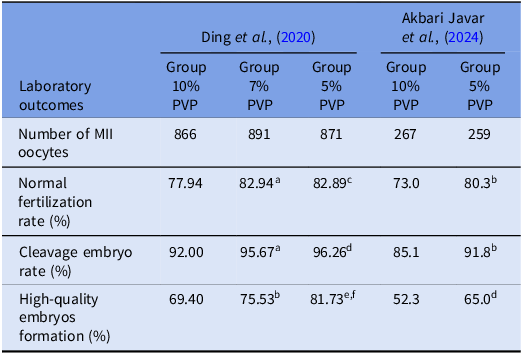
There was no defined deterministic effect of PVP on embryo prior to implantation stage (Mizuno et al., Reference Mizuno, Hoshi and Huang2002). The difference in the rate of cleavage could be attributed to differences in the used concentration of PVP (Arcidiacono et al., Reference Arcidiacono, Walt, Campana and Balerna1983; Dozortsev et al., Reference Dozortsev, Rybouchkin, De Sutter and Dhont1995). Ding et al investigated the effects of three concentrations of PVP (10, 7 and 5%) in 210 ICSI treatment cycles. The findings indicated that the high-quality cleavage embryo rate was significantly higher in 5% compared to the 7% group. Moreover, high-quality embryo and blastocyst rates were significantly higher in the 5% group. These 210 ICSI cycles were divided into two subgroups (high-/intermediate-quality semen group poor-quality semen group). They reported that in the low-quality semen group, 5% PVP significantly increased the cleavage rate of high-quality embryos and blastocysts. Also, in the high/intermediate quality semen group, the rate of fertilization, cleavage, high quality embryo and blastocyst in the 5% PVP group was higher than the 10% group. Therefore, it was concluded that 5% PVP increased embryo development in ICSI cycles (Ding et al., Reference Ding, Wang, Li, Chen, Zou, Ji, Hao, Xue, Zou and Wei2020). Similarly, Akbari and associates evaluated the effects of two concentrations of PVP (10 and 5%) on clinical ICSI Outcomes. They demonstrated that fertilization and high-quality embryo formation rates were increased in 5% PVP concentration. Also, they stated that the pregnancy outcomes in the lower PVP concentrations group were on increasing trend. Furthermore, time lapse analysis showed that the kinetics of embryos were beneficial in the low concentration of PVP. The abnormal cleavage patterns, such as the rates of fragmentation, uneven blastomeres, and reverse cleavage were significantly lower in the embryos derived from 5% PVP (Akbari Javar et al., Reference Akbari Javar, Aflatoonian, Talebi, Dehghanpour, Khalili and Montazeri2024). The effect of PVP on fertilization and clinical outcomes is illustrated in Table 1.
Studies on PVP effects on clinical ICSI outcomes

Table 1 Long description
The table compares the effects of different polyvinylpyrrolidone (PVP) concentrations on intracytoplasmic sperm injection (ICSI) outcomes in two studies by Ding et al. (2020) and Akbari Javar et al. (2024). It has four rows and six columns. The columns are labeled with the studies and PVP concentrations: Ding et al., (2020) with groups 10% PVP, 7% PVP, and 5% PVP, and Akbari Javar et al., (2024) with groups 10% PVP and 5% PVP. The rows are labeled with laboratory outcomes: Number of MII oocytes, Normal fertilization rate (percentage), Cleavage embryo rate (percentage), and High-quality embryos formation (percentage). The table shows that in Ding et al., (2020), the 5% PVP group had higher normal fertilization rates, cleavage embryo rates, and high-quality embryo formation rates compared to the 7% and 10% PVP groups. Similarly, in Akbari Javar et al., (2024), the 5% PVP group had higher normal fertilization rates and high-quality embryo formation rates compared to the 10% PVP group. The data suggests that lower concentrations of PVP may improve ICSI outcomes.
PVP: Polyvinylpyrrolidone, ICSI: Intracytoplasmic sperm injection. Different symbols within columns and different letters within columns and within rows indicate significant differences.
a P < 0.01, compared with 10% PVP group;
b,c P < 0.05, compared with 10% PVP group;
d P < 0.001, compared with 10% PVP group;
e P < 0.0001, compared with 10% PVP group;
f P < 0.01, compared with the 7% PVP group (Ding et al., Reference Ding, Wang, Li, Chen, Zou, Ji, Hao, Xue, Zou and Wei2020).
Potential risk of PVP on oocyte
Since, PVP has a large molecular weight, it is more difficult to diffuse out of the oocyte membrane and digested by lysosomal enzymes (Dozortsev et al., Reference Dozortsev, Rybouchkin, De Sutter and Dhont1995). Cells in contact with PVP secrete mucin which degrades cell-to-cell adhesion in oocyte cytoplasm (Jean et al., Reference Jean, Mirallié, Boudineau, Tatin and Barrière2001). Although, no significant differences were reported between natural ICSI and IVF pregnancies, it has been suggested that the relatively higher abortion rate observed in ART- pregnancies may be partially associated with exposure to PVP or its metabolic byproducts. However, this association has not been conclusively established and requires further investigation (Huang et al., Reference Huang, Lee, Lu, Chen, Li and Hwu2015). Interestingly, while PVP is often associated with adverse effects on gametes and embryos, some evidence suggests that it may play a protective mechanical role during micromanipulation. In Piezo-assisted ICSI (Piezo-ICSI), where a piezoelectric pulse is used to penetrate the oolemma, the use of PVP has shown to reduce the risk of oocyte membrane rupture. This is likely due to PVP’s ability to increase the viscosity of the medium, thus stabilizing the sperm and reducing shear stress at the injection point. As a result, PVP can prevent oolemma lysis caused by abnormal rupture of the plasma membrane during Piezo-ICSI, potentially improving oocyte survival post-injection (Mizuno et al., Reference Mizuno, Okitsu, Goto, Kusuhara and Kusuhara2024). These findings highlight the context-dependent effects of PVP, emphasizing the importance of concentration, timing, and technique in determining its impact on ART outcomes.
Hyaluronic acid and its comparison with PVP
PVP solution is used in the ICSI technique, which facilitates sperm immobilization, aspiration, and injection into the ooplasm (Kimura and Yanagimachi, Reference Kimura and Yanagimachi1995). But, concerns during the ICSI process are related to bypassing the natural barriers to sperm selection. Therefore, the use of a physiologically and more friendly alternative sperm handling/injection medium can alleviate the concerns (Hirao et al., Reference Hirao, Itoh, Shimizu, Iga, Aoyagi, Kobayashi, Kacchi, Hoshi and Takenouchi2004).
HA and PVP are both used as media for sperm selection and immobilization during ICSI. Unlike PVP, which is a synthetic polymer with potential cytotoxic effects, HA is a natural component of the cumulus matrix surrounding the oocyte and can selectively bind mature sperm with intact DNA and membrane integrity. Therefore, comparing HA with PVP highlights the benefits of a more physiological and less harmful alternative for sperm handling in ART. (Gianaroli et al., Reference Gianaroli, Plachot, van Kooij, Al-Hasani, Dawson, DeVos, Magli, Mandelbaum, Selva and van Inzen2000; Parmegiani et al., Reference Parmegiani, Cognigni, Bernardi, Troilo, Ciampaglia and Filicori2010). Mature spermatozoa that have HA receptors can reach the oocyte and fertilize it (Hlinka et al., Reference Hlinka, Herman, Vesela, Hredzak, Horvath and Pacin1998; Gianaroli et al., Reference Gianaroli, Plachot, van Kooij, Al-Hasani, Dawson, DeVos, Magli, Mandelbaum, Selva and van Inzen2000). Liu and co-workers reported that PVP might be toxic to human gametes and embryos. However, HA causes less fragmentation on DNA molecules; so, it is expected to cause less damage on spermatogenesis and oogenesis in comparison to PVP. Liu et al. evaluated the effect of sperm selection and ICSI on subsequent fertilization and embryo development using the HA-based Sperm. They showed normal fertilization rate increased in the HA-ICSI group compared to the PVP-ICSI group. But, the proportion of good quality embryos was similar in both groups (Hirao et al., Reference Hirao, Itoh, Shimizu, Iga, Aoyagi, Kobayashi, Kacchi, Hoshi and Takenouchi2004; Liu et al., Reference Liu, Feenan, Chapple, Roberts and Matson2019). Different studies concluded that HA significantly improves embryo quality and implantation (Huang et al., Reference Huang, Lee, Lu, Chen, Li and Hwu2015). But, the beneficial effects of HA-based selection of spermatozoa for ICSI are still controversial. Further studies are needed to categorize patients that might benefit from such a method. One study identified couples with previous ICSI failure as a category of infertile patients that may benefit from HA sperm selection before ICSI (Hamano et al., Reference Hamano, Li, Qian, Funauchi, Furudate and Minato1999).
Canepa and associates randomly injected seven oocytes by conventional PVP-ICSI and seven oocytes by HA-ICSI. The findings showed that HA-ICSI increased the proportion of oocytes with 2PN after injection (Hlinka et al., Reference Hlinka, Herman, Vesela, Hredzak, Horvath and Pacin1998; Canepa et al., Reference Canepa, Casciano, De Leo, Massarotti, Anserini, Remorgida and Scaruffi2019). Considering that the HA-based sperm selection technique is more expensive and time consuming, does not support using it as a routine method (Gianaroli et al., Reference Gianaroli, Plachot, van Kooij, Al-Hasani, Dawson, DeVos, Magli, Mandelbaum, Selva and van Inzen2000; Tzur-Tseva et al., Reference Tzur-Tseva, Weiss, Ermoshkin, Goldman, Geslevich and Beck2018). In Table 2, fertilization, clinical pregnancy and implantation rates are compared between HA and PVP Groups.
The risk of PVP and HA during ICSI treatments in reviewed studies

Table 2 Long description
A table comparing fertilization, clinical pregnancy, and implantation rates between HA and PVP groups across four studies. The table has four rows and five columns. The rows are labeled with outcomes: fertilization rate, clinical pregnancy rate, and implantation rate. The columns are labeled with the names of the studies: Kato et al, Jean et al, Fridrisch et al, and Meng et al. Each study compares two groups: Group HA and Group PVP. The fertilization rate for Kato et al is seventy-two point six percentage for Group HA and seventy-four point six percentage for Group PVP. For Jean et al, the fertilization rate is seventy percentage for Group HA and seventy-five percentage for Group PVP. Fridrisch et al shows a fertilization rate of ninety-one point six percentage for Group HA and eighty-five point eight percentage for Group PVP. Meng et al reports a fertilization rate of ninety-three point four percentage for Group HA and eighty-seven point one percentage for Group PVP. The clinical pregnancy rate for Kato et al is fifty percentage for Group HA and thirty-eight point five percentage for Group PVP. Jean et al shows a clinical pregnancy rate of forty-one point seven percentage for Group HA and forty-three point two percentage for Group PVP. Fridrisch et al reports a clinical pregnancy rate of twenty-four point eight percentage for Group HA and twenty point nine percentage for Group PVP. Meng et al shows a clinical pregnancy rate of thirty-two point eight percentage for Group HA and twenty-one point six percentage for Group PVP. The implantation rate for Kato et al is eighteen point six percentage for Group HA and fourteen percentage for Group PVP. Jean et al shows an implantation rate of eighteen point one percentage for Group HA and nineteen point one percentage for Group PVP. Fridrisch et al reports an implantation rate of twelve point four percentage for Group HA and ten point two percentage for Group PVP. Meng et al shows an implantation rate of seventeen point one percentage for Group HA and ten point three percentage for Group PVP.
PVP: Polyvinylpyrrolidone, HA: Hyaluronic Acid.
Conclusion
Application of conventional 10% PVP can be harmful in ICSI by triggering acrosome reaction and lowering fertilization and embryo development process. In addition, PVP can be maintained in embryonic cells for a long time and result in reducing blastocyst formation. Therefore, ART-aided pregnancies with lower concentration of PVP (5%) are more successful.
Funding
There was no funding.
Competing interests
The authors declare none.