Introduction
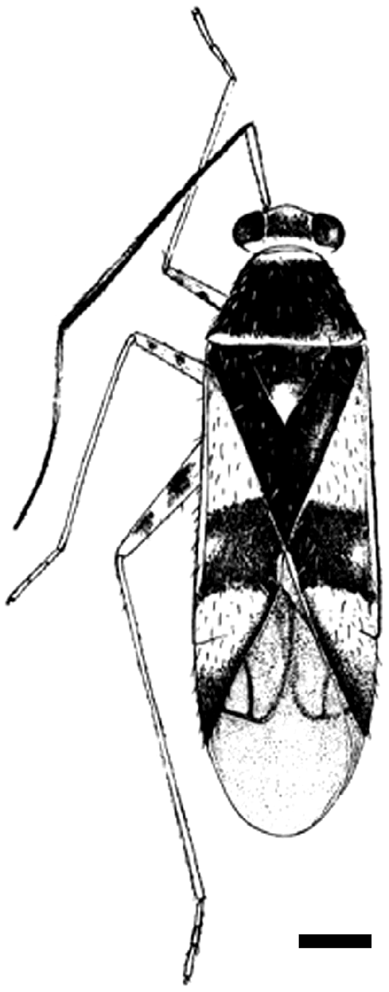
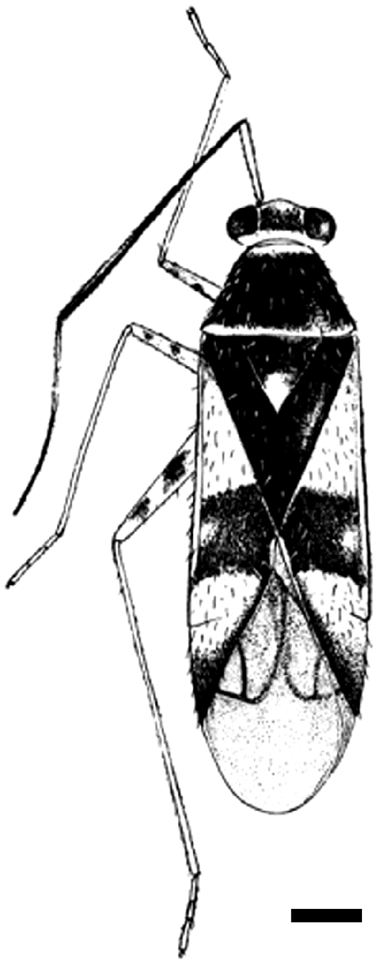
Adparaproba gabrieli Carvalho (Miridae: Orthotylinae) is a phytophagous bug (4.0 mm in length), with the following diagnostic characters: general colouration pale with a black transverse spot on the corium, pale only next to the embolium; black abdomen and the distinct characteristics of male genitalia (Fig. 1; Carvalho, Reference Carvalho1987). The Orthotylinae subfamily has been the object of several projects, the most important being the Plant Bug Planetary Biodiversity Inventory (http://research.amnh.org/pbi/), a global study that brings together biodiversity information on two closely related Miridae subfamilies, Orthotylinae and Phylinae. One of the goals of this study is to increase the number of known host plants as most Orthotylinae and Phylinae are host specific. Until now, knowledge about A. gabrieli has been restricted to its description and geographical distribution, in Juiz de Fora and Cataguases counties, Minas Gerais state, Brazil (Carvalho, Reference Carvalho1987), but data about host plants, behaviour and internal morphology are lacking. Therefore, in this study, we describe the digestive and reproductive tracts of male and female A. gabrieli, and possible identification of its host plant.
Schematic drawing of the dorsal view of Adparaproba gabrieli. Bar = 0.5 mm
Materials and methods
Fifteen adults of A. gabrieli, six males and nine females, were collected at Universidade Federal de Viçosa (20°45′S; 42°52′W; 680 m above sea level) from a Piper sp. (Piperaceae) in a semideciduous seasonal forest. Voucher specimens were deposited in Viçosa's Regional Museum of Entomology (UFVB).
The specimens were transferred to Zamboni fixative solution (Stefanini et al., Reference Stefanini, De Martino and Zamboni1967) for 24 h. After the removal of wings and legs, the insects were dehydrated in a graded ethanol series (70, 80, 90 and 95%). Subsequently, the insects were embedded in historesin and slices (5 μm thick) were stained with haematoxylin and eosin. Some adults were dissected for the anatomical analysis of the organs.
Results and Discussion
The male reproductive tract of A. gabrieli had a pair of testes, two short vasa defentia that joined in a muscular ejaculatory duct with two pairs of accessory glands. This morphology is commonly found in males of Hemiptera (Andersen, Reference Andersen1997; Ahmad and Mcpherson, Reference Ahmad and Mcpherson1998; Freitas et al., Reference Freitas, Santos-Mallet, Serrão, Lorosa and Gonçalves2007b; Lemos et al., Reference Lemos, Ribeiro, Ramalho, Serrão and Zanuncio2010).
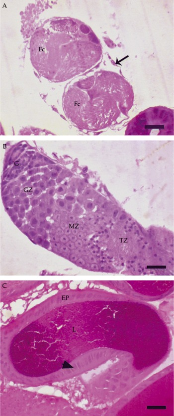
The testes of A. gabrieli had two follicles, each lined by a thin peritoneal sheath (Fig. 2A). The testis follicles are recognized in the following regions from the apex to the base: the germarium, with spermatogonia; the growth zone, in which the spermatogonia divide and increase in size to form spermatocytes; the maturation and reduction zone, in which each spermatocyte undergoes the two meiotic divisions to produce spermatids; the transformation zone, in which spermatids develop into spermatozoa (Fig. 2B). This structural organization is similar to that found in other insects (Barth, Reference Barth1958b; Chapman, Reference Chapman1998), but the low number of testis follicles is an intriguing finding, since Hemiptera usually have four to seven testis follicles (Lemos et al., Reference Lemos, Serrão, Ramalho, Zanuncio and Lacerda1995, Reference Lemos, Ribeiro, Ramalho, Serrão and Zanuncio2010; Freitas et al., Reference Freitas, Santos-Mallet, Serrão, Lorosa and Gonçalves2007b, Reference Freitas, Santos-Mallet, Costa, Souza, Serrão and Gonçalves2008). The occurrence of two testis follicles in A. gabrieli may be a specific feature for Miridae, whereas Asopinae have six (Lemos et al., Reference Lemos, Serrão, Ramalho, Zanuncio and Lacerda1995, Reference Lemos, Ribeiro, Ramalho, Serrão and Zanuncio2010) and Triatominae have seven (Freitas et al., Reference Freitas, Santos-Mallet, Serrão, Lorosa and Gonçalves2007b, Reference Freitas, Santos-Mallet, Costa, Souza, Serrão and Gonçalves2008); however, studies on more species of Miridae are necessary to validate this hypothesis.
Histological sections of the male reproductive tract of Adparaproba gabrieli. A, transverse section of a testis showing two follicles (Fc). B, longitudinal section of a testis follicle showing the germarium (G), growth zone (GZ), maturation zone (MZ) and transformation zone (TZ). C, section of an accessory gland showing the well-developed secretory epithelium (EP) and the lumen (L) filled with an amorphous substance. Arrows, nucleus. Bars = 10 μm (a colour version of this figure can be found online at journals.cambridge.org/jti).

The male accessory glands are lined by a single-layered columnar epithelium, with a nucleus containing decondensed chromatin (Fig. 2C). The gland content is characterized by a basophilic secretion (Fig. 2C). The occurrence of male accessory glands is common in Hemiptera (Barth, Reference Barth1958a; Pires et al., Reference Pires, Ferreira, Guedes and Serrão2006; Freitas et al., Reference Freitas, Gonçalves, Serrão and Santos-Mallet2007a). Because the male accessory glands of A. gabrieli are not lined by a cuticle, they are mesodermic in origin and can be classified as mesadenia (Chapman, Reference Chapman1998). The products of these glands have a variety of functions, such as sperm protection, storage and activation, sperm competition, female behaviour, fecundity, ovulation, oviposition and protection of laid eggs (Davey, Reference Davey1958; Chen, Reference Chen1984; King et al., Reference King, Eubel, Millar and Baer2011).
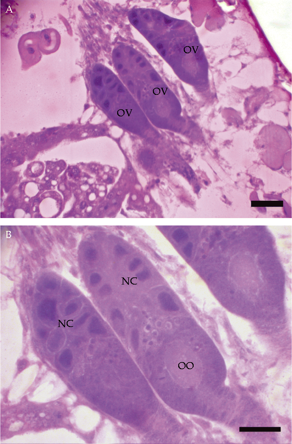
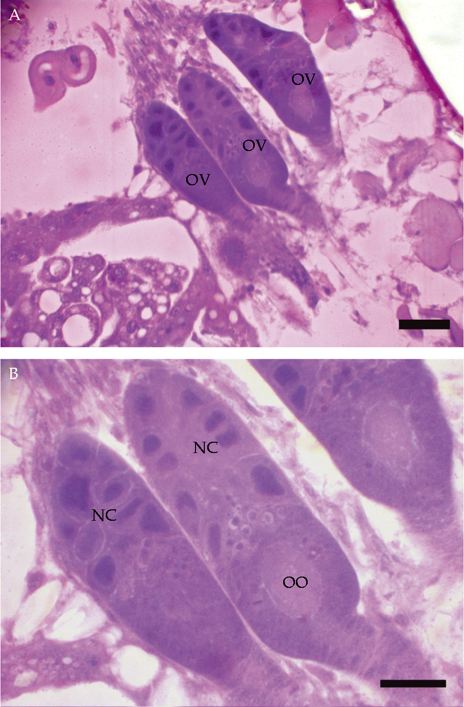
Ovaries of A. gabrieli are symmetrical with three ovarioles each joined in the distal portion by the terminal filament, forming a pair of compact structures (Fig. 3A). The ovarioles of A. gabrieli are meroistic telotrophic (Fig. 3B) with oocytes in different degrees of developmental stages. Our findings show that ovaries of A. gabrieli are similar to other Hemiptera (Büning, Reference Büning and Büning1994; Simiczyjew et al., Reference Simiczyjew, Ogorzalek and Stys1998; Lemos et al., Reference Lemos, Ramalho, Serrão and Zanuncio2005). In Hemiptera, the number of ovarioles of each ovary varies from 2 to 17 (Schuh and Slater, Reference Schuh and Slater1995).
Histological sections of the female reproductive tract of Adparaproba gabrieli. A, longitudinal section of an ovary showing three ovarioles (OV). B, longitudinal section of ovarioles showing the nurse chamber (NC) and developing oocytes (OO). Bars = 10 μm (a colour version of this figure can be found online at journals.cambridge.org/jti).
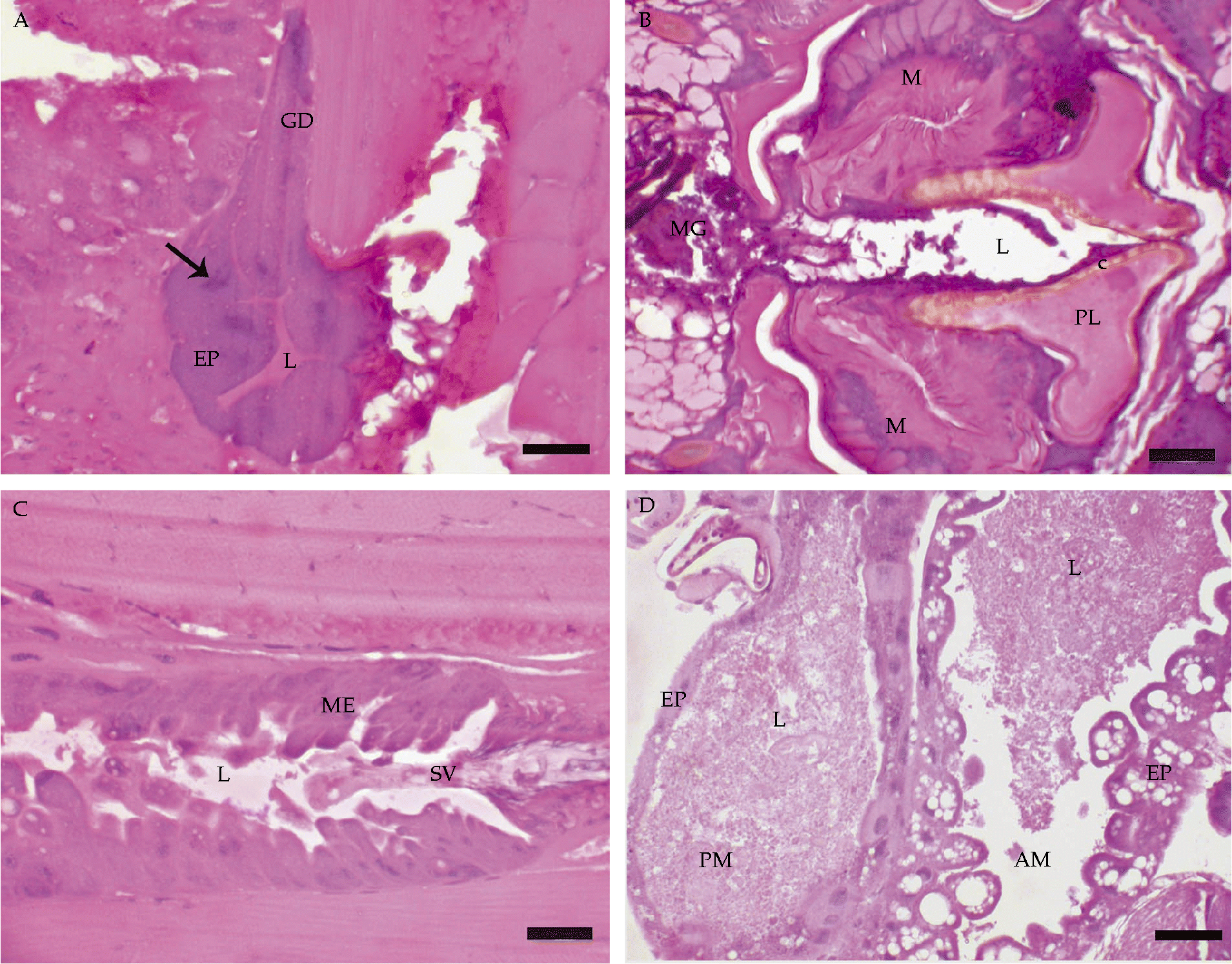
The salivary gland of A. gabrieli is represented by a single pair of sac-like structures that project to the prothorax. The gland wall is lined with a single layer of columnar cells with a granular cytoplasm and well-developed nucleus with a predominance of decondensed chromatin (Fig. 4A). The occurrence of a single pair of salivary glands in A. gabrieli is intriguing because the salivary glands of Hemiptera generally have a complex array of two to four lobes plus a pair of accessory glands (Baptist, Reference Baptist1941; Milles, Reference Milles1960; Roma et al., Reference Roma, Camargo-Mathias, Arrigoni and Marin-Morales2003; Azevedo et al., Reference Azevedo, Zanuncio, Zanuncio, Martins, Marques-Silva, Sossai and Serrão2007; Serrão et al., Reference Serrão, Castrillon, Santos-Mallet, Zanuncio and Gonçalves2008). The presence of a single pair of salivary glands in A. gabrieli may be because this insect feeds on a specific host plant. Salivary glands with different lobes and accessory glands produce different substances in these compartments and are found in generalist predators and plant-feeding Hemiptera (Roma et al., Reference Roma, Camargo-Mathias, Arrigoni and Marin-Morales2003; Pires et al., Reference Pires, Ferreira, Guedes and Serrão2006; Azevedo et al., Reference Azevedo, Zanuncio, Zanuncio, Martins, Marques-Silva, Sossai and Serrão2007).
Histological sections of Adparaproba gabrieli. A, the sac-like salivary gland showing well-developed secretory epithelium (EP) and a thin glandular duct (GD); arrow, nucleus of the secretory cell; L, lumen. B, longitudinal section of the proventriculus showing two proventricular lips (PL) lined by a thick cuticle (c) and well-developed muscles (M); L, lumen; MG, midgut. C, transition fore-midgut showing the stomodeal valve (SV) protruding into the midgut lumen (L) and the columnar midgut epithelium (ME). D, longitudinal section of the midgut showing the anterior midgut (AM) with columnar cells in the epithelium (EP) and the posterior midgut (PM) with flattened cells in the epithelium (EP); L, lumen. Bars = 10 μm (a colour version of this figure can be found online at journals.cambridge.org/jti).
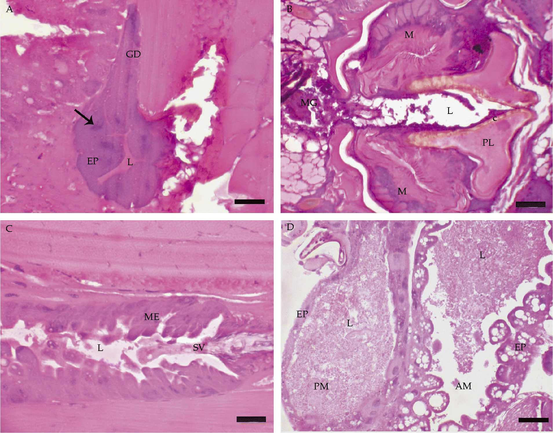
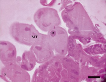
The digestive tract of A. gabrieli has a short pharynx followed by an oesophagus that is a simple narrowed tubular canal lined by a single layer of flattened cells. The oesophagus ends in a proventriculus characterized by a folded wall forming a proventriculus lip lined by a thick cuticle on the luminal surface and well-developed muscle layer (Fig. 4B), although a differentiated crop was lacking. The transition fore-midgut is characterized by a short stomodeal valve that is double-walled and protrudes into the midgut lumen (Fig. 4C). The midgut of A. gabrieli is divided into two regions: the anterior midgut, with thick walls lined by a vacuolated epithelium and a posterior midgut, with a thin wall lined by flattened cells with a condensed nucleus (Fig. 4D). The lumen was filled with a homogeneous fluid (Fig. 4D). The hindgut is a short region of the digestive tract with an ileum and a sac-like rectum. The transition from the midgut to the hindgut is characterized by the insertion of the Malpighian tubules, which are long and narrow. The Malpighian tubules of A. gabrieli are formed by a single layer of cubic cells, which have a homogeneous cytoplasm and nucleus with a predominance of condensed chromatin (Fig. 5). The midgut occupies a large part of the abdomen of Hemiptera and is divided, depending on the species, into two or three chambers (Billingsley and Downe, Reference Billingsley and Downe1983, Reference Billingsley and Downe1989; Guedes et al., Reference Guedes, Zanuncio, Ramalho and Serrão2006; Pires et al., Reference Pires, Ferreira, Guedes and Serrão2006; Fialho et al., Reference Fialho, Zanuncio, Neves, Ramalho and Serrão2009). The occurrence of the fluid in the midgut lumen supports the hypothesis that A. gabrieli is phytophagous.
Histological sections of the Malpighian tubules (MT) of Adparaproba gabrieli with a well-developed nucleus (n). Bar = 10 μm (a colour version of this figure can be found online at journals.cambridge.org/jti).

The current study revealed some aspects of the internal morphology of A. gabrieli, its male and female reproductive and digestive tracts, and provided some interesting data about its biology, such as the putative host plant and feeding habit.
Adults and nymphs together were collected from Piper sp., indicating that this is the host plant of A. gabrieli. The feeding process was not observed. However, the presence of a single pair of lobes in the salivary gland, differing from most Hemiptera with generalist or predacious feeding habits, the well-developed muscular layers of the proventriculus, as usually found in sucking insects, and the homogeneous fluid content found in the midgut, suggest the phytophagus habits of A. gabrieli.
The male reproductive tract of A. gabrieli has its structural organization, in general, very similar to other insects. The lower number of testis follicles in this species (two) is interesting, in contrast with what is commonly found in other Hemiptera (from four to seven testis follicles). It may be a specific feature of Miridae, but more studies are needed to corroborate this hypothesis. The female reproductive tract is similar to other Hemiptera.
This is the first record of A. gabrieli in the region of Viçosa, Minas Gerais state, and is endemic to this part of Brazil.
Acknowledgements
This research was supported by the Brazilian research agencies CNPq and Fapemig.