Introduction
The whitefly, Bemisia tabaci (Gennadius) (Hemiptera; Aleyrodidae), is one of the very few species of whitefly described, which is considered to be a major agricultural pest. Bemisia tabaci gained prominence globally, particularly in the 1990s, as a serious pest of vegetable, fibre and root crops due to its wide geographical distribution, polyphagous nature of some biotypes and the diverse ways that it damages crops (Duffus, Reference Duffus1987; Polston & Anderson, Reference Polston and Anderson1997; Varma & Malathi, Reference Varma and Malathi2003). Bemisia tabaci is a vector of numerous plant viruses belonging to the family genus Begomovirus and family Geminiviridae, and its heavy infestations can cause phytotoxic symptoms of squash silverleaf in cucurbits and uneven ripening of tomato (Costa & Brown, Reference Costa and Brown1991; Costa et al., Reference Costa, Ullman, Johnson and Tabashnik1993b; Brown et al., Reference Brown, Frohlich and Rosell1995; Secker et al., Reference Secker, Bedford, Markham and William1998; Jones, Reference Jones2003).
In the 1950s, the presence of biotypes was recognised when it was discovered that morphologically indistinguishable populations of B. tabaci exhibited variation in biological and ecological traits (Bird, Reference Bird1957; Costa & Russell, Reference Costa and Russell1975). Since then, many biotypes have been identified and characterized to different degrees (Burban et al., Reference Burban, Fishpool, Fauquet, Fargette and Thovenel1992; Brown et al., Reference Brown, Frohlich and Rosell1995; De Barro et al., Reference De Barro, Liebregts and Carver1998, Reference De Barro, Driver, Trueman and Curran2000, Reference De Barro, Trueman and Frohlich2005; Brown, Reference Brown2001; Perring, Reference Perring, Cooper, Rodriguez, Farrar and Bellows2001). Amongst them, the B biotype has attracted greatest attention because of the unprecedented losses that it has caused to global agriculture (Costa & Brown, Reference Costa and Brown1991; Brown et al., Reference Brown, Costa and Laemmlen1992; Perring et al., Reference Maruthi, Colvin, Gibson, Thwaites, Banks and Seal1993; Varma & Malathi, Reference Varma and Malathi2003). B biotype and associated begomovirus diseases in the southern states of the USA in the early 1990s were estimated to have caused combined annual losses of 500 million USD to the winter vegetable crops (Perring et al., Reference Maruthi, Colvin, Gibson, Thwaites, Banks and Seal1993). The economic importance of the B biotype became immediately evident when it was first detected in the Kolar district of South India in 1999, which was associated with a 1000-fold increase in the number of whiteflies on tomato, cabbage and other vegetable crops that were seldom infested by local whiteflies (Banks et al., Reference Banks, Colvin, Chowda Reddy, Maruthi, Muniyappa, Venkatesh, Kiran Kumar, Padmaja, Beitia and Seal2001). The most significant damage was due to an epidemic of tomato leaf curl virus disease, which has since continued to affect tomato production in India.
The ability of B biotype to induce silverleaf symptoms on cucurbits has been used as a marker for its identification (Costa & Brown, Reference Costa and Brown1991; Brown et al., Reference Brown, Costa and Laemmlen1992; Costa et al., Reference Costa, Ullman, Johnson and Tabashnik1993b; Secker et al., Reference Secker, Bedford, Markham and William1998). Subsequently, a number of non-specific esterases and randomly amplified polymorphic DNA (RAPD) markers were developed (De Barro & Driver, Reference De Barro and Driver1997; Guirao et al., Reference Guirao, Beitia and Cenis1997; Brown et al., Reference Brown, Pering, Cooper, Bedford and Markham2000; Calvert et al., Reference Calvert, Cuervvo, Arroyave, Constantino, Belloti and Frohlich2001; Maruthi et al., Reference Maruthi, Colvin and Seal2001). RAPD markers are convenient since the samples can be preserved in alcohol prior to analysis, and relatively large numbers of samples can be processed rapidly. However, the RAPD patterns are complex and difficult to reproduce and interpret reliably. Sequence characterized amplified regions (SCARs) were, therefore, developed for simple PCR-based detection of B. tabaci biotypes (Khasdan et al., Reference Khasdan, Levin, Rosner, Morin, Kontsedalov, Maslenin and Horowitz2005). DNA sequence data from mitochondrial cytochrome oxidase I and ribosomal genes have also been used to differentiate biotypes and to estimate phylogenetic relationships both within and between B. tabaci and other whitefly species (Clark et al., Reference Clark, Baumann, Munsom and Campbell1992; Frohlich et al., Reference Frohlich, Torres-Jerez, Bedford, Markham and Brown1999; Brown, Reference Brown2001; De Barro et al., Reference De Barro, Driver, Trueman and Curran2000, Reference De Barro, Trueman and Frohlich2005; Maruthi et al., Reference Perring2004; Rekha et al., Reference Rekha, Maruthi, Muniyappa and Colvin2005). Investigations on the genetic diversity of Indian B. tabaci biotypes, however, are limited, and only recently has the distribution of the B biotype in South India been recorded (Rekha et al., Reference Rekha, Maruthi, Muniyappa and Colvin2005). The aims of the study were to develop and optimise silverleaf assay, esterase patterns, RAPD polymorphisms and a SCAR marker in order to detect and further monitor the spread of B biotype in India.
Materials and methods
Whitefly colonies and sample collections
Colonies of B biotype and indigenous B. tabaci were established at the Virology Laboratory, University of Agricultural Sciences (UAS), Bangalore as described previously (Rekha et al., Reference Rekha, Maruthi, Muniyappa and Colvin2005). Colonies were maintained separately on cotton plants placed in nylon mesh cages, and the colony purity was checked periodically by silverleaf assay and RAPD-PCR (Costa & Brown, Reference Costa and Brown1991; Rekha et al., Reference Rekha, Maruthi, Muniyappa and Colvin2005). Additional B. tabaci samples were collected in 70% ethanol from a range of crops in several regions of India from 2004 to 2006 (Appendix 1).
Silverleaf assay on pumpkin
A number of parameters that can influence the development of silverleaf symptoms by whitefly feeding on cucurbitaceous plants were investigated (Costa & Brown, Reference Costa and Brown1991). Five pumpkin, two squash and one gherkin genotypes grown popularly in and around the Bangalore area were tested for their susceptibility to silverleaf symptoms upon B biotype feeding. A mixture of 50 female and male B biotype adults infested each of the 15 seedlings for 48 h, and the plants were subsequently kept in a glasshouse for silverleaf symptom expression. In order to determine the number of B biotype nymphs required to induce silverleafing, groups of 1, 2, 3, 5, 20 and 30 B biotype females were exposed to each of the 15 pumpkin seedlings (cv. Big, the most susceptible variety) using leaf cages. Adults were allowed to feed and oviposit for 48 h, after which they were removed. The exposed seedlings were maintained in insect-proof cages for the development of nymphs, which induced silverleaf symptoms. Plants not exposed to B biotype served as controls. After three weeks, the numbers of nymphs present on leaves and silverleaf symptom severity were recorded on a 0–5 scale (Costa et al., Reference Costa, Ullman, Johnson and Tabashnik1993b).
The length of feeding time by adults determines the number of eggs laid and surviving nymphs which can induce silvering. This was determined by leaving a group of 20 B biotype adults separately for durations of 6, 12, 24, 48 and 72 h on pumpkin plants. Plants were maintained in nylon mesh cages and scored for silverleaf symptom severity after three weeks.
The role of B biotype nymphs, as opposed to adults, that induce silverleaf symptoms was assessed by leaving a group of 25 females on each of the 30 pumpkin seedlings to oviposit for 48 h on the first true leaf confined by leaf cages. Fifteen inoculated plants were maintained in insect-proof cages for the development of nymphs (Costa et al., Reference Costa, Ullman, Johnson and Tabashnik1993b). The exposed leaves of the remaining 15 plants were dipped four times (5, 7, 9 and 11 days after exposure) in an insecticidal solution (Methomyl 2 ml l−1) for 10 min to kill whitefly eggs and nymphs. Ten plants not exposed to B biotype adults were also dipped in the insecticide and used as control.
In order to assess the current distribution and presence of the B biotype in Karnataka, South India, live B. tabaci adults were obtained from many locations in Bangalore, Bijapur, Dharwad, Gulbarga and Kolar districts and assayed by exposing to pumpkin plants (appendix 1). Adults of mixed sex were confined (15–20) to leaves using leaf cages and allowed to feed and oviposit for 48 h. Plants were further maintained in cages in a glasshouse for the development of nymphs and observed for silverleaf symptoms.
Esterase electromorph analysis
Napthyl esterase banding patterns of the indigenous and B biotype adults were analysed after separation on 7.5% polyacrylamide gels following the protocol of Byrne & Devonshire (Reference Byrne and Devonshire1991), which is described briefly below. Samples were prepared by homogenizing five individual whiteflies separately in a microfuge tube in 25 μl of protein extraction buffer (0.05 m Tris-HCl, pH 6.8, containing 10% sucrose and 0.1% Triton X-100). The homogenates were transferred into wells of the stacking gel (3.0%) superposed on the 7.5% resolving gel. The reservoir contained 0.06% m Tris, 0.37 m glycine buffer, pH 8.2. Gels were run at 15 mA for 15 min and then at 19 mA for 2 h. Bromophenol blue was used as a running marker. Esterase was stained by placing gels in substrate solution for 1 h in 0.4% (w/v) fast blue RR salt after incubation for 30 min in 0.02% (w/v) solution of 1-naphthyl acetate and 2-naphthyl acetate in 0.2 m phosphate buffer (pH 6.5), which contained 1% acetone. All staining reactions were quenched by placing the gel in the destaining solution (25 ml acetic acid+150 ml methanol+325 ml distilled water). In addition to this general protocol, three more esterase-based techniques were developed for quick and reliable detection of B biotype using its distinct feature of high esterase activity.
Filter paper dot blot test
A single female adult B biotype was homogenized in a microfuge tube in 15 μl of protein extraction buffer, as described above (Byrne & Devonshire, Reference Byrne and Devonshire1991). The sample was applied onto Whatman filter paper (125 mm) using a micropipette, air dried for a few minutes and incubated in 15 ml of substrate solution (napthyl acetate and fast blue RR) for 30 min at 30°C. The membrane was destained using the destaining solution with occasional shaking, air dried and observed for variation in colour intensity between the indigenous (faint blue) and B biotype (intense blue) samples.
Field-based dot blot test
This method was developed to detect B biotype adults instantly in the field by modifying the above described filter paper dot blot technique. Individual indigenous or B biotype adults were homogenized separately in 25 μl of ice-cold esterase protein extraction buffer (Byrne & Devonshire, Reference Byrne and Devonshire1991) on a glass slide. A Whatman filter paper disc of 5 mm diameter was placed on the homogenate to absorb the contents. The discs were then stained using 25 μl of substrate solution (napthyl acetate and fast blue RR) for 10 min and observed for differences in colour development.
Test tube method
Groups of five indigenous or B biotype adults were homogenized separately in 50 μl of 0.04 m phosphate buffer (pH 7.0) and the volume was made up to 1 ml with ice-cold phosphate buffer. The sample was transferred to a test tube containing 5 ml of substrate solution 3×10⁻⁴m 1-naphthyl acetate and incubated at 30°C for 30 min. The reaction was arrested by the addition of 1 ml of DBLS reagent (Diazo Blue Laurate solution −5% SDS +1% Diazo blue salt in 5:2). Variation in colour intensity of solutions was recorded after 15 min.
Nucleic acid-based detection techniques
RAPD-PCR analysis
Total DNA was extracted from individual female adult whiteflies according to the protocol of De Barro & Driver (Reference De Barro and Driver1997). Conditions for RAPD-PCR were those described earlier (Maruthi et al., Reference Maruthi, Colvin and Seal2001; Rekha et al., Reference Rekha, Maruthi, Muniyappa and Colvin2005) using the primer OpB11 (Operon Technologies, Huntsville, USA). Clear and reproducible polymorphisms, generated using OpB11, were used for the separation of biotypes. Each set of PCR contained at least one known sample each of indigenous and B biotype as controls.
Development of a B biotype-specific SCAR marker
A band of ~800 bp, obtained using OpB11 unique to the B biotype, was purified using a gel extraction kit (Qiagen, GmbH, Hilden, Germany) and cloned into the plasmid pTZ57R/T (MBI, Fermentas) following the manufacturer's instructions. A sequence of the insert was determined at the MWG Biotech Pvt Ltd, Bangalore, India. The identity of the sequence was confirmed by BLAST search, which was most similar to the sequences of the tenecin 3 precursor gene of Tenebrio molitor (Coleoptera; Tenebrionidae) and on the chromosome 2R gene of Drosophila melanogaster (Diptera; Drosophilidae).
In order to develop a SCAR marker specific to B biotype, the oligonucleotide set BB-1 (forward primer 5′ ACCCGTTGAATCCTATAGAC 3′ and reverse primer 5′ TGTTATGCTTACCCGGAAC 3′) was designed to amplify the ~550 bp PCR product. Specificity of the primers to B biotype was tested using DNA samples of known B biotype and indigenous B. tabaci. PCR was performed in a 25 μl reaction mix containing 6.0 mm Tris-HCl, pH 8.8, 1.5 mm MgCl2, 0.2 mm dNTPs, 0.4 μm forward and reverse primers, 2.5 U of Taq DNA polymerase (Bangalore Genie Pvt Ltd, Bangalore) and 2 μl whitefly DNA sample. PCR reactions were carried out with the following amplification program: 1 cycle of 2 min at 94°C followed by 35 cycles of 1 min at 94°C, 1 min at 58°C and 1 min at 72°C, followed by a final cycle of 10 min at 72°C. The SCAR marker was used to detect B biotype in field-collected whitefly samples.
Sequencing mtCOI gene of selected B. tabaci samples
Partial mtCOI gene sequences of a total of seven B. tabaci samples were sequenced (Frohlich et al., Reference Frohlich, Torres-Jerez, Bedford, Markham and Brown1999; Maruthi et al., Reference Perring2004; Rekha et al., Reference Rekha, Maruthi, Muniyappa and Colvin2005). Four samples were from brinjal collected at Hosadurga (Chitradurga district), Bellary, Coimbatore and Hyderabad, and one from pumpkin, New Delhi (appendix 1). The remaining two sequences were of the indigenous and B biotype colonies maintained at UAS, Bangalore. Unambiguous sequences of 689 bases were analyzed by parsimony and distance methods (Swofford, Reference Swofford2002) along with those published previously (Rekha et al., Reference Rekha, Maruthi, Muniyappa and Colvin2005). The heuristic and neighbour-joining method of search was employed for reconstructing phylogenetic trees (Swofford, Reference Swofford2002) using the software PAUP version 4.0 b10 for Mac.
Results
Silverleaf assay
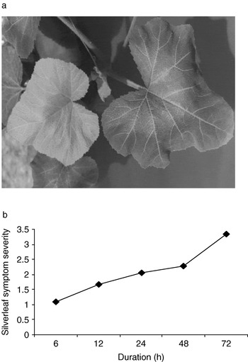
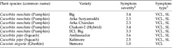
All the pumpkin, gherkin and squash genotypes expressed silvering symptoms, to varying degrees, about three weeks after the feeding of B biotype (table 1). Prominent vein clearing was the first visible symptom followed by silvering of leaves, except in gherkin, which developed faint vein clearing and mild silvering symptoms. Pumpkin hybrid Chakori-2 and variety Big were the most susceptible with the severest symptoms (fig. 1a) and expressed symptoms typical to those of squash cv. Ambassador (Costa & Brown, Reference Costa and Brown1991). For this reason and the lack of the ready availability of Ambassador seeds in India, the variety Big was used in subsequent silverleaf assays.
(a) Silverleaf symptoms on pumpkin plant variety Big upon feeding by B biotype nymphs. (b) The effect of the feeding period of B biotype on the induction of silverleaf symptoms measured by using a 0–5 scale.

Sliver leaf symptom severity on popularly grown cucurbit varieties in South IndiaFootnote 1.

1 Ten plants of each variety were infested with 25 female B biotype adults per plant for 48 h.
2 Symptom severity was recorded three weeks after inoculation using a 0–5 point scale, where 0=no symptoms and 5=severest symptoms.
3 VCL, vein clearing; SL, silvering of leaf.
Silverleaf symptoms were not seen on pumpkin plants exposed to B biotype females and subsequently dipped in insecticide solution. They also had no immature stages of B. tabaci developing on them. Plants that were not treated with insecticides had many eggs and nymphs and expressed silverleafing.
A minimum of 6 h feeding and ovipositing on pumpkin by B biotype adults was essential to lay eggs, which later developed into nymphs to induce silverleaf symptoms. Exposure of more than 12 h resulted in more nymphs feeding on plants and induced 100% silverleaf incidence, often with severe symptoms. Silverleaf symptom severity increased with an increase in the exposure period (fig. 1b), which was directly correlated to the number of nymphs feeding on the plant.
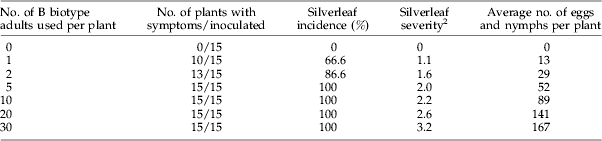
A single B biotype female laid enough eggs, which subsequently developed into nymphs and induced silverleafing on 67% plants, while nymphs developed from eggs laid by five or more females caused 100% incidence. The most severe symptoms (rating 3.2) were observed when nymphs developed from eggs laid by 30 females fed on each plant (table 2).
Estimation of the number of B biotype female adults required for the induction of silverleaf symptoms on the pumpkin variety BigFootnote 1.
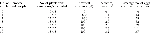
1 Each group of insects inoculated each plant for 48 h.
2 Symptom severity was recorded three weeks after inoculation using a 0–5 point scale, where 0=no symptoms and 5=severest symptoms.
Esterase patterns of B and indigenous biotypes
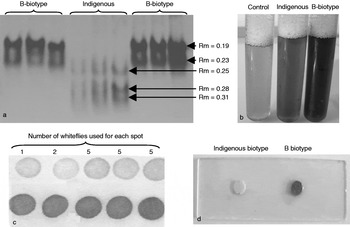
Separation of non-specific napthyl esterases by PAGE helped to distinguish indigenous and B biotype populations effectively. Both quantitative and qualitative differences were found in esterase patterns of the two biotypes. Two intensely stained esterases, Est-1 (relative mobility, Rm 0.19) and Est-2 (Rm 0.23), were detected only from the B biotype samples; while three faintly-stained fast-moving esterases, Est-3 (Rm 0.25), Est-4 (Rm 0.28) and Est-5 (Rm 0.31), were detected in indigenous biotypes (fig. 2a).
(a) Relative mobility of non-specific napthyl esterases of Indian indigenous and B biotype B. tabaci on a polyacrylamide gel. Detection of B biotype adults based on esterases using (b) test tube method, (c) filter paper dot blot method and (d) field-based dot blot method.

Filter paper dot blot test
This technique involved applying whitefly extractions onto Whatman filter paper, staining with napthyl esterase substrates and recording differences in colour intensity between the biotypes. The B biotype samples produced a deep blue colour while the indigenous samples produced a faint blue colour. This method was found suitable for esterase analysis of both individual and groups of adults (fig. 2c).
Field-based dot blot test
The filter paper dot blot technique was modified for the instant detection of the individual adult B biotype in the field. Whiteflies were crushed on a glass slide and the esterases were soaked into a membrane disc which was then stained. B and indigenous biotypes developed deep blue and faint blue colours, respectively (fig. 2d).
Test tube method
This method involved testing for differences in colour intensity in a test tube containing esterases of whiteflies and staining reaction mixture. Similar to the above tests, the B biotype developed a deep blue colour compared to indigenous biotypes. This method was suitable for testing samples of five adults or more (fig. 2b).
RAPD-PCR and the development of a SCAR marker
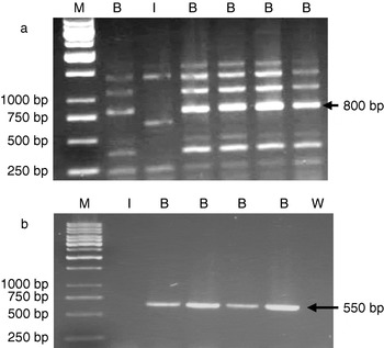
RAPD-PCR polymorphisms, generated using the primer OpB11, were used to detect and monitor the spread of the B biotype, mainly in Karnataka state where the B biotype was first introduced, but also from Andhra Pradesh, Kerala, Maharashtra and Tamilnadu, all from South-Central India, and in New Delhi and Punjab, from North India. Three diagnostic bands of sizes ~350 bp, 800 bp and 1 kb, unique to the B biotype samples, were consistently reproduced using the primer OpB11 (fig. 3a) (Rekha et al., Reference Rekha, Maruthi, Muniyappa and Colvin2005). Polymorphisms obtained from the indigenous biotypes using OpB11 were variable, but three bands of sizes ~600 bp, 700 bp and 750 bp were consistently reproducible, which were used as diagnostic markers. The Indian and Israeli B biotypes shared identical banding patterns, thus allowing its easy identification.
(a) RAPD-PCR polymorphisms of B biotype populations obtained using the primer OpB11. The 800 bp size PCR product used to develop SCAR marker is indicated by an arrow, and the sizes of 1 kb molecular weight marker used are also shown. (b) PCR amplification of the ~550 bp SCAR marker from B biotype samples. M, molecular weight marker; B, B biotype; I, indigenous biotype; w, water control.

In order to develop a simple and quick PCR-based identification protocol for B biotype, the 800 bp band specific to B biotype was cloned and sequenced. Based on sequence information, PCR primers (BB-1) were designed and used to amplify DNA from indigenous and B biotype samples. The SCAR marker primer set BB-1, which was designed to amplify a fragment of 550 bp, amplified expected PCR products from B biotype samples only (fig. 3b).
Mitochondrial COI gene sequences
Based on mtCOI gene sequence analysis, six of the seven sequences obtained in this study (the EMBL accession numbers were from AM408895 to AM408901) were indigenous and belonged to three separate genotypes within the Asia group (figure not shown) that have been identified previously (Simon et al., Reference Simon, Cenis, Beitia, Khalid, Moreno, Fraile and Arenal2003; Rekha et al., Reference Rekha, Maruthi, Muniyappa and Colvin2005). Bemisia tabaci populations from pumpkin, New Delhi, and brinjal, Coimbatore, belonged to genotype I; brinjal, Chitradurga, to genotype II; and brinjal, Hyderabad and Bellary, and cotton, Bangalore (the indigenous colony), to genotype III.
The mtCOI sequences of the B biotype colony maintained at UAS, Bangalore, grouped with reference B biotype sequences from India, Israel and USA, and shared >99% sequence identities with them. The indigenous B. tabaci colony shared >99% nucleotide identities with many field-collected populations in Karnataka. It also shared highest nucleotide identities (99%) with brinjal, Pakistan, and cucumber, Thailand, further indicating the widespread occurrence of the Indian indigenous B. tabaci across Asia.
Distribution of B biotype in India
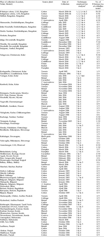
A total of 639 whitefly samples were analyzed to estimate the distribution of B biotype in India based on silverleaf assay, esterase patterns, RAPD polymorphisms, mtCOI gene sequences or SCAR marker. The majority of the samples were drawn from Karnataka, where the B biotype was first reported, and a few from Andhra Pradesh, Tamil Nadu, Kerala and Maharashtra, Punjab and New Delhi. The B biotype was present in all parts of southern Karnataka and adjacent areas in Tamil Nadu and Andhra Pradesh (appendix 1). However, the proportion of B biotype to indigenous B. tabaci varied between locations and host species. The number of B biotype adults was high (>90%) at many locations of Kolar and adjoining districts of southern Karnataka compared to northern Karnataka where they were low (~20%) and were considered to have spread relatively recently. The B biotype was absent in Kerala, Maharashtra in the South, and Punjab and New Delhi in the North of India.
Discussion
The B biotype was first recorded in the Kolar district of Karnataka state, South India, during the summer growing season (March–June) of 1999 (Banks et al., Reference Banks, Colvin, Chowda Reddy, Maruthi, Muniyappa, Venkatesh, Kiran Kumar, Padmaja, Beitia and Seal2001), and it was shown to be spreading based on its detection using RAPD polymorphisms and mtCOI sequences (Rekha et al., Reference Rekha, Maruthi, Muniyappa and Colvin2005). These techniques require substantial capital investment and skilled personnel; simple, cost-effective alternative detection protocols were therefore developed in this study. Although three distinct genetic groups of B. tabaci have been shown to induce silverleafing on squash and zucchini plants (De Barro et al., Reference De Barro, Trueman and Frohlich2005; Sseruwagi et al., Reference Sseruwagi, Legg, Maruthi, Colvin, Rey and Brown2005), the silverleaf assay has been used to detect B biotype worldwide as it is the most invasive of the biotypes; and, therefore, the likelihood of the individual being B is very high (Costa & Brown, Reference Costa and Brown1991; Schuster et al., Reference Schuster, Kring and Price1991; Brown et al., Reference Brown, Costa and Laemmlen1992; Costa et al., Reference Costa, Ullman, Johnson and Tabashnik1993b; Rekha et al., Reference Rekha, Maruthi, Muniyappa and Colvin2005; De Barro et al., Reference De Barro, Bourne, Khan and Brancatini2006). However, the cultivation of squash and zucchini is limited in South India, and their seeds are difficult to obtain; whereas other cucurbits, such as pumpkin, are popular and grown extensively throughout the year. For these reasons, the ability of the B biotype to induce silverleafing on pumpkin genotypes was investigated. The pumpkin variety Big and hybrid Chakori-2 were highly susceptible and expressed silverleaf symptoms similar to those on squash plants. The variety Big is readily available in Indian markets and is cheap to obtain; and, hence, it is suitable to use for silverleaf bioassays in India.
Further, several factors that may influence the development of silverleaf symptoms on pumpkin were optimized to detect the B biotype reliably. Ovipositing by a single female B biotype for 48 h, and the subsequent development of nymphs induced silverleafing, although the severest symptoms were recorded by groups of five or more adults. This is consistent with the findings of Bharathan et al. (Reference Bharathan, Narayanan and McMillan1992). While feeding on plant leaves, B. tabaci females lay eggs, which develop into nymphs. The feeding of large numbers of nymphs, but not adults, caused symptoms (Yokomi et al., Reference Yokomi, Hoelmer and Osborne1990; Costa & Brown, Reference Costa and Brown1991). This was further confirmed in this study, where the removal of nymphs with an insecticide treatment prevented silvering, but those infested plants left untreated developed symptoms within three weeks. In conclusion, the parameters that influenced the development of silverleaf symptoms on pumpkin were similar to those of squash and zucchini (Costa & Brown, Reference Costa and Brown1991; Schuster et al., Reference Schuster, Kring and Price1991), hence the pumpkin variety Big can also be used in silverleaf bioassays.
Esterase polymorphism is another marker used extensively for separating B. tabaci biotypes and, based on them, over 20 were identified (Prabhakar et al., Reference Prabhaker, Coudriet and Meyerdick1987; Costa & Brown, Reference Costa and Brown1991; Perring et al., Reference Maruthi, Colvin, Gibson, Thwaites, Banks and Seal1993; Byrne et al., Reference Byrne, Bedford, Devonshire and Markham1995; Brown et al., Reference Brown, Pering, Cooper, Bedford and Markham2000). Our results showed that esterase profiles of the Indian indigenous and B biotype adults were different, which allowed easy discrimination of the two biotypes. Moreover, esterase levels were considerably greater (3–4-fold) in B biotype compared to the indigenous adults (Byrne et al., Reference Byrne, Gorman, Cahill, Denholm and Devonshire2000). Taking advantage of this unique property, three low-cost, simple and quick techniques were developed to detect B biotype. The filter paper dot blot method was developed for easy identification of single B biotype adults without the need to use the expensive and laborious PAGE technique, which also involves the use of toxic chemicals. However, the filter paper dot blot method still required bench space (a laboratory), considerable expertise in the handling of reagents and about 1–2 h to obtain the results. In order to develop an even quicker and field-based detection protocol, the filter paper dot blot test was modified to develop a field-based dot blot test where the esterase analysis was carried out on a glass slide with minimal quantities of reagents and the results were obtained in about 15 min. This test was found reliable to detect single B biotype adults and can be performed by people with minimum training.
As an alternative to PAGE and filter paper dot blot test, the test tube method was developed, which was based on the intensity of colour development in the reaction mixture. In this test, when less than five adults were used per reaction, the difference in colour intensity was insufficient to differentiate B and indigenous biotypes by the naked eye. This was further confounded by the mixtures of biotypes present; therefore, the test tube method is not suitable for detecting biotypes reliably.
RAPD polymorphisms, using the primer OpB11, have been shown useful to detect the B biotype in India and elsewhere (Guirao et al., Reference Guirao, Beitia and Cenis1997; Rekha et al., Reference Rekha, Maruthi, Muniyappa and Colvin2005). OpB11 produced consistently reproducible diagnostic bands for B biotype populations from many countries and can still be used to differentiate B. tabaci biotypes. However, RAPDs, in general, are increasingly perceived to be less reliable because of the inherent difficulties within the technique. RAPD-PCR relies on random annealing of short oligonucleotides to the target DNA at low annealing temperatures, which may produce unspecific, similar-sized PCR products but of different content from different populations. In order to overcome these constraints and for greater technical reliability and simplicity, a simple PCR-based SCAR marker was developed. The SCAR marker utilizes species/biotype-specific oligonucleotides and amplifies a single region of genome, which makes interpretation of results much simpler than RAPD. The SCAR primer set BB-1 developed in this study amplified the ~550 bp PCR product and was highly specific to the B biotype. Using SCAR markers, the distribution of B and Q biotypes was mapped in southern Europe and the Middle East (Khasdan et al., Reference Khasdan, Levin, Rosner, Morin, Kontsedalov, Maslenin and Horowitz2005).
Using a combination of detection protocols, such as silverleaf assay, esterases, RAPD polymorphisms and SCAR marker, the B biotype was detected in samples collected during the survey from 2004–2006. Results indicated that B biotype has spread from the introduction foci (Kolar district) to northern parts of Karnataka and the neighbouring states of Andhra Pradesh and Tamil Nadu. These results were concordant with an earlier survey conducted in 2000–2002, where the B biotype had spread ~650 km in about three years since its first detection in 1999 (Rekha et al., Reference Rekha, Maruthi, Muniyappa and Colvin2005). However, much to our surprise and for reasons unknown, the spread of the B biotype appears to have halted with no further spread being observed in the 3–4-year period after 2002. We did not find B biotype in samples collected in Maharashtra, Kerala, Punjab and New Delhi in 2006 from the places located beyond the 650 km radius from Kolar. Kolar district continued to be the hub of vegetable production and export to many cities in India. Bemisia tabaci nymphs were found on leafy vegetables such as cabbage and cauliflower that were transported to northern India, yet the B biotype remains restricted to some parts of southern India. This is unexpected, considering that the B biotype swept through much of the Caribbean, North America and the Mediterranean regions of Europe shortly after its introduction there, which also resulted in the displacement of indigenous biotypes in the USA and the Caribbean (Costa et al., Reference Costa, Brown, Sivasubramaniam and Bird1993a; Frohlich et al., Reference Frohlich, Torres-Jerez, Bedford, Markham and Brown1999; Brown, Reference Brown2001). One possible reason may be the negative biotype interactions (e.g. competition for food and inter-biotype mating barriers), which can prevent one biotype from spreading into areas dominated by another (De Barro et al., Reference De Barro, Bourne, Khan and Brancatini2006). It has been shown that two distinct groups of B. tabaci genotypes exist in the north and south of Karnataka (Rekha et al., Reference Rekha, Maruthi, Muniyappa and Colvin2005), and whether the northern populations have suppressed the spread and establishment of B biotype forms the basis of future investigations. These require the further monitoring of the spread of B biotype throughout India, and the diagnostic techniques developed in this study are designed to aid such studies easily.
Acknowledgements
The Indian Council of Agricultural Research, New Delhi, India, funded the research. Drs A. Sharma, N. Kulkarni and V.B. Nargund helped in collecting whitefly samples. Profs V. Muniyappa and J. Colvin are acknowledged for their initial contributions to the project development.
Detection of B biotype in field-collected B. tabaci samples in India.

* 1, Silverleaf assay; 2, PAGE esterase analysis; 3, RAPD-PCR analysis; 4, SCAR marker; 5, mtCOI gene sequencing.