Coronary sinus abnormalities are rare, underdiagnosed congenital conditions that comprise approximately 80 cases reported in world literature. Hiroshima et al. identified three adult patients with coronary sinus atresia and a group of 3577 patients undergoing electrophysiologic study for treatment of supraventricular tachycardia.Reference Hiroshima, Goya and Nagashima1 Discovered incidentally, or during autopsy, coronary sinus anomalies may complicate electrophysiologic procedures and cardioplegia administration during cardiac surgery.Reference Shah, Teague and Lu2 These malformations are frequently missed during routine cardiac catheterisations should direct angiography of the coronary sinus be omitted in patients with normal coronary artery angiography. Coronary sinus stenosis and coronary sinus atresia frequently occur in association with CHD and rarely occur as an isolated diagnosis. We present three interesting cases of patients with isolated coronary sinus obstruction as the primary cause to their exertional angina.
Case Report 1
An 11-year-old male presented with exertional dyspnoea and new onset dull, non-radiation, mid-sternal exertional chest pain. There were no other associated symptoms except for dizziness. A prior echocardiogram was “normal,” and past medical history was unremarkable. Physical examination was normal. Electrocardiography showed left ventricular hypertrophy and a non-specific intraventricular conduction delay without ST segment abnormality. Echocardiography demonstrated a structurally normal heart, with trivial mitral and mild tricuspid valvar regurgitation with an estimated right ventricular systolic pressure of 35 mmHg. There was no ventricular hypertrophy, and the anatomy of the coronary arteries appeared normal. A cardiolyte treadmill stress test demonstrated no electrocardiogram evidence of ischaemia, but showed a stress-induced reversible abnormality in perfusion of the anterior wall. Left ventricular ejection fraction was 67%. Pulmonary function studies were normal.
Cardiac catheterisation demonstrated normal oximetric and haemodynamic data. The cardiac index estimated by the Fick method was 4.351 L/min/M2. A probe patent oval foramen was found with no left atrial desaturation. Angiography demonstrated normal left ventricular systolic function without mitral regurgitation. Selective coronary angiography was normal. The coronary sinus was dilated secondary to atresia at its mouth with venous collaterals empty directly into the right atrium and ventricle without unroofing (Fig. 1).
Selective angiography of the left coronary artery in an 11-year-old male demonstrating complete obstruction of the coronary sinus os resulting in a dilated coronary sinus.

Surgical intervention involved unroofing the coronary sinus to the left atrium. The coronary sinus was opened along much of its length to allow free drainage to the left atrium. The foramen ovale was closed, and a small persistent left superior vena cava was ligated to prevent the development of a significant right-to-left shunt. Post-operative course was complicated by atrial flutter requiring cardioversion and short-term antiarrhythmic therapy.
Repeat cardiolyte stress testing showed mildly decreased exercise tolerance, no arrhythmias, and no evidence of ischaemia. Coronary perfusion and systolic function were normal and patient symptoms resolved.
Case Report 2
A 13-year-old athletic male experienced persistent exertional chest pain during soccer. Pain was localised to the mid-sternal and left chest areas, associated with dizziness, occasional palpitations, and shortness of breath. Physical examination was unremarkable with normal vital signs. Electrocardiogram was abnormal with left ventricular hypertrophy and inferior lateral ST T-wave abnormality. Transthoracic echocardiography demonstrated a structurally normal heart with a prominent left coronary system, normal coronary colour flow mapping, without ventricular systolic dysfunction or wall motion abnormality. Chest x-ray was normal. Treadmill stress testing was remarkable for inducible chest pain without ST segment abnormality, but he had isolated premature ventricular contractions late in stress. Nuclear stress testing showed normal coronary perfusion, no wall motion abnormalities, and a normal left ventricular ejection fraction.
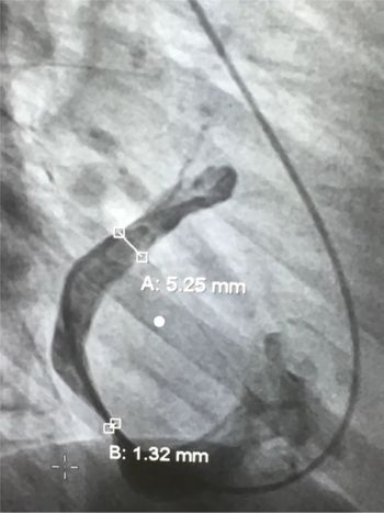
He underwent cardiac catheterisation demonstrating normal oximetry and haemodynamics. Mixed venous oxygen saturation was 78%, a cardiac index of 4.2 L/min/M2, and a left ventricular end-diastolic pressure of 5–7 mmHg. The coronary arteries were normal; however, the coronary sinus drainage suggested impaired egress and mild coronary sinus dilatation (Fig. 2). Coronary sinus cannulation demonstrated a peak coronary sinus pressure between 10 and 12 mmHg with a mean pressure of 6 mmHg and a gradient of 4 mmHg across the ostium. To better evaluate the effect of this stenosis during exercise, the patient was started on an Isuprel infusion to obtain a heart rate of roughly 150% of his baseline heart rate. This resulted in a severe gradient across the distal coronary sinus roughly 1 cm before the orifice. The coronary sinus distal pressure increased with increasing heart rate to a peak of 20–21 mmHg and pullback gradient of 19 mmHg. The area of stenosis measured 1.4 mm by angiography, and balloon angioplasty was unsuccessful.
Angiography of the coronary sinus in a 13-year-old male demonstrating impaired egress due to coronary sinus stenosis and mild dilation of the coronary sinus proximal to the site of obstruction.

Surgical intervention involved unroofing of the coronary sinus with patch closure of the coronary sinus os. No coronary sinus obstruction was seen post-operatively, and the patient’s exertional symptoms resolved. His electrocardiography abnormalities also improved.
Case Report 3
A 14-year-old male presented with persistent exertional and resting chest pain associated only with tachycardia. Physical examination was unremarkable. His initial electrocardiogram demonstrated sinus rhythm with normal cardiac intervals, left ventricular hypertrophy, and lateral T-wave abnormality. Echocardiography was suggestive of mild diastolic dysfunction with preserved ventricular systolic function. Holter monitor demonstrated occasional isolated premature ventricular contractions with ventricular bigeminy. Chest x-ray was normal.
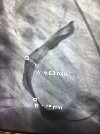
Nuclear cardiac stress testing revealed inducible chest pain and isolated premature ventricular contractions with occasional ventricular bigeminy. Nuclear assessment demonstrated normal ventricular systolic function (left ventricular ejection fraction: 82%) without perfusion abnormalities. Cardiac catheterisation demonstrated a slightly decreased mixed venous saturation of 72%, left ventricular end-diastolic pressure was 8 mmHg with a cardiac index by Fick of 2.7 L/min/M2. Coronary sinus cannulation showed an elevated coronary sinus peak pressure of 15 mmHg with a mean coronary sinus pressure of 7 mmHg and a pullback gradient of 3 mmHg. Coronary arterial angiography was normal; however, there was an area of discrete narrowing in the coronary sinus roughly 2 cm before the os of the coronary sinus. The distal portion of the coronary sinus was dilated. The most narrowed area measured 1.7 mm with a distal coronary sinus measurement of 5.8 mm (Fig. 3). Attempted balloon dilatation of this area using 6 mm diameter inflated to 4 atm did show some angiographic improvement in size; however, the pressure gradient was still elevated.
Angiography prior to balloon dilation of coronary sinus stenosis in a 14-year-old male demonstrating venous outflow obstruction proximal to the coronary sinus os.

The patient’s data were presented for potential surgical intervention, and due to improvement in the angiographic size of the narrowing, the surgeons recommended continued observation for now.
Discussion
We present three paediatric cases of coronary sinus stenosis or atresia that resulted in persistent exertional chest pain. In coronary sinus atresia, the sinus has normal structure; however, its opening into the right atrium ends in a blind sac. There are several outflow pathways that can compensate for the lack of direct communication between the coronary sinus and the right atrium allowing for coronary sinus decompression.Reference Mantini, Grodin, Lillehei and Edwards3 Frequently, a persistent left superior vena cava can decompress the coronary sinus into the left brachiocephalic vein in a retrograde direction. Stenosis of this pathway will result in patient’s symptoms similar to our first case. In coronary sinus atresia, “congenital unroofing,” typically occurring along the lateral aspect of the coronary sinus, allows patients to remain asymptomatic. In patients without a persistent left superior vena cava or unroofing, a hypoplastic coronary sinus may be present. In these patients, the great, middle, and small cardiac veins have impaired egress of venous drainage and require decompression through anatomic connections to the cardiac venous or atrial systems.Reference Adatia and Gittenberger4
Coronary sinus stenosis may be present at the ostium, lumen, or origin of the coronary sinus. Coronary sinus stenosis that occurs at the origin may be due to formation of the left atrial oblique vein from the left superior vena cava, which joins the coronary sinus at its junction with the great cardiac vein, approximately 3 cm from the coronary sinus ostium in the right atrium. Rarely, the vieussens valve may be present, covering the opening of the persistent left superior vena cava with the coronary sinus, causing haemodynamic obstruction. Coronary sinus stenosis is associated with coronary sinus dilation, which may be noted by echocardiography when the lumen of the coronary sinus increases to 12 mm. Stenotic diameters are indicated from 1 to 4 mm.Reference Song, Qiao, Ren, Chen and Zhou5,Reference Hidestrand, Kirkpatrick and Mitchell6
In patients with coronary sinus stenosis, we expect this pressure gradient difference to dramatically increase under conditions of higher cardiac output, such as during exercise or cardiac stress.Reference Chou, Wu, Chen, Lee and Tzeng7 In normal conditions, as the cardiac output increases, compliance across a vessel will allow the venous return to the heart to gradually increase in order to compensate for increased blood delivered to the vessel. However, in the cases, the degree of stenosis does not allow the vessel to comply with the increased blood delivery to the coronary sinus, and the degree of egress does not proportionally increase. Thus, we see a larger pressure gradient across a stenotic vessel and an increase in coronary sinus pressure, resulting in decreased coronary reserve and decreased coronary blood flow. This was demonstrated at the time of cardiac catheterisation in our second case during Isuprel infusion. This technique should be employed when concerned about patients with mild angiographic obstruction but exertional symptoms.
We theorise that in instances of coronary obstruction at the level of venous outflow, myocardial congestion will lead to significant morbidity and mortality. Dilation and obstructed blood flow proximal to the site of obstruction can lead to damages to capillary beds that increase arterial capillary resistance and alter myocardial oxygen supply. The left heart is more sensitive than the right heart to coronary sinus obstruction due to the fact the right coronary system has multiple potential drainage pathways. Approximately 75% of the blood from the left coronary artery is drained into the coronary sinus and enters the right atrium. Approximately 20% of the blood from the right coronary artery will enter the coronary sinus, while the rest of blood from this territory enters into the right ventricle through anterior cardiac veins. This makes the right ventricle seemingly resistant to coronary sinus obstruction.Reference Suarez, Sundararaghavan and Bove8
The presentation of persistent exertional chest pain requires an extensive invasive evaluation after non-invasive diagnostic methods have been exhausted. The lack of ischaemia on cardiolyte stress testing is not uncommon in patients with coronary artery abnormalities or coronary venous abnormalities, exemplified by second and third cases. Due to the lack of stress testing abnormalities and the difficulty in visualisation of the coronary sinus during echocardiography, coronary sinus stenosis and atresia can be a difficult diagnosis that is often discovered post-mortem. The nature of symptoms during exercise and not at rest urges invasive diagnostic methods such as cardiac catheterisation.
Due to the rarity of coronary sinus stenosis, the most prudent intervention remains to be seen. Surgical intervention remains the most likely therapeutic and invasive modality. There is not enough experience to know the long-term impact of balloon angioplasty of stenotic lesions involving the coronary sinus. Endovascular stent placement might seem a viable alternative for resistant lesions; however, potential for in-stent neointimal hyperplasia or in-stent thrombosis may limit this potential intervention. Our lack of understanding of this anatomic substrate warrants further exploration of non-surgical intervention in patients with coronary sinus stenosis. As stent therapy evolves, the possibility of biodegradable stents or stents impregnated with materials to avoid in-stent stenosis, neointimal hyperplasia, or thrombosis may provide a non-surgical option for this rare vascular event.
Conclusion
Persistent exertional chest pain in paediatrics requires an extremely thorough evaluation frequently involving invasive diagnostic testing. We have demonstrated three patients that exemplify impaired coronary blood flow due to isolated congenital atresia or stenosis of the CS. In these cases, symptoms developed when patients could no longer decompress the coronary sinus adequately during exercise, resulting in impaired coronary reserve and angina. We strongly recommend the use of diagnostic cardiac catheterisation and coronary angiography, paying particular attention to the coronary sinus phase to identify this uncommon aetiology for exertional chest pain. Additionally, the use of an Isuprel infusion as a pharmacologic stress helps to reproduce what may be happening during exercise and demonstrate the true haemodynamics effect of coronary sinus stenosis. The latter identifies enhanced obstruction associated with an increased cardiovascular state as opposed to rest. Although surgery proves to be very effective at alleviating symptoms, the best therapeutic maneuver is still undetermined.
Financial support
This research received no specific grant from any funding agency, commercial, or not-for-profit sectors.
Conflicts of interest
None.